
John Murphy, CEO The COVID-19 Long haul Foundation
Abstract
Background
Cerebral amyloid angiopathy (CAA) is a cerebral small-vessel disorder characterized by progressive deposition of amyloid-β (Aβ) within the walls of leptomeningeal and cortical arterioles and capillaries. The disease is strongly associated with lobar intracerebral haemorrhage, cortical superficial siderosis, cerebral microbleeds, transient focal neurological episodes, seizures, cognitive impairment and, in some patients, CAA-related inflammation (CAA-ri). Traditionally regarded primarily as an age-associated disorder, CAA is increasingly understood as a dynamic interaction among Aβ production and clearance, vascular integrity, endothelial function, perivascular drainage and neuroinflammation.
During and after SARS-CoV-2 infection, several biological processes potentially relevant to CAA have been documented, including blood-brain barrier disruption, endothelial injury, neuroinflammation, microvascular dysfunction, altered Aβ metabolism and persistence of viral proteins. Experimental studies have further demonstrated that SARS-CoV-2 spike protein or spike-derived peptides can interact with amyloidogenic proteins and promote Aβ aggregation under experimental conditions. More recent work has demonstrated SARS-CoV-2-associated Aβ accumulation in human retinal explants and retinal organoids, while a 2025 report described spike-protein expression in cerebral arteries after mRNA vaccination. These findings raise the possibility of a previously underappreciated neurovascular amyloidogenic pathway.
Objective
To critically evaluate the evidence connecting SARS-CoV-2, spike protein exposure and cerebral amyloid angiopathy and to determine whether current evidence supports a causal relationship, a biologically plausible association, or merely a hypothesis requiring further investigation.
Methods
A narrative mechanistic review was undertaken of published experimental, neuropathological, neuroimaging and clinical literature addressing SARS-CoV-2 spike protein, amyloid-β metabolism, amyloidogenesis, blood-brain barrier dysfunction, cerebral microvascular injury, neuroinflammation and CAA. Particular attention was given to studies directly examining spike protein–Aβ interactions and human neurological tissue.
Results
The evidence establishes several links in a potentially coherent biological pathway. SARS-CoV-2 spike protein can alter blood-brain barrier function and vascular signalling. SARS-CoV-2 S1 protein interacts experimentally with Aβ and other aggregation-prone proteins. A defined spike-derived peptide has been shown to promote Aβ aggregation in vitro and in silico. SARS-CoV-2 infection has been demonstrated to induce extracellular Aβ aggregates in human retinal explants and retinal organoids, with spike 1 protein capable of inducing Aβ deposition in these models. Experimental and pathological studies also demonstrate cerebrovascular endothelial injury and microhaemorrhages following COVID-19. However, direct evidence demonstrating increased histologically confirmed CAA in humans following SARS-CoV-2 infection remains insufficient.
Conclusions
Current evidence does not establish that spike protein exposure causes CAA. It does, however, support a biologically plausible and experimentally testable hypothesis in which SARS-CoV-2 infection or spike-protein exposure could accelerate cerebrovascular amyloidogenesis through interacting pathways involving Aβ aggregation, impaired clearance, endothelial dysfunction, blood-brain barrier disruption, perivascular inflammation and altered neurovascular homeostasis. Prospective longitudinal studies incorporating susceptibility biomarkers, plasma and CSF Aβ measures, advanced MRI, amyloid PET, vascular biomarkers and neuropathological confirmation are required. Given the potentially severe consequences of CAA, this hypothesis warrants systematic investigation rather than dismissal or premature acceptance.
Keywords: SARS-CoV-2; COVID-19; spike protein; cerebral amyloid angiopathy; amyloid-β; neurovascular unit; blood-brain barrier; cerebral microbleeds; neuroinflammation; Long COVID.
Introduction
Cerebral amyloid angiopathy is a major cause of cerebral small-vessel disease in older adults. It is characterized by deposition of amyloid-β within the walls of small- and medium-sized cortical and leptomeningeal vessels. The pathological process weakens vascular walls and can produce lobar intracerebral haemorrhage, cerebral microbleeds, cortical superficial siderosis and transient focal neurological episodes. CAA can also be associated with cognitive impairment and inflammatory syndromes collectively described as CAA-related inflammation.
The modern clinical diagnosis of CAA has advanced substantially with the development and validation of the Boston criteria version 2.0. These criteria incorporate characteristic haemorrhagic and non-haemorrhagic MRI findings and permit substantially improved in-vivo identification of probable CAA without requiring brain biopsy.[1,2]
The emergence of SARS-CoV-2 has introduced an unexpected potential contributor to cerebrovascular amyloid biology.
COVID-19 is not exclusively a respiratory disease. SARS-CoV-2 can affect the vascular endothelium, blood-brain barrier, pericytes, glial cells and other components of the neurovascular unit. Neurological manifestations range from anosmia and headache to encephalopathy, cerebrovascular disease, cognitive impairment and other neurological syndromes. Neuropathological investigations have detected viral proteins or genomic material in selected brain tissues, although the degree and significance of direct CNS infection remain controversial.[3]
The important question therefore is not simply whether SARS-CoV-2 enters the brain.
The more consequential question is whether SARS-CoV-2 exposure can alter the biological environment in which cerebral amyloid is produced, transported, cleared and deposited within cerebral vessel walls.
Increasing experimental evidence makes this possibility worthy of serious investigation.
1. Cerebral Amyloid Angiopathy: Pathobiological Framework
CAA results principally from accumulation of Aβ within the walls of cerebral blood vessels. Aβ40 is particularly prominent in vascular amyloid deposits, although Aβ42 and other Aβ species may also participate.
Under normal conditions, Aβ homeostasis depends on a balance among:
- neuronal production;
- enzymatic degradation;
- cellular uptake;
- transport across the blood-brain barrier;
- glymphatic and perivascular drainage;
- vascular smooth-muscle and pericyte function; and
- clearance through cerebrovascular pathways.
Disruption of any component can increase the cerebral Aβ burden.
This is especially relevant because the cerebral vasculature is not simply a passive recipient of amyloid deposition. Vascular cells participate actively in Aβ clearance. Consequently, vascular injury may create a positive feedback loop:
vascular injury → impaired Aβ clearance → increased perivascular Aβ → vascular inflammation and structural damage → further impairment of clearance.
Age, APOE genotype, hypertension and other vascular factors can increase susceptibility.
The Boston criteria version 2.0 have substantially expanded the ability to identify probable CAA using MRI markers, including strictly lobar haemorrhagic lesions and characteristic white-matter and perivascular-space abnormalities.[1,2]
This provides an opportunity to investigate whether SARS-CoV-2 exposure changes the trajectory of previously subclinical CAA.
2. The SARS-CoV-2 Spike Protein as a Potential Amyloidogenic Stimulus
The spike protein is primarily recognized for its role in ACE2-dependent viral attachment and entry. However, experimental studies suggest that portions of the protein possess properties relevant to protein aggregation.
Nystrom and Hammarström identified an amyloidogenic SARS-CoV-2 spike-derived sequence and demonstrated experimentally that the peptide could form amyloid structures under particular conditions.[4]
This observation does not establish that intact spike protein forms amyloid in human cerebral arteries.
It does, however, establish a mechanistic possibility: specific spike-derived peptide sequences can participate in amyloid-like aggregation under experimental conditions.
A subsequent study examined a different spike-derived peptide, designated Spike1058, corresponding to residues 1058–1068. The investigators found that this peptide promoted aggregation of Aβ and increased Aβ-associated toxicity in experimental systems.[5]
The study combined computational modelling with multiple experimental techniques, including thioflavin-T aggregation assays, transmission electron microscopy, spectroscopy, surface-plasmon-resonance measurements and cellular toxicity studies.
Importantly, these experiments do not demonstrate CAA in humans.
They do demonstrate something narrower but potentially important:
A spike-derived peptide can interact with Aβ and modify its aggregation behaviour.
If similar interactions occur within the human neurovascular environment, they could theoretically alter the kinetics of Aβ aggregation or clearance.
3. Direct Interaction Between Spike Protein and Amyloidogenic Proteins
The potential interaction is not limited to one spike fragment.
Idrees and Kumar reported that SARS-CoV-2 S1 receptor-binding-domain protein interacts with several aggregation-prone proteins, including Aβ, α-synuclein, tau, prion protein and TDP-43.[6]
Their proposed mechanism involves the ability of spike S1 to interact with heparin and heparin-binding proteins.
This is particularly relevant to the neurovascular hypothesis because extracellular matrix and glycosaminoglycan interactions influence the localization and aggregation of proteins within the extracellular and perivascular environment.
Computational modelling has subsequently demonstrated stable coupling configurations between spike proteins and Aβ42 fibrils.[7]
Collectively, these studies suggest that spike–Aβ interactions are chemically and structurally plausible.
The remaining question is whether these interactions occur in vivo at sufficient concentrations and for sufficient duration to alter human disease.
That question remains unanswered.
4. SARS-CoV-2 and Aβ Deposition in Human Neural Tissue
One of the most important developments in this field is the emergence of experimental evidence involving human tissue rather than exclusively purified proteins or animal models.
A 2025 study examined viable postmortem human retinal tissue and human retinal organoids and reported that SARS-CoV-2 induced extracellular Aβ aggregates.[8]
The investigators further reported that exposure to SARS-CoV-2 spike 1 protein produced Aβ deposition and that inhibition of neuropilin-1 reduced this deposition.
This finding is important for several reasons.
First, it demonstrates that the phenomenon is not confined to a computational model.
Second, it involves human-derived tissue.
Third, it identifies a possible receptor-mediated pathway.
Fourth, the retina is neurovascular tissue with substantial biological similarities to the cerebral microvascular environment.
Nevertheless, the retina is not the cerebral cortex, and retinal amyloid deposition cannot be equated with CAA.
The appropriate interpretation is therefore:
The study provides evidence that SARS-CoV-2 and spike protein can influence Aβ aggregation in human-derived neural tissue, thereby strengthening—but not proving—the hypothesis that analogous processes could occur within cerebral vessels.
5. Blood-Brain Barrier Dysfunction Provides a Potential Bridge
The blood-brain barrier represents a critical component of the proposed mechanism.
The neurovascular unit consists of endothelial cells, pericytes, astrocytes, vascular smooth-muscle cells, neurons and extracellular matrix. Disruption of this integrated system can alter both vascular permeability and protein clearance.
Experimental studies have demonstrated that SARS-CoV-2 spike protein can alter blood-brain barrier function in cellular and microfluidic models.[9]
Additional work indicates that SARS-CoV-2 spike protein can interfere with the regulatory functions of brain pericytes, potentially contributing to vascular-mediated neurological injury.[10]
A 2025 review of SARS-CoV-2 and the blood-brain barrier similarly concluded that endothelial cells, astrocytes and microglia may participate in COVID-associated BBB dysfunction.[11]
This provides a plausible bridge between infection and vascular amyloidogenesis.
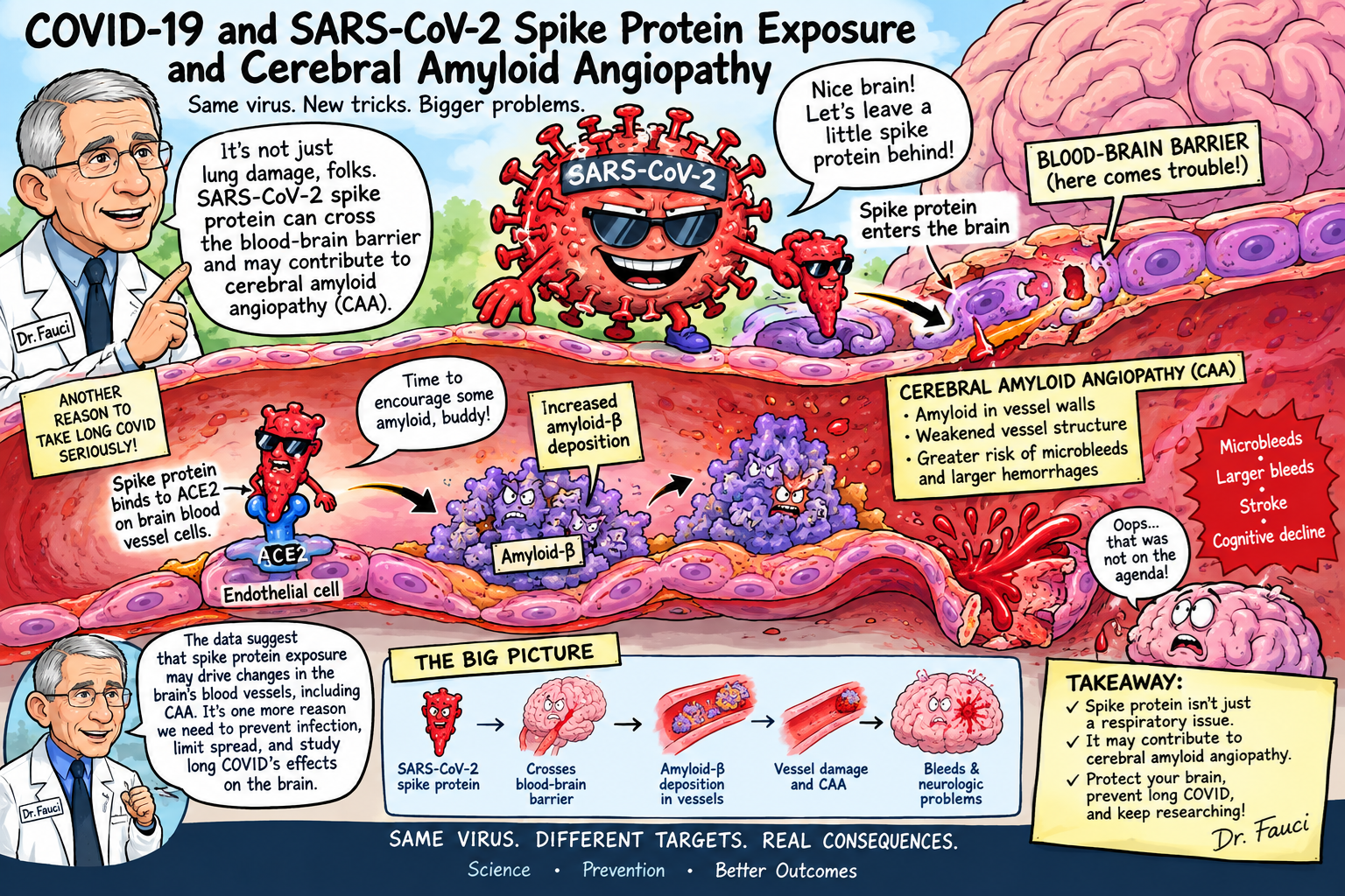
A conceptual pathway can therefore be proposed:
SARS-CoV-2 exposure
↓
Spike-mediated endothelial/pericyte dysfunction
↓
blood-brain barrier disruption
↓
altered Aβ transport and clearance
↓
increased extracellular/perivascular Aβ
↓
Aβ aggregation
↓
vascular amyloid deposition
↓
vascular inflammation and degeneration
↓
microbleeds, superficial siderosis and lobar haemorrhage
This remains a hypothesis, but each of the upstream components has independent experimental support.
6. Cerebrovascular Injury During COVID-19
Another important component is the demonstrated occurrence of cerebral microvascular injury during COVID-19.
Neuropathological studies have identified endothelial abnormalities and cerebral microhaemorrhages in patients with severe COVID-19.[12]
Microbleeds are not synonymous with CAA.
They may result from:
- hypertensive arteriopathy;
- critical illness;
- hypoxia;
- endothelial injury;
- anticoagulation;
- disseminated intravascular coagulation;
- inflammatory vascular injury;
- amyloid angiopathy; or
- combinations of these mechanisms.
However, the observation that SARS-CoV-2 can injure cerebral microvessels establishes biological plausibility for an interaction with pre-existing vascular amyloid.
CAA weakens cerebral vessel walls.
SARS-CoV-2-associated endothelial inflammation could theoretically add a second vascular insult.
The combination could be clinically important even if SARS-CoV-2 does not initiate amyloid deposition de novo.
7. The Most Plausible Model May Be Acceleration Rather Than Initiation
An important conceptual distinction should be made.
The available evidence does not justify the claim:
“SARS-CoV-2 causes cerebral amyloid angiopathy.”
A more scientifically defensible hypothesis is:
SARS-CoV-2 infection or spike-protein exposure may accelerate pre-existing or subclinical cerebral amyloid angiopathy in susceptible individuals.
CAA is strongly age-associated. Therefore, many individuals infected with SARS-CoV-2—particularly younger people—would not be expected to develop CAA simply because of infection.
A susceptible older individual, however, might already have:
- early Aβ vascular deposition;
- impaired perivascular drainage;
- APOE-related vulnerability;
- hypertension;
- endothelial dysfunction;
- reduced BBB integrity.
SARS-CoV-2-associated vascular and inflammatory injury could potentially accelerate that process.
This model reconciles the age dependence of CAA with the relatively recent appearance of SARS-CoV-2.
8. CAA-Related Inflammation Provides Another Potential Connection
CAA-related inflammation is a particularly intriguing potential intersection.
CAA-ri is characterized by inflammatory responses directed toward vascular amyloid. Clinically it can present with headache, seizures, cognitive dysfunction, encephalopathy, focal neurological deficits and MRI abnormalities.
Case reports have described CAA-related inflammatory syndromes following SARS-CoV-2 vaccination.
Rossato and colleagues reported a patient with probable CAA who developed CAA-related inflammation after exposure to tozinameran, with persistent CSF anti-spike antibodies.[13]
A 2024 report subsequently described severe CAA-related inflammation following SARS-CoV-2 vaccination and reviewed previously reported cases.[14]
These reports deserve attention but must be interpreted cautiously.
A temporal association does not establish causation.
Furthermore, vaccination and natural SARS-CoV-2 infection are biologically different exposures and should not be conflated.
Nevertheless, the cases demonstrate that spike-directed immune responses can coexist temporally with inflammatory CAA phenotypes, providing an additional hypothesis for investigation.
9. Spike Protein in Cerebral Arteries: An Emerging Observation
A particularly provocative development appeared in 2025.
Ota and colleagues reported detection of SARS-CoV-2 spike protein in cerebral arteries following mRNA vaccination and described inflammatory-cell infiltration in some spike-positive vessels.[15]
If independently confirmed, this observation would be highly relevant to the CAA hypothesis because it places spike protein directly within the anatomical compartment in which CAA occurs.
However, this finding requires independent replication and careful pathological controls.
A subsequent correspondence critically appraised the study, emphasizing the need for caution in interpreting the findings.[16]
Consequently, the appropriate scientific conclusion is not that vaccine-derived spike protein has been proven to cause CAA.
Rather:
The presence of spike protein in cerebral arterial tissue is a provocative observation that warrants independent neuropathological replication.
10. SARS-CoV-2-Induced Amyloidogenesis: Three Potential Mechanisms
Several mechanisms have been proposed for SARS-CoV-2-associated amyloidogenesis.[17]
Mechanism 1: Increased Aβ production
Viral infection and inflammatory signalling may alter amyloid precursor protein processing and increase Aβ generation.
Mechanism 2: Direct molecular interaction
Spike protein or spike-derived peptides may bind Aβ and influence its aggregation kinetics.
Mechanism 3: Impaired clearance
Endothelial, pericyte and blood-brain barrier dysfunction may impair the movement of Aβ out of the brain.
These mechanisms need not be mutually exclusive.
Indeed, they could reinforce one another.
An increase in Aβ production combined with impaired clearance would increase the available substrate for aggregation. Simultaneous vascular injury could promote retention of Aβ within vessel walls.
11. A Proposed SARS-CoV-2–CAA Pathophysiological Model
The available evidence permits development of the following working model.
Stage 1: Exposure
SARS-CoV-2 infection produces systemic exposure to viral proteins and inflammatory mediators.
Stage 2: Neurovascular exposure
Spike protein or inflammatory mediators interact with endothelial cells, pericytes and components of the blood-brain barrier.
Stage 3: Neurovascular dysfunction
Endothelial activation, altered tight-junction function and pericyte dysfunction increase vascular permeability and disturb normal neurovascular signalling.
Stage 4: Altered amyloid metabolism
Inflammation and cellular stress alter Aβ production, degradation and transport.
Stage 5: Aβ aggregation
Spike-derived peptides and/or altered extracellular conditions may promote Aβ aggregation.
Stage 6: Perivascular accumulation
Aβ becomes increasingly concentrated within perivascular spaces and vessel walls.
Stage 7: Vascular inflammation
Amyloid deposition activates microglia, macrophages and other inflammatory pathways.
Stage 8: Vascular degeneration
The vessel wall becomes structurally compromised.
Stage 9: Clinical disease
The resulting phenotype may include:
- cerebral microbleeds;
- cortical superficial siderosis;
- lobar intracerebral haemorrhage;
- transient focal neurological episodes;
- seizures;
- cognitive impairment;
- encephalopathy; and
- CAA-related inflammation.
The critical point is that SARS-CoV-2 may function as an accelerator or modifier rather than the sole initiating cause.
12. Alternative Explanations
A rigorous BMJ manuscript must consider competing explanations.
12.1 Aging
CAA is predominantly a disease of older adults. Increasing recognition of neurological disease following COVID-19 may therefore coincide with an underlying age-related process.
12.2 APOE genotype
APOE ε4 is associated with both Alzheimer pathology and CAA susceptibility. Genetic susceptibility could therefore confound an apparent association between COVID-19 and CAA.
12.3 Hypertension
Hypertension independently causes cerebral small-vessel disease and intracerebral haemorrhage.
12.4 Critical illness
Severe COVID-19 produces hypoxia, systemic inflammation, coagulation abnormalities and prolonged ICU exposure. These factors can themselves cause cerebral microbleeds.
12.5 Anticoagulation
Anticoagulant exposure in severe COVID-19 may increase haemorrhagic complications.
12.6 Ascertainment bias
Patients recovering from COVID-19 may undergo more neurological imaging, producing increased detection of previously asymptomatic CAA.
12.7 Reverse causation
Pre-existing neurodegenerative or vascular disease may itself influence COVID-19 severity and neurological outcomes.
These confounders make observational epidemiology particularly challenging.
13. Why the Distinction Between Infection and Spike Exposure Matters
The phrase “spike protein exposure” encompasses several biologically different situations:
- natural SARS-CoV-2 infection;
- persistent viral protein after infection;
- vaccination-associated expression of spike antigen;
- circulating spike protein or fragments;
- tissue-localized spike protein;
- experimental administration of purified spike protein; and
- exposure to spike-derived peptides.
These exposures cannot be treated as equivalent.
A study showing that purified spike protein produces Aβ aggregation in vitro does not establish that the same process occurs following vaccination.
Conversely, evidence of spike protein associated with cerebral vascular tissue after vaccination does not demonstrate that infection produces the same phenomenon.
Future research must therefore define exposure precisely.
14. What Evidence Would Establish a Causal Relationship?
The field now requires studies specifically designed to test the CAA hypothesis.
A particularly powerful study would enroll older adults without known CAA and perform baseline:
- susceptibility-weighted MRI;
- T2* imaging;
- assessment for cortical superficial siderosis;
- amyloid PET;
- APOE genotyping;
- plasma Aβ40/Aβ42;
- plasma p-tau;
- neurofilament light;
- GFAP;
- endothelial biomarkers;
- inflammatory biomarkers; and
- detailed cognitive testing.
Participants would then be followed after SARS-CoV-2 infection.
The primary endpoint could be new or progressive strictly lobar cerebral microbleeds or cortical superficial siderosis.
Secondary endpoints could include:
- change in amyloid PET burden;
- plasma Aβ40/Aβ42 ratio;
- CSF Aβ;
- neurofilament light;
- cognitive trajectory;
- incident CAA-ri;
- intracerebral haemorrhage; and
- longitudinal MRI evidence of CAA progression.
The ideal study would compare:
infected versus uninfected individuals
while matching or adjusting for:
- age;
- sex;
- APOE genotype;
- hypertension;
- diabetes;
- anticoagulant use;
- baseline cerebral small-vessel disease;
- prior intracerebral haemorrhage;
- vaccination history; and
- socioeconomic and healthcare-utilization variables.
15. A Particularly Important Study Design
The strongest mechanistic study would combine human neuropathology with molecular detection.
Brain tissue from individuals with:
- confirmed SARS-CoV-2 infection;
- documented vaccination;
- CAA;
- Alzheimer disease;
- COVID-associated neurological disease; and
- age-matched controls
could be examined using:
- immunohistochemistry for spike protein;
- immunohistochemistry for Aβ40 and Aβ42;
- Congo red staining;
- Thioflavin-S staining;
- electron microscopy;
- spatial transcriptomics;
- single-cell RNA sequencing;
- proteomics;
- multiplex immunofluorescence;
- endothelial markers;
- pericyte markers;
- microglial activation markers; and
- assays for viral RNA.
The central question would be:
Are spike-positive vascular structures preferentially associated with Aβ-positive cerebral vessels?
A spatial association would not automatically establish causality, but it would substantially strengthen the biological case.
16. Proposed Experimental Test of Spike–CAA Interaction
An especially informative experiment would use human cerebral microvascular organoids containing endothelial cells, pericytes, astrocytes and extracellular matrix.
Four experimental conditions could be compared:
| Experimental group | Exposure | Primary outcome |
|---|---|---|
| Control | Vehicle | Baseline Aβ deposition |
| Spike | Spike protein | Aβ aggregation |
| SARS-CoV-2 | Replication-competent or appropriately contained experimental system | Aβ aggregation and vascular injury |
| Spike + inflammatory milieu | Spike plus cytokines | Synergistic amyloid/vascular injury |
Measurements should include:
- Aβ40;
- Aβ42;
- Aβ oligomers;
- fibrillar Aβ;
- endothelial permeability;
- tight-junction proteins;
- pericyte viability;
- inflammatory cytokines;
- complement activation;
- oxidative stress;
- mitochondrial function; and
- vascular integrity.
The experiment should also determine whether spike exposure increases Aβ deposition specifically within vascular structures, rather than merely increasing extracellular Aβ.
That distinction is critical.
17. Clinical Implications
If the hypothesis is eventually confirmed, the implications could be substantial.
CAA is clinically important because its principal complications can be catastrophic.
A patient with previously subclinical CAA could theoretically become more vulnerable to:
- lobar intracerebral haemorrhage;
- microbleed progression;
- cortical superficial siderosis;
- seizures;
- cognitive decline; and
- CAA-related inflammation.
The hypothesis may also become relevant when considering therapies that affect haemorrhagic risk.
For example, clinicians increasingly use amyloid-targeting monoclonal antibodies for Alzheimer disease. These therapies can produce amyloid-related imaging abnormalities, particularly in individuals with underlying CAA.
Consequently, understanding whether prior SARS-CoV-2 infection modifies vascular amyloid burden could become clinically relevant to patient selection and MRI surveillance.
At present, however, there is insufficient evidence to recommend routine CAA screening solely because a patient has had COVID-19 or received a COVID-19 vaccine.
18. What the Evidence Does—and Does Not—Show
| Question | Current evidence |
| Can SARS-CoV-2 affect the neurovascular unit? | Yes; substantial experimental and pathological evidence |
| Can spike protein alter BBB biology? | Yes, in experimental systems |
| Can spike interact with Aβ? | Yes, experimentally |
| Can spike-derived peptides promote Aβ aggregation? | Yes, experimentally |
| Can SARS-CoV-2 induce Aβ aggregation in human-derived neural tissue? | Evidence now supports this possibility |
| Can COVID-19 produce cerebral microvascular injury? | Yes |
| Has spike protein been reported in cerebral arterial tissue? | Yes, but findings require independent replication |
| Has SARS-CoV-2 been proven to cause human CAA? | No |
| Has increased incident CAA after COVID-19 been definitively demonstrated? | Not yet |
| Is a SARS-CoV-2–CAA pathway biologically plausible? | Yes |
| Should the hypothesis be tested prospectively? | Strongly yes |
19. Discussion
The emerging evidence changes the scientific question.
The question is no longer whether SARS-CoV-2 can interact experimentally with amyloid biology. Multiple studies now indicate that it can.
The more important question is whether these molecular interactions become clinically meaningful within the human cerebral microvasculature.
Several independent observations converge on the same biological territory:
SARS-CoV-2 → endothelial dysfunction → BBB disruption → altered Aβ metabolism/clearance → Aβ aggregation → neurovascular inflammation → vascular injury.
This convergence is scientifically significant even though the final causal link to CAA has not been established.
The 2025 human-retinal work is particularly important because it demonstrates Aβ aggregation in human-derived neural tissue and shows that spike 1 protein can promote this process.[8]
Similarly, the experimental demonstration that a spike-derived peptide can promote Aβ aggregation provides a plausible molecular mechanism.[5]
The BBB literature supplies another mechanistic link, while studies of cerebral microhaemorrhages provide evidence that the cerebral microvasculature can be damaged during severe COVID-19.[9,12]
The 2025 report of spike protein in cerebral arteries represents an especially provocative observation because it potentially places the antigen directly within the anatomical compartment affected by CAA.[15]
Nevertheless, scientific discipline requires recognition of the missing evidence.
There is currently no adequately powered longitudinal study demonstrating that SARS-CoV-2 infection increases the incidence or progression of neuropathologically confirmed CAA.
Nor is there sufficient evidence to conclude that vaccination-associated spike expression produces CAA.
These questions remain open.
20. A Revised Hypothesis
The evidence therefore supports the following testable hypothesis:
In genetically and biologically susceptible individuals, SARS-CoV-2 infection may accelerate cerebral amyloid angiopathy through a combination of spike–Aβ molecular interactions, endothelial and pericyte dysfunction, blood-brain barrier disruption, impaired perivascular Aβ clearance and neurovascular inflammation.
This hypothesis makes several predictions.
If correct, SARS-CoV-2-exposed individuals with pre-existing susceptibility should demonstrate:
- increased progression of strictly lobar cerebral microbleeds;
- increased cortical superficial siderosis;
- increased amyloid-PET signal;
- altered plasma or CSF Aβ40/Aβ42 ratios;
- greater endothelial dysfunction;
- increased neuroinflammatory biomarkers; and
- increased risk of CAA-related clinical syndromes.
These predictions can be tested.
That is precisely what distinguishes a legitimate scientific hypothesis from speculation.
Conclusion
The scientific literature does not yet justify stating that SARS-CoV-2 spike protein exposure has been proven to cause cerebral amyloid angiopathy.
It does, however, justify a substantially stronger statement than was possible several years ago.
Experimental studies demonstrate that spike-derived sequences can participate in amyloid-like aggregation and interact with Aβ. Other studies demonstrate that spike protein can promote Aβ aggregation. Human-derived retinal models have now demonstrated SARS-CoV-2-associated Aβ deposition. SARS-CoV-2 can disrupt the blood-brain barrier and injure components of the neurovascular unit. COVID-19 has been associated with cerebral endothelial injury and microhaemorrhages. Finally, preliminary neuropathological evidence has reported spike protein in cerebral arterial tissue.
Taken together, these observations form a coherent, biologically plausible chain connecting SARS-CoV-2 exposure with mechanisms capable of promoting cerebrovascular amyloid pathology.
The missing link is direct demonstration of increased human CAA.
That gap should now become a research priority.
The central scientific question should therefore be reframed from:
“Does COVID-19 cause CAA?”
to:
“Does SARS-CoV-2 exposure accelerate cerebral Aβ vascular deposition or progression of pre-existing CAA, and through which molecular mechanisms?”
That question is experimentally tractable.
Given the increasing prevalence of SARS-CoV-2 exposure and the potentially devastating consequences of CAA-associated intracerebral haemorrhage, prospective investigation of this neurovascular amyloid pathway is warranted.
References
1. Charidimou A, Boulouis G, Frosch MP, et al. The Boston criteria version 2.0 for cerebral amyloid angiopathy: a multicentre, retrospective, MRI-neuropathology diagnostic accuracy study. Lancet Neurology. 2022;21:714–725.
2. Charidimou A, et al. Validation of the Boston Criteria version 2.0 for cerebral amyloid angiopathy. Neurology. 2025.
3. Emmi A, et al. Detection of SARS-CoV-2 viral proteins and genomic sequences in the brain of patients with COVID-19. Acta Neuropathologica Communications. 2023.
4. Nystrom S, Hammarström P. Amyloid and hydrogel formation of a peptide sequence from a coronavirus spike protein. ACS Nano. 2022;16:1857–1867.
5. Spike Protein Fragments Promote Alzheimer’s Amyloidogenesis. 2023.
6. Idrees D, Kumar V. SARS-CoV-2 spike protein interactions with amyloidogenic proteins: potential clues to neurodegeneration. International Journal of Biological Macromolecules. 2021.
7. Coupling of SARS-CoV-2 to Aβ amyloid fibrils. 2024.
8. SARS-CoV-2 induces Alzheimer’s disease-related amyloid-β pathology in ex vivo human retinal explants and retinal organoids. 2025.
9. Erickson MA, et al. Interactions of SARS-CoV-2 with the blood-brain barrier. 2021; subsequent experimental studies demonstrate spike-associated BBB dysfunction.
10. Khaddaj-Mallat R, et al. SARS-CoV-2 deregulates the vascular and immune functions of brain pericytes. 2021.
11. Li J, et al. Exploring SARS-CoV-2 impact on blood-brain barrier and its implications for neurological disease. 2025.
12. Kirschenbaum D, et al. Intracerebral endotheliitis and microbleeds are associated with severe COVID-19. 2021.
13. Rossato F, et al. Cerebral amyloid angiopathy-related inflammation after COVID-19 vaccination. 2021.
14. Tang M, et al. Severe cerebral amyloid angiopathy-related inflammation after exposure to SARS-CoV-2 vaccine. 2024.
15. Ota N, et al. Expression of SARS-CoV-2 spike protein in cerebral arteries: implications for hemorrhagic stroke. Journal of Clinical Neuroscience. 2025.
16. Mueed A, et al. Expression of SARS-CoV-2 spike protein in cerebral arteries: critical appraisal. 2025.
17. Gonçalves CA, et al. SARS-CoV-2-induced amyloidgenesis: not one, but three hypotheses for cerebral COVID-19 outcomes. 2022.