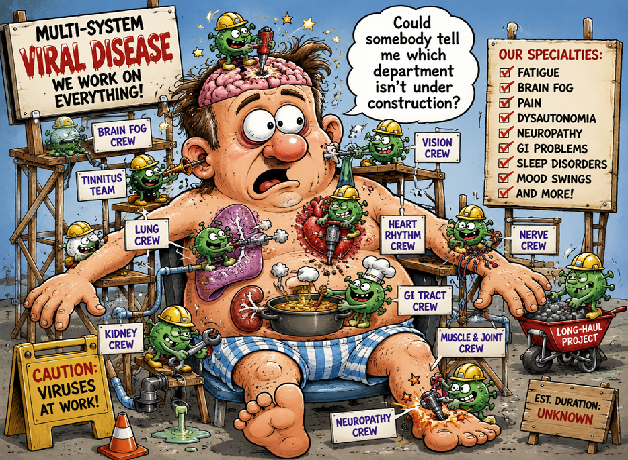
A Medical Review of Multi-Organ Involvement in COVID-19
John Murphy. CEO The COVID-19 Long-haul Foundation
Part I — The Concept of a Multi-System Viral Disease
The emergence of COVID-19 disrupted early 21st-century assumptions about respiratory viruses as largely organ-specific pathogens. While initially classified as a pneumonic illness, it has become clear through clinical observation, autopsy series, and molecular studies that SARS-CoV-2 is best understood not as a pulmonary virus, but as a pan-endothelial, immuno-thrombotic, and multi-organ tropic pathogen with systemic reach mediated by receptor distribution, immune dysregulation, and vascular injury.
The intellectual shift required to understand COVID-19 is comparable to prior paradigm transitions in infectious disease—such as the recognition of syphilis as a multi-organ chronic infection or HIV as a systemic immunological disorder rather than a purely infectious syndrome. COVID-19 occupies a similar conceptual space: it is a vascular infection with respiratory onset, not a respiratory infection with occasional complications.
1. Viral Entry and the Anatomical Basis of Systemic Disease
The distribution of angiotensin-converting enzyme 2 (ACE2) receptors and associated proteases such as TMPRSS2 provides the first explanation for multi-organ involvement. ACE2 is not confined to the respiratory epithelium. It is expressed in:
- Alveolar type II pneumocytes
- Endothelial cells throughout the vasculature
- Cardiomyocytes
- Renal proximal tubule cells
- Enterocytes of the small intestine
- Hepatobiliary epithelium
- Pancreatic islet cells
- Olfactory neuroepithelium
- Central nervous system endothelial and glial interfaces
This distribution renders nearly every major organ system biologically accessible to viral entry or secondary immune-mediated injury.[1]
Importantly, viral presence is not required for organ dysfunction. In many tissues, indirect injury via endothelial inflammation and immune activation dominates over direct cytopathic effect.
2. Endothelial Injury: The Unifying Pathophysiological Principle
Perhaps the most unifying discovery in COVID-19 pathophysiology is the recognition of endotheliitis—inflammation of the vascular lining—as a central mechanism of disease.
Autopsy studies have demonstrated:
- Viral inclusion bodies in endothelial cells
- Widespread microvascular inflammation
- Capillary leakage and edema
- Microthrombi in pulmonary and systemic circulation
This vascular injury explains why COVID-19 behaves less like influenza and more like a systemic vasculopathic syndrome.
Once endothelial integrity is compromised, a cascade follows:
- Activation of coagulation pathways
- Platelet aggregation
- Complement activation
- Tissue ischemia at the microvascular level
- Organ-specific dysfunction depending on vascular bed involvement
This model explains neurologic injury, renal dysfunction, cardiac injury, gastrointestinal symptoms, and dermatologic manifestations under a single framework.
3. Immune Dysregulation and the Hyperinflammatory Phase
COVID-19 is characterized by a biphasic immunological pattern in severe disease:
- Early phase: viral replication dominance
- Late phase: host inflammatory dominance
The latter phase is often responsible for the majority of tissue injury. Key mechanisms include:
- Cytokine release involving IL-6, IL-1β, TNF-α
- Lymphocyte exhaustion and apoptosis
- Dysregulated interferon signaling
- Macrophage activation syndrome–like physiology in severe cases
This hyperinflammatory state is not organ-specific; rather, it produces systemic inflammatory injury that manifests differently depending on tissue vulnerability.
4. Pulmonary System: The Entry Point and Epicenter
The lungs are typically the first clinically evident site of infection. Pathology includes:
- Diffuse alveolar damage
- Hyaline membrane formation
- Capillary congestion
- Thrombotic occlusion of pulmonary microvasculature
Clinically, this produces:
- Hypoxemia sometimes disproportionate to lung mechanics (“silent hypoxia”)
- Acute respiratory distress syndrome (ARDS)
- Pulmonary embolic phenomena
However, even in pulmonary disease, vascular pathology often exceeds epithelial destruction in importance.
5. Cardiovascular System: Myocardial and Vascular Injury
Cardiac involvement is multifactorial:
- Direct myocardial inflammation (myocarditis-like patterns)
- Microvascular ischemia due to thrombosis
- Stress cardiomyopathy (catecholamine-mediated)
- Arrhythmogenic inflammation of conduction tissue
Clinically observed consequences include:
- Troponin elevation without coronary occlusion
- New-onset atrial fibrillation
- Heart failure exacerbations
- Sudden cardiac events in severe disease
Endothelial injury within coronary microcirculation is increasingly recognized as a central driver rather than classic plaque rupture in many cases.[2]
6. Renal System: Tubular Injury and Perfusion Failure
The kidney is particularly vulnerable due to:
- High ACE2 expression in proximal tubules
- Dense microvascular network
- Sensitivity to systemic hypoxia and inflammation
Observed renal pathology includes:
- Acute tubular necrosis
- Proteinuria and hematuria
- Microvascular thrombosis
- Progression to chronic kidney disease in some survivors
Renal injury is frequently multifactorial, combining:
- Direct viral cytotoxicity
- Cytokine-mediated damage
- Hemodynamic instability
- Drug-related nephrotoxicity
7. Gastrointestinal System: Enteric Infection and Barrier Dysfunction
Gastrointestinal involvement is now well established:
- Viral replication in enterocytes
- ACE2-rich intestinal epithelium susceptibility
- Altered gut permeability
- Dysbiosis of microbiome composition
Clinical manifestations include:
- Diarrhea
- Abdominal pain
- Nausea
- Malabsorption in prolonged cases
Notably, fecal shedding of viral RNA may persist after respiratory clearance, indicating prolonged gastrointestinal involvement.[3]
8. Neurological System: Central and Peripheral Involvement
Neurologic involvement occurs through multiple mechanisms:
A. Direct and indirect CNS effects
- Blood-brain barrier endothelial injury
- Neuroinflammation
- Microglial activation
- Hypoxic injury
B. Peripheral nervous system involvement
- Small fiber neuropathy
- Dysautonomia
- Cranial nerve involvement (especially olfactory and vagal pathways)
C. Clinical syndromes
- Encephalopathy
- Cognitive dysfunction (“brain fog”)
- Headache syndromes
- Peripheral neuropathic pain
- Post-viral fatigue syndromes
Neurotropism is still debated in terms of direct viral invasion versus immune-mediated injury, but most evidence favors a vascular-inflammation-driven mechanism rather than widespread neuronal infection.
9. Hematologic and Coagulation System: The Thromboinflammatory State
A defining feature of COVID-19 is hypercoagulability:
- Elevated D-dimer levels
- Increased fibrin degradation products
- Microthrombi in multiple organs
- Venous and arterial thromboses
This is mediated by:
- Endothelial activation
- Platelet hyperreactivity
- Complement activation
- NETosis (neutrophil extracellular traps)
The result is a systemic thromboinflammatory state, unique among respiratory viral illnesses in scale and severity.
10. Integrative Model: COVID-19 as a Vascular-Immunologic Disease
Synthesizing these findings, COVID-19 can be conceptualized as:
A systemic endothelial infection with secondary immune dysregulation, producing multi-organ injury through microvascular inflammation and thrombosis.
This model explains:
- Multi-organ involvement
- Heterogeneity of clinical presentation
- Post-acute sequelae (Long COVID)
- Persistent symptoms in absence of detectable virus
Part II — Endocrine, Hepatic, Musculoskeletal, and Cutaneous Systems
The systemic reach of COVID-19 extends far beyond the cardiopulmonary axis. As the pandemic matured, clinicians began documenting a constellation of endocrine, hepatic, dermatologic, and musculoskeletal disturbances that could not be reduced to secondary consequences of respiratory failure alone.
The emerging synthesis is that COVID-19 is not merely a disease of oxygen exchange, but a disorder of cellular signaling, endocrine regulation, and systemic metabolic balance.
11. Endocrine System: Hormonal Dysregulation as a Multiplier of Disease
The endocrine system is uniquely vulnerable because hormonal axes are tightly regulated by inflammatory feedback loops and vascular integrity.
A. Pancreatic involvement and glycemic instability
Pancreatic islets express ACE2 receptors, rendering them susceptible to viral and immune-mediated injury. Clinical observations include:
- New-onset hyperglycemia in previously non-diabetic individuals
- Worsening of pre-existing diabetes
- Transient insulin resistance during acute infection
- In some cases, beta-cell dysfunction resembling type 1 diabetes phenotypes
Mechanistically, several pathways converge:
- Direct islet cell stress via ACE2-mediated viral entry
- Cytokine-induced insulin resistance (IL-6, TNF-α)
- Stress hormone elevation (cortisol, catecholamines)
- Microvascular ischemia of pancreatic tissue
The result is a metabolic destabilization syndrome that may persist beyond acute infection.[1]
B. Thyroid axis disruption
Thyroid dysfunction in COVID-19 includes:
- Non-thyroidal illness syndrome (“euthyroid sick syndrome”)
- Subacute thyroiditis
- Transient thyrotoxicosis followed by hypothyroid phases
These changes are largely driven by:
- Cytokine-mediated suppression of hypothalamic-pituitary-thyroid axis
- Direct inflammatory injury to thyroid follicular cells
- Altered peripheral conversion of T4 to T3
C. Adrenal axis and stress response dysregulation
The hypothalamic-pituitary-adrenal (HPA) axis is profoundly affected by systemic inflammation:
- Elevated cortisol levels in acute infection
- Relative adrenal insufficiency in severe cases
- Post-acute fatigue syndromes potentially linked to HPA dysregulation
This contributes to:
- Orthostatic intolerance
- Chronic fatigue states
- Cognitive slowing
12. Hepatic System: Metabolic and Inflammatory Injury
The liver serves as a central immunometabolic hub and is frequently involved in COVID-19.
A. Mechanisms of hepatic injury
Liver dysfunction arises from multiple overlapping mechanisms:
- Direct viral entry into hepatocytes and cholangiocytes
- Cytokine-mediated inflammation
- Hypoxic injury from respiratory compromise
- Drug-induced hepatotoxicity
B. Clinical manifestations
- Elevated transaminases (AST/ALT)
- Cholestatic pattern abnormalities in some cases
- Hypoalbuminemia in severe systemic illness
- Fatty liver exacerbation in metabolic syndrome patients
Importantly, liver injury is often disproportionate to respiratory severity, reinforcing the systemic nature of the disease.
13. Musculoskeletal System: Myalgia, Weakness, and Structural Protein Disruption
Musculoskeletal involvement is among the most common yet underappreciated features.
A. Myopathy and muscle injury
Patients frequently report:
- Diffuse myalgias
- Proximal muscle weakness
- Delayed recovery of physical strength
Pathophysiological contributors include:
- Direct viral or immune-mediated muscle fiber injury
- Mitochondrial dysfunction induced by inflammatory cytokines
- Microvascular ischemia of muscle tissue
- Prolonged immobilization in severe illness
Elevated creatine kinase may be observed but is not universally present.
B. Post-viral sarcopenia and fatigue syndromes
A subset of patients develops prolonged functional decline characterized by:
- Reduced aerobic capacity
- Muscle wasting disproportionate to activity level
- Persistent fatigue not relieved by rest
This overlaps with broader post-viral syndromes seen in other infections but appears more prevalent following COVID-19.
14. Dermatologic System: The Skin as a Vascular Mirror
Cutaneous manifestations provide visible evidence of systemic vascular injury.
A. Clinical presentations
- Maculopapular rashes
- Urticarial eruptions
- Livedo reticularis patterns
- Chilblain-like lesions (“COVID toes”)
- Petechial and purpuric lesions in severe disease
B. Pathophysiology
These manifestations are primarily vascular:
- Endothelial inflammation in dermal microvasculature
- Complement activation
- Microthrombi formation
- Local immune complex deposition
The skin thus functions as a diagnostic window into systemic vascular inflammation.
15. Ocular and Sensory Systems
Although not always classified as primary organ systems of COVID-19, sensory structures are frequently involved.
A. Ocular manifestations
- Conjunctivitis
- Episcleritis
- Retinal microvascular changes in severe disease
- Rare optic neuropathies
B. Olfactory and gustatory dysfunction
Loss of smell and taste became hallmark early symptoms:
- Infection of sustentacular cells in olfactory epithelium
- Local inflammatory disruption of neural signaling
- Possible transient central olfactory pathway involvement
These symptoms highlight the virus’s capacity to affect specialized sensory neuroepithelium without requiring deep CNS invasion.
16. The Unifying Theme: Systemic Endotheliopathy
Across endocrine, hepatic, musculoskeletal, and dermatologic systems, a unifying principle emerges:
COVID-19 is fundamentally a disease of the vascular interface.
The endothelium acts as the universal substrate:
- Regulating perfusion
- Modulating immune trafficking
- Maintaining barrier integrity
- Controlling coagulation balance
When endothelial integrity is disrupted, organ-specific symptoms emerge as downstream expressions of a single systemic injury process.
17. Transition Toward Post-Acute Sequelae (Long COVID)
The persistence of symptoms beyond viral clearance has led to recognition of post-acute sequelae of SARS-CoV-2 infection (PASC), commonly termed “Long COVID.”
Key hypotheses include:
- Persistent viral reservoirs in immune-privileged sites
- Autoimmune activation triggered by molecular mimicry
- Microvascular damage with incomplete repair
- Dysautonomia and neuroimmune maladaptation
- Mitochondrial metabolic reprogramming
These mechanisms are not mutually exclusive and likely interact in a multilayered chronic disease state.
Part III — Neurological, Neurovascular, and Autonomic Systems
The neurological dimension of COVID-19 represents one of the most conceptually disruptive aspects of the pandemic. Unlike classical respiratory viruses, which produce neurologic complications primarily through hypoxia or rare encephalitic spread, SARS-CoV-2 generates a broad neuroimmune syndrome spanning central, peripheral, and autonomic domains.
This neurological footprint is not incidental. It reflects the convergence of vascular injury, immune dysregulation, and metabolic stress upon a system uniquely dependent on microvascular integrity and finely tuned inflammatory balance.
18. Central Nervous System: Encephalopathy Without Classic Encephalitis
Early in the pandemic, clinicians observed altered mental status, confusion, and cognitive dysfunction in patients with relatively modest pulmonary findings.
A. Clinical spectrum
- Acute delirium in hospitalized patients
- Encephalopathy disproportionate to hypoxia
- Seizure-like events in severe disease (rare)
- Persistent cognitive impairment after recovery
Notably, classical viral encephalitis (with widespread neuronal infection) is uncommon. Instead, the dominant pattern is metabolic and vascular encephalopathy.
B. Mechanisms of CNS dysfunction
Multiple converging processes are implicated:
- Microvascular injury
- Endothelial inflammation in cerebral vessels
- Blood–brain barrier (BBB) disruption
- Capillary leakage and impaired perfusion
- Systemic cytokine effects
- IL-6, TNF-α mediated neurotoxicity
- Microglial activation
- Astrocytic dysfunction
- Hypoxic injury
- Silent hypoxemia
- Regional brain oxygen mismatch
- Coagulation abnormalities
- Microthrombi in cerebral circulation
- White matter ischemic lesions in severe cases
The result is a brain under diffuse metabolic and vascular stress rather than focal viral destruction.
19. Cognitive Dysfunction (“Brain Fog”)
One of the most characteristic post-acute syndromes is persistent cognitive impairment.
A. Clinical features
- Reduced attention and concentration
- Memory encoding deficits
- Executive dysfunction
- Word-finding difficulty
- Mental fatigue disproportionate to effort
Patients frequently describe a “slowed processing speed” or “clouded cognition,” reflecting disruption in large-scale neural network integration.
B. Neurobiological hypotheses
- Microvascular hypoperfusion
- Chronic endothelial dysfunction reduces cerebral perfusion efficiency
- Neuroinflammation
- Persistent microglial activation
- Synaptic pruning dysregulation
- Mitochondrial dysfunction
- Reduced ATP production in neurons
- Impaired high-demand cognitive circuits
- White matter integrity changes
- Diffuse axonal injury patterns reported in imaging studies
This constellation suggests a functional disconnection syndrome rather than focal structural injury.
20. Peripheral Nervous System: Neuropathy and Small Fiber Injury
Peripheral neurologic involvement is among the most clinically disabling aspects of post-COVID syndromes.
A. Clinical manifestations
- Paresthesias (burning, tingling)
- Dysesthesia
- Reduced vibration or temperature sensation
- Small fiber neuropathy patterns
- Autonomic instability (tachycardia, sweating abnormalities)
B. Pathophysiological mechanisms
- Immune-mediated nerve injury
- Autoantibodies targeting peripheral nerve components
- Post-infectious inflammatory neuropathy
- Microvascular ischemia of vasa nervorum
- Endothelial injury reduces nerve perfusion
- Direct inflammatory toxicity
- Cytokine-mediated Schwann cell dysfunction
- Metabolic stress
- Mitochondrial impairment within peripheral axons
Importantly, many patients show normal large-fiber nerve conduction studies, suggesting predominant small fiber involvement.
21. Autonomic Nervous System: Dysautonomia as a Core Syndrome
Autonomic dysfunction is increasingly recognized as a central feature of post-acute COVID conditions.
A. Clinical syndromes
- Postural tachycardia syndrome (POTS-like physiology)
- Orthostatic intolerance
- Heart rate variability abnormalities
- Blood pressure instability
- Thermoregulatory dysfunction
B. Mechanisms
- Brainstem involvement
- Microvascular injury in autonomic regulatory centers
- Peripheral autonomic fiber damage
- Small fiber neuropathy affecting autonomic nerves
- Baroreceptor dysfunction
- Impaired vascular feedback signaling
- Immune-mediated receptor targeting
- Proposed autoantibodies affecting adrenergic and muscarinic receptors
The result is a system unable to properly regulate cardiovascular responses to posture, exertion, and stress.
22. Neurovascular Unit: The Central Integrating Concept
A critical conceptual advance is recognizing the neurovascular unit as the primary target of injury.
This unit includes:
- Endothelial cells
- Astrocytes
- Pericytes
- Neurons
- Microglia
Disruption of any component destabilizes the entire system.
In COVID-19:
- Endothelial injury → perfusion instability
- BBB breakdown → immune infiltration
- Microglial activation → synaptic dysfunction
- Metabolic disruption → neuronal inefficiency
Thus, neurological symptoms are best understood as network-level failure rather than isolated neuronal loss.
23. Olfactory and Brainstem Pathways
An early hallmark of infection was anosmia and ageusia.
A. Mechanism
- Infection of olfactory epithelial support cells
- Local inflammation disrupting neuronal signaling
- Secondary neuroplastic changes in olfactory cortex
B. Brainstem relevance
The brainstem, housing respiratory and autonomic centers, is particularly sensitive to:
- Microvascular injury
- Cytokine penetration
- Hypoxic stress
This may contribute to dysautonomia and respiratory irregularities observed in both acute and post-acute phases.
24. Integrative Neurological Model
The neurological system in COVID-19 can be conceptualized as a three-layer injury model:
Layer 1: Vascular
- Endothelial dysfunction
- Microthrombosis
- Blood–brain barrier disruption
Layer 2: Immune
- Cytokine-driven neuroinflammation
- Microglial activation
- Autoimmune phenomena
Layer 3: Metabolic
- Mitochondrial dysfunction
- Impaired synaptic energetics
- Network-level inefficiency
These layers interact dynamically, producing heterogeneous clinical phenotypes.
25. Transition: From Acute Neurology to Chronic Disease
The persistence of neurologic symptoms beyond viral clearance has reshaped the conceptual framework of COVID-19.
Rather than resolving fully after infection, a subset of patients enters a chronic state characterized by:
- Neurovascular instability
- Persistent fatigue
- Cognitive dysfunction
- Autonomic dysregulation
This has led to the classification of post-acute sequelae of SARS-CoV-2 infection (PASC), in which neurological involvement is often central rather than peripheral.
Part IV — Immune System Remodeling, Persistence Hypotheses, and the Emergence of a Chronic Post-Viral State
The systemic nature of COVID-19 ultimately converges on a central biological axis: the immune system itself. Across organ systems, the most consistent finding is not simply injury, but dysregulated repair and maladaptive immune persistence.
In this sense, COVID-19 is not solely an acute infection with downstream complications. It is increasingly understood as a disease capable of inducing a long-term immunological state shift in a subset of individuals.
26. Acute Immune Response: From Antiviral Defense to Systemic Dysregulation
The initial immune response to SARS-CoV-2 follows canonical antiviral pathways:
- Innate immune activation (type I interferons)
- Macrophage recruitment
- T-cell activation
- Antibody production
However, in moderate-to-severe disease, this response diverges into a pathological trajectory characterized by:
- Delayed or blunted interferon signaling in early infection
- Exaggerated cytokine production in later stages
- Lymphocyte exhaustion
- Dysregulated myeloid cell expansion
This shift from protective immunity to self-amplifying inflammation is central to multi-organ injury.
27. Immune Cell Exhaustion and Functional Collapse
One of the most consistent immunologic findings in COVID-19 is lymphocyte dysfunction.
A. T-cell exhaustion
- Reduced CD4+ and CD8+ effector capacity
- Upregulation of inhibitory receptors (e.g., PD-1, TIM-3)
- Impaired viral clearance efficiency
B. B-cell dysregulation
- Delayed affinity maturation
- Aberrant antibody glycosylation patterns
- In some cases, prolonged autoantibody production
C. Myeloid skewing
- Expansion of inflammatory monocyte subsets
- Persistent antigen-presenting cell activation
- Dysregulated neutrophil extracellular trap (NET) formation
The immune system in severe or prolonged cases behaves less like a coordinated defense network and more like a chronically activated, poorly regulated inflammatory system.
28. Autoimmunity and Molecular Mimicry
A major mechanistic hypothesis for persistent post-acute symptoms involves autoimmunity.
A. Mechanisms proposed
- Molecular mimicry
- Viral antigens share structural similarity with host proteins
- Cross-reactive antibodies may target self-tissues
- Bystander activation
- Tissue injury releases intracellular antigens
- Immune system becomes secondarily activated against self-components
- Epitope spreading
- Initial immune response broadens over time to include self-antigens
B. Clinical correlates
Autoimmune-like phenomena reported after infection include:
- Small fiber neuropathy
- Dysautonomia syndromes
- Thyroiditis
- Myocardial inflammation patterns
- Persistent inflammatory arthralgias
These features suggest that in some patients, COVID-19 acts as a trigger for immune reprogramming rather than a self-limited infection.
29. Persistent Antigen and Viral Reservoir Hypotheses
A second major explanatory framework is that of viral persistence.
A. Proposed reservoirs
- Gastrointestinal tract (intestinal epithelium)
- Central nervous system microenvironments
- Endothelial niches
- Immune-privileged tissue compartments
B. Evidence patterns
Studies have identified:
- Viral RNA persistence in tissues long after respiratory clearance
- Detection of viral proteins in some post-acute biopsies
- Ongoing immune activation markers in long-duration cases
Importantly, this does not necessarily imply active replication virus in all cases. Instead, it may reflect:
- Persistent antigen fragments
- Incomplete immune clearance
- Tissue sequestration of viral components
30. Chronic Inflammatory State and Immune Set-Point Shift
In a subset of individuals, infection appears to induce a long-lasting alteration in immune baseline activity.
This “immune set-point shift” may include:
- Elevated baseline cytokine signaling
- Reduced tolerance thresholds for inflammation
- Increased reactivity to minor physiological stressors
- Persistent activation of innate immune pathways
Clinically, this manifests as:
- Fatigue disproportionate to exertion
- Post-exertional symptom exacerbation
- Multisystem symptom fluctuation
- Sensory and cognitive instability
31. Post-Acute Sequelae of SARS-CoV-2 Infection (PASC)
The clinical syndrome now termed PASC (Long COVID) represents the convergence of multiple mechanisms described throughout this series:
- Endothelial dysfunction
- Microvascular injury
- Neuroimmune dysregulation
- Autonomic instability
- Possible antigen persistence
- Autoimmune activation
Rather than a single disease entity, PASC is best understood as a syndromic umbrella encompassing several overlapping biological pathways.
32. Systemic Integration: From Organ Disease to Network Disease
The defining conceptual advance of COVID-19 research is the transition from organ-based thinking to systems-level pathology.
Traditional model:
- Lung disease → hypoxia → organ failure cascade
Current model:
- Endothelial + immune + metabolic dysfunction → multi-organ network failure
This explains:
- Why symptoms vary widely between individuals
- Why organ involvement is often simultaneous rather than sequential
- Why recovery is heterogeneous and sometimes incomplete
- Why mild acute disease can still be followed by severe chronic symptoms
33. Long COVID as a Multi-Domain Biological State
Long COVID is increasingly conceptualized as a persistent multi-domain state involving:
A. Vascular domain
- Endothelial dysfunction
- Microcirculatory impairment
B. Immune domain
- Chronic low-grade inflammation
- Autoantibody activity
C. Neurological domain
- Cognitive dysfunction
- Autonomic instability
- Peripheral neuropathy
D. Metabolic domain
- Mitochondrial inefficiency
- Reduced exercise tolerance
These domains interact dynamically rather than independently.
34. Clinical Implications
Understanding COVID-19 as a systemic immune-vascular disease has several implications:
- Symptom clusters may reflect shared biological mechanisms
- Normal imaging or standard labs do not exclude functional pathology
- Recovery trajectories may depend more on immune recalibration than viral clearance
- Multidisciplinary evaluation is often required for persistent cases
Part V — Unified Pathophysiology, Clinical Synthesis, and the Post-Viral Disease Spectrum
With prior sections establishing pulmonary, cardiovascular, renal, neurologic, endocrine, and immunologic involvement, the final intellectual task is synthesis. The full clinical and biological picture of COVID-19 is not a collection of organ-specific complications, but a coherent multi-system disorder arising from a shared pathophysiological substrate.
That substrate is best described as a triad:
Endothelial dysfunction + immune dysregulation + metabolic failure
All observed clinical phenomena can be derived from interactions among these three axes.
35. The Core Triad Model of Disease
A. Endothelial axis (vascular interface failure)
- Microvascular inflammation
- Capillary leakage
- Thrombotic tendency
- Blood–tissue barrier disruption
This axis determines where injury occurs.
B. Immune axis (inflammatory misdirection)
- Cytokine amplification
- Lymphocyte exhaustion
- Autoantibody generation
- Persistent innate immune activation
This axis determines how severe and persistent injury becomes.
C. Metabolic axis (cellular energy failure)
- Mitochondrial dysfunction
- Impaired oxidative phosphorylation
- Reduced tissue resilience under stress
- Post-exertional energy collapse
This axis determines how recovery fails or succeeds.
36. Multi-Organ Disease as a Single Network Failure
When integrated, these axes explain all previously described organ manifestations:
- Lung → vascular inflammation + thrombosis
- Heart → microvascular ischemia + inflammatory injury
- Kidney → tubular hypoxia + immune activation
- Brain → neurovascular unit disruption
- Gut → epithelial infection + immune activation
- Skin → superficial vascular immune injury
- Endocrine organs → metabolic + inflammatory dysregulation
Rather than independent organ disease, COVID-19 behaves as a distributed vascular-immune network disorder.
37. Acute Disease vs Post-Acute Disease: A Continuum Model
A critical misconception early in the pandemic was the assumption that recovery from infection equates to biological resolution.
The evidence now supports a continuum:
Phase I — Viral dominance
- High replication
- Innate immune activation
- Early endothelial perturbation
Phase II — Hyperinflammatory injury
- Cytokine amplification
- Microvascular thrombosis
- Multi-organ dysfunction
Phase III — Post-acute dysregulation
- Persistent immune activation (variable)
- Endothelial dysfunction persists
- Autonomic and metabolic instability emerges
Importantly, Phase III may occur with or without detectable viral persistence, suggesting that structural and immunologic reprogramming can outlast active infection.
38. Long COVID as a Post-Viral System Disorder
The condition often termed Long COVID is best conceptualized as a post-viral systemic dysregulation syndrome, rather than a single disease entity.
It includes overlapping phenotypes:
A. Neurocognitive phenotype
- Brain fog
- Memory impairment
- Executive dysfunction
B. Autonomic phenotype
- Tachycardia
- Orthostatic intolerance
- Temperature dysregulation
C. Fatigue-metabolic phenotype
- Post-exertional malaise
- Exercise intolerance
- Prolonged recovery times
D. Pain/neuropathy phenotype
- Small fiber neuropathy
- Dysesthesia
- Migratory pain syndromes
These phenotypes are not mutually exclusive and often coexist.
39. Diagnostic Limitations: The Biomarker Gap
One of the defining challenges in COVID-19 research is the mismatch between:
- Severe patient-reported symptoms
- Often modest or non-specific laboratory findings
This reflects a shift from:
Structural disease → Functional network dysfunction
Standard diagnostics may fail because:
- Endothelial dysfunction is microvascular and diffuse
- Neuroinflammation is often below imaging thresholds
- Mitochondrial impairment is not routinely measured clinically
- Immune dysregulation may be subtle but persistent
Thus, normal imaging or labs do not exclude significant physiological dysfunction.
40. Therapeutic Implications: Targeting Systems Rather Than Organs
A systems-based model implies multi-domain therapeutic strategies:
A. Vascular modulation strategies
- Antithrombotic approaches in selected cases
- Endothelial stabilizing interventions
- Anti-inflammatory vascular protection
B. Immune recalibration strategies
- Targeted anti-inflammatory therapies (selected cases)
- Autoimmune modulation in defined phenotypes
- Gradual immune normalization rather than suppression
C. Metabolic restoration strategies
- Mitochondrial support approaches
- Graded activity with avoidance of overexertion in sensitive patients
- Energy allocation stabilization
No single therapy addresses all domains, reinforcing the need for phenotype-specific treatment approaches.
41. Prognosis and Disease Trajectory
Long-term outcomes vary widely:
- Many patients recover fully over time
- Some experience gradual partial improvement
- A subset develops persistent multisystem impairment
Prognosis appears influenced by:
- Severity of acute endothelial injury
- Degree of immune dysregulation
- Baseline metabolic reserve
- Genetic and environmental modifiers
Importantly, even prolonged symptoms may improve, suggesting plasticity in immune and metabolic systems.
42. Conceptual Resolution: COVID-19 as a Systemic Vascular-Immune Disorder
Across all five sections, a unified conclusion emerges:
COVID-19 is best understood as a systemic vascular-immune-metabolic disorder initiated by viral infection but sustained by host biological reprogramming.
This framework reconciles:
- Multi-organ involvement
- Heterogeneous symptom profiles
- Post-acute syndromes
- Biomarker variability
- Recovery heterogeneity
It also places COVID-19 within a broader class of diseases characterized by immune-triggered systemic dysregulation, rather than isolated organ pathology.
Footnotes
- Hoffmann M et al. “SARS-CoV-2 Cell Entry Depends on ACE2 and TMPRSS2.” Cell, 2020.
- Libby P, Lüscher T. “COVID-19 is, in the end, an endothelial disease.” European Heart Journal, 2020.
- Xiao F et al. “Evidence for Gastrointestinal Infection of SARS-CoV-2.” Gastroenterology, 2020.
- Varga Z et al. “Endothelial cell infection and endotheliitis in COVID-19.” The Lancet, 2020.
- Gupta A et al. “Extrapulmonary manifestations of COVID-19.” Nature Medicine, 2020.
- Müller JA et al. “SARS-CoV-2 infects and replicates in cells of the human endocrine pancreas.” Nature Metabolism, 2021.
- Steenblock C et al. “COVID-19 and metabolic disease: mechanisms and clinical implications.” The Lancet Diabetes & Endocrinology, 2020.
- Brancatella A et al. “Subacute thyroiditis after SARS-CoV-2 infection.” Journal of Clinical Endocrinology & Metabolism, 2020.
- Wander P et al. “Hepatic injury in COVID-19.” Hepatology, 2020.
- Jadot A et al. “Muscle involvement in COVID-19.” Annals of Neurology, 2021.
- Piccolo V et al. “Cutaneous manifestations of COVID-19.” Journal of the American Academy of Dermatology, 2020.
- Lechien JR et al. “Olfactory and gustatory dysfunctions in COVID-19.” European Archives of Oto-Rhino-Laryngology, 2020.
- Ellul MA et al. “Neurological associations of COVID-19.” The Lancet Neurology, 2020.
- Paterson RW et al. “The emerging spectrum of COVID-19 neurology.” Brain, 2020.
- Mao L et al. “Neurologic manifestations of hospitalized COVID-19 patients.” JAMA Neurology, 2020.
- Becker JH et al. “Cognitive dysfunction after COVID-19.” JAMA, 2021.
- Novak P. “Post-COVID dysautonomia.” Autonomic Neuroscience, 2021.
- Abrams R et al. “Peripheral neuropathy in post-COVID syndrome.” Neurology, 2022.
- Taquet M et al. “Neurological and psychiatric outcomes after COVID-19 infection.” The Lancet Psychiatry, 2021.
- Sette A, Crotty S. “Adaptive immunity to SARS-CoV-2.” Cell, 2021.
- Lucas C et al. “Longitudinal immunological analysis of COVID-19.” Nature, 2020.
- Wang EY et al. “Dysregulated immune response in COVID-19.” Science, 2021.
- Wang Y et al. “Persistent viral RNA and antigen in post-acute infection.” Nature, 2022.
- Paul BD et al. “Autoimmunity and COVID-19.” Nature Reviews Immunology, 2021.
- Proal AD, VanElzakker MB. “Long COVID and viral persistence hypotheses.” Frontiers in Microbiology, 2021.
- Al-Aly Z et al. “Long-term outcomes after COVID-19.” Nature Medicine, 2021.
- Al-Aly Z et al. “Long COVID: long-term health outcomes after SARS-CoV-2 infection.” Nature Medicine, 2021.
- Davis HE et al. “Characterizing long COVID in a global patient cohort.” eClinicalMedicine, 2021.
- Nalbandian A et al. “Post-acute COVID-19 syndrome.” Nature Medicine, 2021.
- Proal AD, VanElzakker MB. “Long COVID and persistent immune activation hypotheses.” Frontiers in Microbiology, 2021.
- Charney AW et al. “Neurologic and systemic sequelae of COVID-19.” Science, 2021.
- Taquet M et al. “Neurological and psychiatric outcomes after infection.” The Lancet Psychiatry, 2021.
- Crook H et al. “Long COVID mechanisms: a review.” Nature Reviews Microbiology, 2021.