Abstract
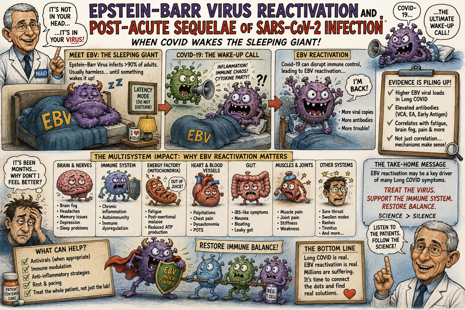
Post-acute sequelae of SARS-CoV-2 infection (PASC), commonly termed Long COVID, encompasses a heterogeneous group of persistent, recurrent, or newly emergent clinical syndromes following acute COVID-19. Although its pathogenesis remains incompletely resolved, convergent evidence implicates several interacting processes, including persistence of SARS-CoV-2 material, immune dysregulation, endothelial and microvascular abnormalities, autonomic dysfunction, altered metabolism, autoimmunity, microbiome perturbation, and reactivation of latent viruses. Among the latter, Epstein–Barr virus (EBV), a ubiquitous γ-herpesvirus capable of lifelong latency in B lymphocytes, has attracted particular attention.
A prospective observational study by Peluso and colleagues provides important evidence that the relationship between EBV and Long COVID is more nuanced than a simple model of persistent EBV viremia. In a cohort of 280 adults followed after SARS-CoV-2 infection, serologic evidence consistent with recent EBV reactivation was independently associated with fatigue and neurocognitive symptoms approximately four months after acute infection, whereas circulating EBV DNA was not associated with these outcomes.¹ This finding is consistent with a model in which transient EBV reactivation during or shortly after acute SARS-CoV-2 infection initiates or amplifies a durable host immune state that may persist after detectable viral replication has subsided.
Subsequent multi-omic studies have strengthened the biological plausibility of this model. Immune profiling has identified altered myeloid and lymphoid compartments and increased antibody responses to latent herpesviruses in patients with Long COVID,² while the large longitudinal IMPACC study, published in Nature in 2026, demonstrated transcriptionally active reactivation of EBV, cytomegalovirus, herpes simplex virus and Anelloviridae during COVID-19, with virus-specific temporal trajectories and associations with inflammatory, transcriptomic and metabolic abnormalities.³ These observations do not establish that EBV causes Long COVID. Rather, they support a testable hypothesis in which SARS-CoV-2 disrupts the host–EBV equilibrium and EBV reactivation acts as a biological amplifier in a subset of susceptible individuals.
The evidence suggests that EBV-associated PASC should be investigated as a potential molecular endotype rather than as a universal explanation for Long COVID. Establishing causality will require prospective studies demonstrating temporal precedence, tissue-specific viral activity, mechanistic linkage to defined symptom clusters, and—most decisively—improvement after selective suppression of EBV-associated biology.
Introduction
Long COVID represents one of the most consequential unresolved problems in postinfectious medicine. The syndrome is characterized not by a single stereotyped illness but by a heterogeneous collection of clinical phenotypes involving the nervous, cardiovascular, pulmonary, gastrointestinal, musculoskeletal, endocrine and autonomic systems. Fatigue, post-exertional malaise, cognitive dysfunction, orthostatic intolerance, dyspnea, sleep disturbance, sensory abnormalities and pain are among its most frequently reported manifestations. Contemporary immunologic studies increasingly suggest that Long COVID comprises multiple biological endotypes rather than one disease mechanism.²,⁴
The etiological problem is consequently not merely to identify a single pathogen but to understand how an acute SARS-CoV-2 infection can produce persistent biological dysfunction after the acute infection has apparently resolved.
Several mechanisms have emerged. SARS-CoV-2 RNA, protein or antigen may persist in selected tissues; immune-cell populations may remain abnormal; autoreactive antibodies may arise; endothelial and platelet physiology may be disturbed; metabolic pathways may become dysregulated; and alterations of the intestinal microbiome may sustain systemic inflammation.⁴⁻⁶ Latent-virus reactivation represents another potentially important component of this network.
EBV is particularly compelling because it combines three properties relevant to postviral disease:
- near-universal prevalence in adults;
- lifelong persistence within the host; and
- the capacity to transition between tightly controlled latency and productive lytic replication.
The central question is therefore not whether an individual has EBV. Most adults do.
The relevant question is whether SARS-CoV-2 infection perturbs EBV latency sufficiently to alter subsequent host physiology.
The evidence increasingly suggests that, in some patients, it may.
I. Epstein–Barr Virus Biology
EBV, formally human herpesvirus 4, is a double-stranded DNA γ-herpesvirus with a genome of approximately 172 kilobases. It establishes lifelong infection after primary exposure and is maintained principally within B-cell populations.⁷
The biology of EBV is fundamentally different from that of an acute respiratory virus.
Following primary infection, EBV enters a latent state characterized by restricted viral gene expression and persistence of the viral episome within host cells. Latency is not biologically inert. Different latency programs involve distinct combinations of EBV nuclear antigens, latent membrane proteins, noncoding RNAs and microRNAs.
The virus can subsequently enter the lytic cycle.
During lytic activation, immediate-early transcription factors initiate a transcriptional cascade leading to viral DNA replication, expression of early viral proteins, structural protein synthesis and production of infectious virions.⁷,⁸
Host immunity ordinarily suppresses this transition.
Consequently, EBV latency represents a dynamic equilibrium:
latent viral genome + infected B cell + antiviral immune surveillance = controlled persistence.
Long COVID raises the possibility that SARS-CoV-2 can disturb this equilibrium.
II. The Peluso Study: The Critical Observation
The most directly relevant study to the present hypothesis was conducted by Peluso and colleagues in a prospective cohort of 280 adults with previous SARS-CoV-2 infection.¹
Participants were evaluated at approximately four months after acute infection.
The investigators examined:
- EBV serologic markers;
- circulating EBV DNA;
- SARS-CoV-2 immune responses;
- clinical symptoms;
- demographic variables;
- comorbidities;
- and other potential confounders.
The principal observation was striking.
Symptoms of Long COVID—particularly fatigue and neurocognitive dysfunction—were associated with serologic evidence suggesting recent EBV reactivation.
By contrast, the presence of circulating EBV DNA was not associated with the same outcomes.¹
This distinction is crucial.
It implies that the biological event potentially relevant to Long COVID may not be persistent systemic EBV replication.
Instead, the clinically important event may be:
transient EBV reactivation → durable immune or tissue consequences → subsequent clinical disease.
The distinction resembles other postinfectious disorders in which the initiating biological event is temporally separated from the mechanism sustaining symptoms.
III. What Constitutes EBV Reactivation?
Interpretation of EBV studies requires considerable caution because “reactivation” is not a single laboratory phenomenon.
Several measurements can be used.
EBV DNAemia
Detection of EBV DNA in blood suggests viral activity but does not necessarily indicate tissue-level productive replication.
EBV Early Antigen
Antibodies against early-antigen components can suggest recent or renewed viral activity.
EBV viral transcripts
RNA transcripts provide more direct evidence that specific viral genes are being expressed.
EBV proteins
Detection of viral proteins provides another level of biological evidence.
Viral load
Quantitative measurements of viral DNA can help characterize the magnitude and duration of replication.
These biomarkers should not be considered interchangeable.
The Peluso study is especially important because it found associations with serologic evidence of recent EBV reactivation without persistent EBV viremia.¹
Thus, a negative EBV-DNA test months later does not necessarily negate the possibility that EBV participated in the disease process earlier.
IV. Temporal Causality
Temporal sequencing is one of the most important problems in determining whether EBV contributes to Long COVID.
There are three possible sequences.
Model 1: EBV precedes Long COVID
SARS-CoV-2 infection induces EBV reactivation.
EBV activity then contributes to persistent symptoms.
This is the strongest causal model.
Model 2: Long COVID induces EBV reactivation
The physiological stress and immune abnormalities of PASC reactivate EBV secondarily.
Here EBV would be a consequence rather than a cause.
Model 3: A common mechanism produces both
SARS-CoV-2 produces persistent immune dysregulation.
That state simultaneously produces Long COVID symptoms and EBV reactivation.
EBV would therefore be a biomarker of the underlying disease rather than its primary driver.
The Peluso study supports the possibility of Model 1 but cannot definitively distinguish it from Models 2 and 3.¹
The newer IMPACC data are important because they provide longitudinal evidence that EBV transcripts can occur early in acute COVID-19 and subsequently decline, while other persistent viruses show different temporal patterns.³
That observation strengthens the temporal argument but still does not establish causation.
V. SARS-CoV-2 as a Perturbation of EBV Latency
How could SARS-CoV-2 provoke EBV reactivation?
The answer probably involves several overlapping pathways.
SARS-CoV-2 produces profound changes in:
- interferon signaling;
- cytokine networks;
- lymphocyte populations;
- B-cell activity;
- T-cell function;
- metabolic physiology;
- cellular stress responses;
- and tissue inflammation.
These pathways are precisely those that participate in maintenance of herpesvirus latency.
A plausible sequence is therefore:
SARS-CoV-2 infection
↓
innate immune dysregulation
↓
altered interferon and cytokine signaling
↓
B-cell/T-cell perturbation
↓
loss of equilibrium controlling EBV
↓
EBV transcriptional activation
↓
secondary immune activation
↓
persistent host dysfunction.
This model does not require continuous EBV replication.
A transient period of viral activity could potentially initiate a much longer-lived immunologic state.
VI. The Molecular Biology of Reactivation
EBV latency is maintained through extensive epigenetic and transcriptional control.
Transition into the lytic cycle involves activation of viral immediate-early transcription factors, particularly BZLF1 and BRLF1.
The resulting transcriptional cascade reorganizes viral chromatin, activates early viral genes, and initiates DNA replication.⁷,⁸
Host cellular physiology participates in this transition.
B-cell differentiation, endoplasmic-reticulum stress, unfolded-protein responses and transcriptional changes can favor EBV lytic activation.
Consequently, systemic inflammatory stress induced by SARS-CoV-2 may affect EBV through several convergent molecular pathways.
The important point is that reactivation need not require profound immunodeficiency.
Indeed, the 2026 IMPACC study found frequent transcriptional evidence of chronic-virus reactivation in hospitalized patients and emphasized that such reactivation occurred in patients who were not conventionally immunosuppressed.³
VII. Immunological Consequences
EBV reactivation could alter the immune system in at least four ways.
1. Antigenic stimulation
New EBV proteins become targets for T-cell and antibody responses.
2. B-cell perturbation
Because EBV resides principally within B cells, reactivation can alter B-cell biology directly.
3. Cytokine amplification
EBV-associated immune responses may increase inflammatory cytokine production.
4. Immune-cell redistribution
The host may expand activated T-cell populations and alter the relative proportions of circulating immune subsets.
The 2026 IMPACC study found associations between viral reactivation and increased inflammatory mediators including IL-6, IL-10, CXCL10 and CXCL11, as well as changes in activated CD4+ and CD8+ T cells.³
These observations provide biological support for the hypothesis that viral reactivation is not merely an inert laboratory finding.
VIII. IL-10 and the EBV–Immune Interface
IL-10 is particularly interesting.
EBV encodes a viral homolog of IL-10, commonly termed viral IL-10, which can modulate immune responses.⁹
The relationship between EBV and human IL-10 is complex and context-dependent.
The 2026 IMPACC analysis found increased IL-10 associated with EBV and CMV reactivation.³
This raises the possibility that EBV activity could modify the balance between inflammatory activation and immune regulation.
The paradox is important.
A virus can simultaneously provoke an immune response and manipulate the immune system designed to eliminate it.
Such a mechanism could theoretically contribute to a state in which inflammation persists while antiviral control becomes incomplete.
IX. B-Cell Biology and Autoimmunity
The connection between EBV and autoimmunity predates COVID-19 by decades.
EBV has been investigated extensively in relation to systemic autoimmune diseases, particularly multiple sclerosis and systemic lupus erythematosus.¹⁰,¹¹
The mechanisms remain incompletely defined but include:
- molecular mimicry;
- aberrant B-cell activation;
- epitope spreading;
- altered antigen presentation;
- cytokine modulation;
- and survival of autoreactive B-cell clones.
SARS-CoV-2 infection could therefore create a particularly favorable environment for these mechanisms.
A conceptual sequence would be:
SARS-CoV-2 inflammation
↓
EBV reactivation
↓
B-cell activation
↓
expansion or activation of autoreactive clones
↓
autoantibody formation
↓
persistent tissue inflammation.
This remains a hypothesis.
Importantly, some carefully controlled Long-COVID studies have not found a universal autoantibody signature, suggesting that autoimmunity is likely an endotype rather than a defining feature of every case.²
X. T-Cell Dysfunction
T-cell biology provides another possible bridge.
Long COVID has been associated with abnormalities in both CD4+ and CD8+ T-cell compartments.
Studies have reported evidence of activated and exhausted T-cell phenotypes, altered trafficking potential and abnormalities in SARS-CoV-2-specific cellular responses.²,¹²
EBV reactivation would impose an additional antigenic burden on the same immune system.
A patient therefore could experience simultaneous stimulation by:
SARS-CoV-2 antigens + EBV antigens + damaged self-antigens.
The immune system would then have to discriminate among three overlapping antigenic environments.
Such persistent antigenic complexity could contribute to the disordered adaptive immune response observed in some Long-COVID cohorts.
XI. Genomics
The interaction between EBV and SARS-CoV-2 is ultimately a problem in three genomes:
- the SARS-CoV-2 genome;
- the EBV genome;
- the human genome.
Host genetic variation may determine susceptibility to viral reactivation through differences in:
- HLA antigen presentation;
- interferon signaling;
- innate viral sensing;
- T-cell activation;
- B-cell regulation;
- NK-cell function;
- and cytokine signaling.
The human HLA system is particularly important because effective control of both SARS-CoV-2 and EBV requires presentation of viral peptides to T cells.
A plausible explanation for interindividual susceptibility is therefore:
host genotype → antiviral immune efficiency → probability of EBV reactivation → probability of a PASC phenotype.
Future studies should integrate host whole-genome sequencing with HLA typing and longitudinal EBV sequencing.
XII. EBV Genomics
EBV itself is genetically heterogeneous.
Different EBV strains exhibit variation in genes encoding:
- latent proteins;
- lytic proteins;
- immune-modulatory molecules;
- microRNAs;
- and regulatory elements.
It is therefore possible that particular viral strains are more capable of reactivation after SARS-CoV-2 infection.
The hypothesis has not been adequately tested.
A sufficiently large study could sequence EBV in patients with and without Long COVID and determine whether specific viral variants, haplotypes or mutational signatures correlate with:
- reactivation;
- viral load;
- symptom phenotype;
- neurocognitive disease;
- fatigue;
- autonomic dysfunction;
- or persistence.
Such an analysis would represent an important step beyond serologic epidemiology.
XIII. Epigenetics
EBV latency is profoundly dependent on epigenetic regulation.
The viral episome undergoes chromatinization, and methylation patterns influence whether latent or lytic transcriptional programs dominate.
Host inflammation can modify epigenetic machinery.
Thus SARS-CoV-2 could theoretically affect EBV reactivation without directly infecting the EBV reservoir.
This possibility is particularly attractive because it explains how a respiratory RNA virus could activate a latent DNA virus residing in B cells.
The interaction would occur through host-cell physiology rather than direct viral coinfection of the same cell.
XIV. Mitochondrial and Metabolic Physiology
Persistent fatigue and post-exertional malaise have generated substantial interest in cellular bioenergetics.
EBV replication is metabolically demanding.
Viral DNA synthesis, protein production and assembly require substantial host resources.
Conversely, metabolic stress may facilitate viral reactivation.
The relationship may therefore be bidirectional:
metabolic stress → EBV reactivation → further metabolic demand.
The 2026 IMPACC study strengthens the plausibility of this concept by identifying metabolomic changes associated with chronic viral reactivation, including abnormalities in amino-acid and lipid metabolism.³
This does not demonstrate mitochondrial disease caused by EBV.
It suggests that viral reactivation occurs within a broader metabolic disturbance.
XV. Oxidative Stress
Reactive oxygen species have been implicated in both viral replication and host inflammatory signaling.
Viral infection can increase oxidative stress through:
- mitochondrial dysfunction;
- altered electron transport;
- inflammatory-cell activation;
- and metabolic reprogramming.
Conversely, oxidative stress can influence cellular signaling pathways involved in viral latency.
A potential feedback loop therefore exists:
SARS-CoV-2 → oxidative stress → EBV activation → inflammation → additional oxidative stress.
Such a loop could provide one mechanism by which an acute infection becomes a persistent physiological disorder.
XVI. Endothelial and Microvascular Effects
Although EBV is not primarily an endothelial virus, its immunologic consequences could influence the vascular system indirectly.
Long COVID has been associated in several studies with:
- endothelial activation;
- altered vascular reactivity;
- platelet abnormalities;
- microvascular dysfunction;
- and coagulation disturbances.⁴,¹³
The 2026 IMPACC study identified associations between viral reactivation and host transcriptional programs involving platelet activation and neutrophil-related pathways.³
A mechanistic chain can therefore be hypothesized:
EBV reactivation
↓
immune activation
↓
cytokine and platelet signaling
↓
endothelial activation
↓
microvascular dysfunction
↓
impaired tissue oxygen and substrate delivery.
Again, this is a mechanistic hypothesis rather than an established causal pathway.
XVII. Neurological Disease
The association of EBV-reactivation markers with fatigue and neurocognitive dysfunction is among the most clinically intriguing findings in the Peluso cohort.¹
The nervous system could be affected indirectly through:
- systemic cytokine signaling;
- endothelial dysfunction;
- altered cerebral perfusion;
- blood–brain-barrier perturbation;
- autonomic dysfunction;
- microglial activation;
- and immune-mediated neural injury.
Direct EBV involvement of the nervous system is also biologically possible, although it should not be presumed in ordinary Long COVID.
The key question is whether a subset of patients with neurocognitive Long COVID exhibits evidence of a persistent neuroimmune phenotype initiated by EBV reactivation.
That hypothesis requires tissue-specific investigation.
XVIII. Fatigue and Post-Exertional Malaise
Fatigue was the symptom most strongly associated with serologic evidence of recent EBV reactivation in the Peluso study.¹
This is biologically notable because profound fatigue is a cardinal feature of both Long COVID and other postinfectious syndromes, including myalgic encephalomyelitis/chronic fatigue syndrome.
Several pathways could converge:
EBV reactivation
→ immune activation
→ cytokine signaling
→ autonomic disturbance
→ metabolic dysfunction
→ impaired exertional physiology
→ fatigue/post-exertional malaise.
The hypothesis should not be reduced to “EBV causes fatigue.”
Fatigue is a final common phenotype generated by many mechanisms.
The important proposition is that EBV may identify a biologically distinct pathway producing the phenotype in some patients.
XIX. Clinical Course
The clinical course of EBV-associated PASC can be conceptualized in four stages.
Stage 1: Acute SARS-CoV-2 infection
The virus produces systemic inflammatory and metabolic stress.
Stage 2: EBV perturbation
Latent EBV transitions toward increased transcriptional or lytic activity.
Stage 3: Immune amplification
EBV antigens, host cytokines and SARS-CoV-2 antigens interact to produce a persistent immune disturbance.
Stage 4: Clinical PASC
The patient develops one or more persistent phenotypes:
- fatigue;
- cognitive dysfunction;
- sleep disturbance;
- dysautonomia;
- pain;
- sensory abnormalities;
- exertional intolerance;
- or multisystem disease.
The critical possibility is that Stage 2 may be transient.
Consequently, by the time the patient presents months later, EBV DNA may be undetectable even though the earlier reactivation contributed to initiating the pathological cascade.
This provides a coherent explanation for the Peluso finding that serologic evidence of recent reactivation was associated with Long-COVID symptoms whereas contemporaneous EBV viremia was not.¹
XX. The 2026 Multi-Omic Evidence
The most important recent development is the 2026 IMPACC investigation of 1,154 hospitalized patients.³
Unlike earlier studies relying primarily upon antibody titers, this investigation used viral transcriptomics and longitudinal multi-omic measurements.
It detected EBV transcripts as well as transcripts from CMV, HSV-1, HSV-2, HHV-6 and Anelloviridae.
Importantly, these viruses exhibited different temporal trajectories.
EBV activity was most prominent early after hospitalization, whereas other viruses displayed later or more persistent patterns.³
This finding argues against a simplistic interpretation of herpesvirus reactivation as merely a nonspecific marker of critical illness.
Instead, the host appears to experience a temporally structured disturbance of the endogenous virome.
XXI. Dysvirosis Rather Than Isolated EBV Reactivation
The 2026 study introduces a broader concept: dysvirosis—disruption of the normal equilibrium among persistent viruses and their host.³
This concept may ultimately prove more important than EBV alone.
A patient may have:
SARS-CoV-2 persistence + EBV reactivation + immune dysregulation
while another may have:
SARS-CoV-2 persistence + CMV reactivation + vascular dysfunction.
A third may have no demonstrable herpesvirus activity but exhibit:
persistent SARS-CoV-2 antigen + autoimmunity + autonomic dysfunction.
Such heterogeneity would explain why empirical antiviral or immunomodulatory treatment has produced inconsistent results across unselected Long-COVID populations.
XXII. A Unified Etiological Model
The evidence can now be assembled into a testable model.
Initiation
SARS-CoV-2 infection
↓
Primary perturbation
interferon/cytokine disruption + tissue injury + immune-cell dysfunction
↓
Viral ecological disturbance
EBV reactivation
↓
Secondary immune stimulation
B-cell activation + T-cell activation + cytokine production
↓
Systemic amplification
endothelial dysfunction + metabolic disturbance + autonomic dysfunction
↓
Organ-specific manifestations
brain + skeletal muscle + cardiovascular system + gastrointestinal tract
↓
Clinical Long COVID.
This model accommodates the fact that EBV is unlikely to explain every case.
It proposes instead that EBV is a secondary amplifier within a subset of mechanistically heterogeneous PASC.
XXIII. Differential Interpretation of the Evidence
The evidence supporting EBV involvement can be ranked according to inferential strength.
Strong evidence
EBV can reactivate during SARS-CoV-2 infection.
EBV-reactivation markers are associated with some Long-COVID phenotypes.
Transcriptionally active EBV has been demonstrated during acute COVID-19.
Moderate evidence
EBV reactivation may occur early enough to contribute to subsequent disease.
EBV-associated immune and metabolic abnormalities may amplify inflammation.
Uncertain
EBV is a primary cause of Long COVID.
EBV persists in tissues responsible for particular Long-COVID symptoms.
EBV eradication improves Long COVID.
The distinction between these categories is essential for rigorous clinical science.
XXIV. Why Antiviral Treatment Is Not Yet Established
A logical therapeutic inference would be to suppress EBV.
Unfortunately, the biology is more complicated.
There is no currently established standard antiviral regimen that reliably eradicates latent EBV.
Drugs such as acyclovir and related nucleoside analogues primarily inhibit productive herpesvirus DNA replication; they do not eliminate the latent EBV reservoir.
Therefore, even if EBV reactivation contributes to Long COVID, conventional antiviral therapy may have limited effects unless active lytic replication is present.
The therapeutic target might instead need to be:
- lytic EBV replication;
- B-cell activation;
- specific immune pathways;
- or the downstream inflammatory consequences of reactivation.
This makes biomarker-guided clinical trials essential.
XXV. The Definitive Causality Experiment
The field now has a clear experimental pathway.
A prospective study should enroll patients shortly after SARS-CoV-2 infection and measure:
- EBV DNA;
- EBV RNA transcripts;
- EBV early-antigen antibodies;
- EBV-specific T-cell responses;
- host transcriptomics;
- cytokines;
- metabolomics;
- autonomic physiology;
- endothelial biomarkers;
- and longitudinal symptoms.
Participants should then be followed for at least 12 months.
Patients demonstrating objective EBV reactivation should be randomized to an intervention capable of suppressing productive EBV replication or otherwise interrupting EBV-associated biology.
The critical endpoints should include:
- fatigue;
- post-exertional malaise;
- cognitive performance;
- autonomic function;
- exercise physiology;
- inflammatory biomarkers;
- EBV transcription;
- and quality-of-life measures.
The decisive result would be:
viral suppression → biological normalization → clinical improvement.
That would provide substantially stronger evidence for causality than serologic association.
XXVI. Implications for Precision Medicine
The emerging evidence suggests that the future of Long-COVID medicine may depend upon biological stratification.
Instead of treating:
“Long COVID”
as one disease, clinicians may eventually diagnose molecular endotypes such as:
- SARS-CoV-2-persistence-associated PASC;
- EBV-reactivation-associated PASC;
- CMV-associated PASC;
- autoimmune PASC;
- endothelial/immunothrombotic PASC;
- autonomic PASC;
- metabolic PASC;
- or mixed phenotypes.
Such classification could transform therapeutic development.
A trial of an EBV-directed therapy should not necessarily enroll every patient with Long COVID.
It should enroll patients with objective evidence of biologically relevant EBV activity.
That is the fundamental lesson of precision medicine.
XXVII. Outstanding Questions
Several questions now require urgent investigation.
First: Does EBV reactivation precede the onset of Long COVID?
Second: Which EBV biomarkers best identify clinically relevant reactivation?
Third: Does tissue EBV activity persist after blood markers disappear?
Fourth: Are particular EBV genotypes associated with PASC?
Fifth: Do particular human HLA or immune-regulatory genotypes predispose to EBV-associated PASC?
Sixth: Does EBV reactivation correlate with specific clinical endotypes rather than Long COVID generally?
Seventh: Does suppression of EBV activity improve those endotypes?
Eighth: Does EBV interact synergistically with persistent SARS-CoV-2 antigen?
Ninth: Are repeated symptom exacerbations associated with recurrent EBV transcription?
Tenth: Can a combination of viral transcriptomics, immune phenotyping and metabolomics produce a clinically useful diagnostic test?
These questions are experimentally tractable.
Conclusion
The hypothesis that EBV participates in Long COVID has evolved substantially.
The earliest observations were largely serologic. They suggested that patients with persistent symptoms had evidence compatible with recent EBV reactivation. The prospective work of Peluso and colleagues subsequently demonstrated that serologic evidence of recent EBV reactivation was independently associated with fatigue and neurocognitive symptoms approximately four months after SARS-CoV-2 infection, whereas contemporaneous EBV viremia was not.¹
That finding is important because it points toward a model of transient initiating viral activity followed by persistent host pathology, rather than simple chronic EBV infection.
The subsequent immune-profiling literature has identified altered lymphoid and myeloid compartments and increased antibody responses against latent herpesviruses among people with Long COVID.² The 2026 IMPACC investigation provides the most extensive evidence to date that SARS-CoV-2 infection can produce a temporally organized disturbance of the human virome, with transcriptionally active EBV and other persistent viruses associated with inflammatory, cellular, transcriptomic and metabolic abnormalities.³
The emerging model is therefore neither that “EBV causes Long COVID” nor that EBV reactivation is irrelevant.
A more defensible hypothesis is that SARS-CoV-2 can destabilize the host–EBV equilibrium and, in a genetically and immunologically susceptible subset of individuals, transient EBV reactivation may amplify immune, metabolic, vascular and neurologic dysfunction sufficiently to initiate or perpetuate a Long-COVID phenotype.
This distinction matters.
If EBV is merely a biomarker, antiviral treatment will have little effect.
If EBV is an amplifier, targeted suppression may benefit a definable subgroup.
If EBV is a primary driver in a still smaller subset, successful treatment could potentially prevent the transition from acute COVID-19 to chronic disease.
The question is therefore no longer whether EBV reactivation occurs after SARS-CoV-2 infection. That proposition has substantial empirical support.
The crucial question is whether EBV reactivation is mechanistically upstream of disease.
Answering that question will require longitudinal virology, host genomics, tissue analysis, systems immunology and randomized intervention.
Until those data exist, EBV should be regarded as a strongly plausible component of a biologically heterogeneous Long-COVID syndrome, but not yet an established universal cause.
Numbered References and Footnotes
1. Peluso MJ, Deveau TM, Munter SE, et al. Chronic viral coinfections differentially affect the likelihood of developing long COVID. J Clin Invest. 2023;133(3):e163669. doi:10.1172/JCI163669. In a prospective cohort of 280 adults, serologic evidence suggesting recent EBV reactivation was independently associated with Long-COVID fatigue and neurocognitive symptoms, whereas ongoing EBV viremia was not.
2. Klein J, Wood J, Jaycox JR, et al. Distinguishing features of Long COVID identified through immune profiling. Nature. 2023;623:139–148. The study identified altered myeloid and lymphoid populations, increased SARS-CoV-2 antibody responses, increased antibody responses against EBV and VZV, and altered hormonal and immune profiles in Long COVID.
3. Maguire C, et al. Virus reactivation in acute and long COVID-19. Nature. 2026. The IMPACC study followed 1,154 hospitalized patients longitudinally and used viral transcriptomics, host transcriptomics, immunophenotyping, cytokine profiling, proteomics and metabolomics. It demonstrated temporally distinct reactivation of Herpesviridae and Anelloviridae and associations with inflammatory, molecular and clinical outcomes.
4. Davis HE, McCorkell L, Vogel JM, Topol EJ. Long COVID: major findings, mechanisms and recommendations. Nat Rev Microbiol. 2023;21:133–146. The review synthesizes evidence for persistent viral material, immune dysregulation, endothelial abnormalities, autoimmunity and latent-virus reactivation.
5. Su Y, Yuan D, Chen DG, et al. Multiple early factors anticipate post-acute COVID-19 sequelae. Cell. 2022;185:881–895. The longitudinal multi-omic study identified early virologic, immunologic and autoantibody features associated with subsequent PASC, including evidence consistent with latent-virus reactivation.
6. Altmann DM, Whettlock EM, Liu S, Arachchillage DJ, Boyton RJ. The immunology of long COVID. Nat Rev Immunol. 2023;23:618–634. The authors describe converging evidence for persistent viral reservoirs, EBV reactivation, immune perturbation, endothelial activation and immunothrombosis.
7. Damania B, Kenney SC, Dittmer DP. Epstein-Barr virus and KSHV. In: Fields Virology. Herpesviridae biology establishes the molecular framework for EBV latency, B-cell persistence and lytic reactivation.
8. Lieberman PM. Chromatin structure and transcriptional control of Epstein-Barr virus latency and reactivation. Curr Opin Virol. Reviews of EBV molecular biology describe epigenetic and transcriptional control of the latent–lytic transition.
9. Hsu H, et al. Viral IL-10 and EBV immune modulation. EBV encodes a viral IL-10 homolog capable of modifying immune responses during infection and reactivation.
10. Lanz TV, Brewer RC, Ho PP, et al. Clonally expanded B cells in multiple sclerosis bind to Epstein-Barr virus EBNA1 and GlialCAM. Nature. 2022;603:321–327. This work provides experimental evidence linking EBV-directed B-cell responses with molecular mimicry relevant to autoimmune neurologic disease.
11. Bjornevik K, Cortese M, Healy BC, et al. Longitudinal analysis reveals high prevalence of Epstein-Barr virus associated with multiple sclerosis. Science. 2022;375:296–301. The study demonstrated a striking temporal relationship between EBV infection and subsequent multiple sclerosis, although the mechanisms linking EBV to autoimmunity remain incompletely defined.
12. Phetsouphanh C, Darley DR, Wilson DB, et al. Immunological dysfunction persists for 8 months following initial mild-to-moderate SARS-CoV-2 infection. Nat Immunol. 2022;23:210–216.
13. Pretorius E, Venter C, Laubscher GJ, et al. Persistent clotting protein pathology in Long COVID/post-acute sequelae of COVID-19. Cardiovasc Diabetol. 2021;20:172. The study contributed to the literature investigating persistent coagulation abnormalities and circulating microaggregates in Long COVID.
14. Tarasco MC, Iacomino N, Mantegazza R, Cavalcante P. COVID-19, Epstein-Barr virus reactivation and autoimmunity: casual or causal liaisons? J Microbiol Immunol Infect. 2025;58:508–516. The review emphasizes that the relationship among SARS-CoV-2, EBV reactivation and autoimmunity remains biologically plausible but causally unresolved.
15. Davis HE, Assaf GS, McCorkell L, et al. Characterizing long COVID in an international cohort: 7 months of symptoms and their impact. EClinicalMedicine. 2021;38:101019.
16. Gold JE, Okyay RA, Licht WE, Hurley DJ. Investigation of long COVID prevalence and its relationship to Epstein-Barr virus reactivation. Pathogens. 2021;10:763. This early study reported an association between EBV serologic markers suggestive of reactivation and Long-COVID symptoms.
17. Peluso MJ, Deitchman AN, Torres L, et al. Evidence of recent Epstein-Barr virus reactivation in individuals experiencing Long COVID. The investigators’ original longitudinal observations provided the foundation for the subsequent peer-reviewed JCI publication.
18. The 2026 IMPACC study is particularly important methodologically because it moves beyond serology toward direct detection of viral transcripts and integrates these findings with host multi-omics. This distinction substantially strengthens the biological evidence for active viral reactivation, although it does not by itself establish clinical causality.
19. The current literature supports a model of Long COVID as a heterogeneous collection of biological endotypes rather than a single pathophysiological entity. EBV reactivation should therefore be investigated as a candidate mechanism within a subset of patients rather than assumed to explain the entire syndrome.
20. The definitive test of the EBV hypothesis is therapeutic: objectively documented EBV reactivation must be linked to a reproducible clinical phenotype, and targeted interruption of EBV-associated biology must improve that phenotype in a randomized trial. Until such evidence exists, the appropriate scientific characterization is association with plausible mechanistic relevance, not established causation.