John Murphy, CEO The COVID-19 Long haul Foundation
Abstract
Coronavirus disease 2019 (COVID-19), caused by severe acute respiratory syndrome coronavirus 2 (SARS-CoV-2), is a systemic disease in which renal involvement ranges from transient urinary abnormalities to severe acute kidney injury (AKI), glomerular disease, persistent loss of renal function, and progression to kidney failure. The kidney is particularly susceptible because of its extraordinary blood flow, highly specialised glomerular microcirculation, metabolically demanding tubular epithelium, extensive endothelial surface, and expression of components of the renin–angiotensin–aldosterone system (RAAS), including angiotensin-converting enzyme 2 (ACE2).
The pathogenesis of COVID-associated kidney injury is multifactorial. Direct viral infection of renal cells has been demonstrated in some investigations, whereas other biopsy studies have failed to identify convincing renal viral infection. The available evidence therefore does not support a simple model of direct viral cytotoxicity. Instead, renal injury appears to arise from interacting mechanisms that include systemic inflammation, cytokine signalling, endothelial dysfunction, complement activation, platelet activation, microvascular thrombosis, hypoxaemia, haemodynamic instability, RAAS perturbation, mitochondrial stress, tubular epithelial injury and maladaptive repair. Acute tubular injury is the predominant pathological lesion identified in autopsy series.
Glomerular disease constitutes a second, distinctive phenotype. Collapsing glomerulopathy and nephrotic-range proteinuria have occurred particularly in genetically susceptible patients carrying high-risk APOL1 variants, supporting a “two-hit” model in which host genetic susceptibility interacts with the inflammatory consequences of SARS-CoV-2 infection.
Diabetes substantially modifies this biology. Pre-existing diabetes and diabetic kidney disease reduce renal reserve and increase vulnerability to severe COVID-19 and AKI. Conversely, COVID-19 has been associated with subsequent development of diabetes, although the magnitude of this association is affected by confounding, illness severity, corticosteroid exposure, previously unrecognised diabetes, and heterogeneous diagnostic definitions. A 2025 meta-analysis of more than 48 million people reported a 41% increased relative risk of new-onset diabetes after COVID-19. A 2026 meta-analysis reported a pooled prevalence of 8.33%, but heterogeneity was extreme, making the pooled prevalence unsuitable as an estimate of individual risk.
Increasing evidence indicates that renal consequences may extend beyond acute infection. A large 2025 OpenSAFELY study found an adjusted hazard ratio of 1.93 for kidney failure after COVID-19 overall and 7.74 among patients requiring hospitalisation; excess risk persisted beyond 180 days. Importantly, no increased kidney-failure risk was observed among non-hospitalised individuals in that cohort.
COVID-19 therefore appears capable of initiating a renal trajectory extending from acute tubular injury to incomplete recovery, chronic kidney disease (CKD), and, in susceptible individuals, kidney failure. The central unresolved question is not whether SARS-CoV-2 can injure the kidney—it clearly can—but which biological mechanisms determine whether renal injury resolves, persists subclinically, or progresses to irreversible nephron loss.
Introduction
The first clinical descriptions of COVID-19 understandably emphasised pneumonia and respiratory failure. Within months, however, it became apparent that SARS-CoV-2 infection produces a multisystem disorder involving the cardiovascular, nervous, gastrointestinal, endocrine, haematological and renal systems.
Among these extrapulmonary manifestations, renal disease is particularly important because the kidney functions simultaneously as a filtration organ, endocrine organ, metabolic organ and regulator of vascular and electrolyte homeostasis.
Renal involvement during acute COVID-19 includes:
- proteinuria;
- albuminuria;
- haematuria;
- acute tubular injury;
- acute kidney injury;
- electrolyte disturbances;
- glomerular disease;
- thrombotic microangiopathic lesions;
- and, in severe cases, the need for kidney-replacement therapy.
The subsequent discovery of persistent renal abnormalities after acute infection has expanded the question from:
“Does COVID-19 cause AKI?”
to the considerably more consequential question:
“Can SARS-CoV-2 initiate a long-term disease process within the kidney?”
The answer increasingly appears to be yes in selected populations, although the magnitude of risk is highly dependent upon disease severity, baseline renal reserve, diabetes, hypertension, age and the occurrence of AKI.
Renal Physiology and the Vulnerability of the Kidney
The kidney receives approximately one-fifth of cardiac output despite representing only a small fraction of total body mass.
This enormous blood supply permits continuous filtration but also exposes renal tissue to circulating inflammatory mediators, immune complexes, complement products, activated platelets and potentially circulating viral components.
The nephron is composed of highly specialised segments:
glomerulus → proximal tubule → loop of Henle → distal tubule → collecting duct.
The glomerulus performs ultrafiltration through a three-layer barrier:
- fenestrated endothelium;
- glomerular basement membrane;
- podocyte slit diaphragms.
The proximal tubule subsequently performs enormous quantities of active transport.
It reabsorbs virtually all filtered glucose and amino acids and the majority of filtered sodium and bicarbonate.
This process is extraordinarily energy-intensive.
Proximal tubular epithelial cells therefore contain abundant mitochondria and operate close to their physiological limits of oxygen consumption.
This creates a fundamental vulnerability:
the kidney requires continuous perfusion and oxygen delivery to maintain filtration and tubular transport.
COVID-19 can disrupt both.
SARS-CoV-2 Biology and the Kidney
SARS-CoV-2 is an enveloped positive-sense RNA virus belonging to the betacoronavirus family.
Its spike protein mediates attachment and cellular entry.
ACE2 is a principal host receptor, while proteases including TMPRSS2 and related pathways facilitate spike-protein activation and membrane entry.
ACE2 is physiologically important within the kidney.
The conventional RAAS pathway is:
angiotensinogen
→ renin
→ angiotensin I
→ ACE
→ angiotensin II
→ AT1 receptor.
Angiotensin II promotes:
- vasoconstriction;
- sodium retention;
- oxidative stress;
- inflammation;
- fibrosis.
ACE2 provides a counter-regulatory pathway by converting angiotensin II into angiotensin-(1–7), which signals through the Mas receptor and generally exerts vasodilatory, anti-inflammatory and antifibrotic effects.
Consequently, ACE2 occupies a dual position in COVID-19.
It is both:
a receptor involved in viral entry
and
a physiological component of a protective renal pathway.
This duality complicates interpretation of renal SARS-CoV-2 biology.
Does SARS-CoV-2 Directly Infect the Kidney?
This question remains one of the most controversial areas of COVID-associated nephropathology.
Evidence supporting renal infection includes detection of SARS-CoV-2 RNA or protein in some renal specimens and studies demonstrating viral-associated molecular signatures in renal tissue.
Conversely, important biopsy series have failed to demonstrate convincing viral infection within kidney cells.
In a landmark biopsy study of 17 patients, 15 had AKI and nine had nephrotic-range proteinuria. The renal lesions included collapsing glomerulopathy, minimal-change disease, membranous glomerulopathy, crescentic transformation of lupus nephritis, anti-GBM nephritis and isolated acute tubular injury. Investigators found no definitive evidence of SARS-CoV-2 within kidney cells and concluded that cytokine-mediated and adaptive immune mechanisms were more likely major drivers of injury.
Conversely, a 2023 systematic review and meta-analysis of 39 autopsy studies involving 954 patients found acute tubular injury in approximately 85% of cases and detected SARS-CoV-2 in renal samples in approximately 48% of sampled specimens.
These apparently contradictory findings are not necessarily incompatible.
The most defensible interpretation is that:
renal tropism occurs in at least a subset of patients, but direct viral cytotoxicity cannot explain the majority of COVID-associated renal injury.
The kidney appears to be damaged through a combination of viral, immune, vascular, metabolic and haemodynamic mechanisms.
Acute Tubular Injury: The Central Renal Lesion
The most reproducible pathological finding in COVID-19 is acute tubular injury.
Histologically, this may include:
- epithelial flattening;
- brush-border loss;
- tubular dilation;
- epithelial degeneration;
- tubular obstruction;
- cellular debris;
- mitochondrial abnormalities;
- epithelial necrosis.
Acute tubular injury can produce a substantial reduction in GFR even when the glomerular architecture remains relatively preserved.
Several pathways converge upon the tubular epithelial cell:
hypoxaemia
↓
reduced renal perfusion
↓
systemic inflammation
↓
endothelial dysfunction
↓
oxidative stress
↓
mitochondrial dysfunction
↓
possible direct viral effects
↓
tubular epithelial injury.
The predominance of acute tubular injury in autopsy studies strongly supports this multifactorial model.
The Renal Microvasculature
The kidney is fundamentally a microvascular organ.
Its filtration function depends upon the integrity of:
- glomerular capillaries;
- afferent and efferent arterioles;
- peritubular capillaries;
- medullary microcirculation.
COVID-19 can activate the vascular endothelium and disturb the endothelial glycocalyx.
Potential consequences include:
- increased vascular permeability;
- altered nitric-oxide signalling;
- vasoconstriction;
- leukocyte adhesion;
- platelet activation;
- complement activation;
- microthrombosis.
The consequence is a reduction in effective microvascular oxygen delivery.
The resulting physiological sequence may be represented as:
endothelial activation
↓
microvascular dysfunction
↓
reduced renal oxygen delivery
↓
tubular metabolic stress
↓
acute tubular injury.
Complement, Platelets and Immunothrombosis
COVID-19 produces an inflammatory state capable of activating both complement and coagulation pathways.
Complement products can amplify endothelial injury and inflammatory signalling.
Activated platelets interact with:
- neutrophils;
- monocytes;
- endothelial cells;
- complement proteins.
Neutrophil extracellular traps may further promote local thrombosis.
The kidney’s dense microvascular architecture makes it particularly vulnerable to this phenomenon.
Importantly, renal microvascular injury need not produce a conventional large-vessel thrombus.
Microscopic disturbances of capillary flow may be sufficient to produce regional hypoxia and tubular injury.
Hypoxaemia and Haemodynamic Stress
Severe COVID-19 can generate several simultaneous renal insults:
- hypoxaemia;
- fever;
- dehydration;
- sepsis;
- vasodilation;
- hypotension;
- shock;
- right-heart dysfunction;
- venous congestion.
The kidney responds by activating sympathetic and RAAS mechanisms.
Initially this response is adaptive.
Renin release increases angiotensin II and aldosterone.
However, sustained RAAS activation can become maladaptive by promoting:
- renal vasoconstriction;
- sodium retention;
- oxidative stress;
- inflammatory signalling;
- fibrogenesis.
Thus, the same mechanisms that preserve blood pressure during acute illness can contribute to chronic renal injury when sustained.
Glomerular Disease
COVID-19-associated glomerular pathology is heterogeneous.
Reported lesions include:
- collapsing glomerulopathy;
- minimal-change disease;
- membranous nephropathy;
- crescentic glomerulonephritis;
- lupus nephritis;
- C3 glomerulopathy;
- thrombotic microangiopathy.
Among these, collapsing glomerulopathy has received particular attention.
Patients may present with:
- abrupt AKI;
- massive proteinuria;
- nephrotic syndrome;
- severe reduction in GFR.
The pathology includes collapse of glomerular capillary loops accompanied by podocyte hypertrophy and hyperplasia.
APOL1 and the “Two-Hit” Model
The association between COVID-19-associated collapsing glomerulopathy and APOL1 high-risk variants provides an important insight into host susceptibility.
In a series of six Black patients with COVID-19-associated collapsing glomerulopathy, all six possessed APOL1 high-risk genotypes. Five required dialysis and two died. No SARS-CoV-2 particles or RNA were detected within the kidney. Investigators proposed that genetic susceptibility combined with cytokine-mediated injury constituted a “two-hit” mechanism.
The conceptual model is:
genetic susceptibility
↓
SARS-CoV-2-induced inflammatory stress
↓
podocyte injury
↓
collapsing glomerulopathy
↓
massive proteinuria + AKI.
This model illustrates an important principle.
SARS-CoV-2 need not directly infect a cell to cause profound cellular injury.
It can act as the initiating stimulus for a host-mediated pathological cascade.
Proteinuria and Haematuria
Proteinuria and haematuria may occur early in COVID-19.
Proteinuria can arise through several mechanisms:
- glomerular barrier disruption;
- podocyte injury;
- tubular reabsorptive failure;
- endothelial dysfunction.
Albuminuria is especially significant because it can represent glomerular barrier dysfunction even when serum creatinine has not yet substantially increased.
Thus:
normal creatinine ≠ normal kidney.
This distinction is especially important during recovery.
Mitochondrial Dysfunction
The proximal tubule has one of the highest mitochondrial energy requirements in the human body.
Its transport systems depend upon ATP generated through oxidative phosphorylation.
COVID-19-associated:
- hypoxia;
- cytokine signalling;
- oxidative stress;
- altered substrate utilisation;
- endothelial dysfunction
can impair mitochondrial function.
Reduced ATP production compromises:
- sodium transport;
- glucose transport;
- bicarbonate reabsorption;
- cellular ion gradients.
This can transform a metabolic disturbance into structural tubular injury.
Mitochondrial dysfunction may also create a bridge between acute kidney injury and persistent post-acute symptoms by sustaining oxidative and inflammatory signalling after the initial infection has resolved.
Diabetes as a Renal Vulnerability State
Diabetes profoundly alters renal physiology before SARS-CoV-2 infection occurs.
Chronic hyperglycaemia produces:
- advanced glycation end products;
- oxidative stress;
- endothelial dysfunction;
- glomerular hyperfiltration;
- mesangial expansion;
- basement-membrane thickening;
- podocyte injury;
- tubulointerstitial inflammation.
The diabetic kidney therefore begins with reduced physiological reserve.
COVID-19 adds:
- systemic inflammation;
- hypoxaemia;
- endothelial injury;
- microvascular dysfunction;
- metabolic stress;
- haemodynamic instability.
The two processes can therefore converge upon the same nephron.
COVID-19 and Hyperglycaemia
Hyperglycaemia during COVID-19 can arise from several mechanisms.
Pre-existing diabetes
Some patients already have established diabetes.
Stress hyperglycaemia
Severe infection activates:
- cortisol;
- catecholamines;
- glucagon;
- inflammatory cytokines.
These promote insulin resistance and hepatic glucose production.
Corticosteroid therapy
Glucocorticoids used appropriately in severe COVID-19 can substantially increase blood glucose.
Persistent metabolic dysfunction
Post-COVID insulin resistance has also been reported.
A recent systematic review and meta-analysis found evidence of persistent abnormalities in HbA1c and HOMA-IR after COVID-19, although the underlying studies were heterogeneous.
New-Onset Diabetes After COVID-19
One of the most consequential observations from the pandemic has been the increased incidence of diabetes following SARS-CoV-2 infection.
A 2025 systematic review and meta-analysis involving 12 studies and more than 48 million participants found a 41% increased relative risk of new-onset diabetes after COVID-19 compared with uninfected controls (RR 1.41, 95% CI 1.07–1.84). Risk was greater among adults and those with more severe COVID-19.
A 2026 systematic review and meta-analysis including 33 studies estimated an overall prevalence of new-onset diabetes of 8.33%, with an estimated prevalence of 8.92% for type 2 diabetes. However, statistical heterogeneity was extraordinarily high (I² approximately 99.7% for combined diabetes and 99.96% for type 2 diabetes). Consequently, the pooled prevalence should not be interpreted as a universal probability that an individual infected with SARS-CoV-2 will develop diabetes.
This distinction is critical.
The evidence supports an association between COVID-19 and subsequent diabetes.
It does not establish that SARS-CoV-2 directly causes diabetes in every affected individual.
Potential Mechanisms of COVID-Associated Diabetes
Several mechanisms have been proposed.
β-cell dysfunction
SARS-CoV-2 may influence pancreatic endocrine cells directly or indirectly, potentially impairing insulin secretion.
Inflammation
Cytokines can interfere with insulin signalling and β-cell function.
Insulin resistance
Systemic inflammation and altered adipose and hepatic metabolism can promote insulin resistance.
Corticosteroids
Glucocorticoid treatment can produce substantial transient or persistent hyperglycaemia.
Unmasking of pre-existing disease
Some patients diagnosed with “new-onset diabetes” may have had previously undiagnosed metabolic disease.
This last possibility is particularly important in epidemiological studies.
The Diabetes–Kidney–COVID Feedback Loop
The relationship among these diseases can be represented as a biological feedback system.
Pre-existing diabetes
hyperglycaemia
↓
endothelial dysfunction + oxidative stress
↓
glomerular and tubular vulnerability
↓
COVID-19 produces greater renal injury
Conversely:
SARS-CoV-2 infection
inflammation + metabolic dysfunction
↓
insulin resistance / β-cell dysfunction
↓
hyperglycaemia
↓
accelerated diabetic renal injury
Thus:
COVID-19 ↔ diabetes ↔ kidney disease
may form a mutually reinforcing pathological network.
Acute Kidney Injury
AKI is one of the most clinically important renal complications of severe COVID-19.
Risk is strongly associated with:
- older age;
- diabetes;
- hypertension;
- CKD;
- cardiovascular disease;
- obesity;
- severe respiratory failure;
- sepsis;
- mechanical ventilation;
- haemodynamic instability.
A systematic review of COVID-associated AKI found that AKI was associated with severe disease, prolonged hospitalisation and worse outcomes.
AKI is not simply a laboratory abnormality.
It represents an abrupt loss of nephron function and can become the initiating event for chronic kidney disease.
From AKI to Chronic Kidney Disease
Renal recovery after AKI is biologically complex.
Tubular epithelial cells can regenerate after injury.
However, repair can become maladaptive.
Persistent injury may result in:
- fibroblast activation;
- extracellular-matrix accumulation;
- peritubular capillary loss;
- tubular atrophy;
- interstitial fibrosis;
- nephron loss.
The resulting sequence is:
acute injury
↓
incomplete repair
↓
persistent tubular stress
↓
interstitial fibrosis
↓
loss of functioning nephrons
↓
CKD.
This provides a plausible biological mechanism through which severe COVID-19 may increase long-term renal risk even after serum creatinine partially recovers.
Long-Term Kidney Outcomes After COVID-19
The strongest recent evidence comes from large longitudinal cohorts.
A 2025 population-based OpenSAFELY matched cohort study examined long-term renal outcomes after COVID-19.
Overall, kidney failure was associated with a hazard ratio of 1.93 after COVID-19.
Among hospitalised patients, the hazard ratio rose to 7.74.
The increased risk remained evident beyond 180 days.
Importantly, there was no evidence of increased kidney-failure risk among individuals who were not hospitalised in this analysis.
This distinction is essential.
It suggests that long-term renal injury is concentrated particularly among patients who experience severe systemic disease rather than being an inevitable consequence of every SARS-CoV-2 infection.
Renal Long COVID
The concept of renal Long COVID should therefore be approached cautiously.
Potential manifestations include:
- persistent reduction in eGFR;
- new albuminuria;
- persistent proteinuria;
- recurrent AKI;
- accelerated CKD progression;
- kidney failure.
However, post-COVID renal dysfunction can be difficult to distinguish from:
- ageing;
- hypertension;
- diabetes;
- pre-existing CKD;
- medication toxicity;
- recurrent infection;
- cardiovascular disease;
- recurrent episodes of AKI.
Longitudinal measurements obtained before infection are therefore essential.
A 2026 Epidemiological Signal
Recent population-level data provide an additional signal of increased CKD recognition.
A 2026 analysis of long-term diagnoses in England reported CKD diagnoses approximately 34.8% above expected levels during the post-pandemic recovery period, corresponding to approximately 359,000 additional diagnoses.
This finding does not establish that SARS-CoV-2 caused all of these additional diagnoses.
Possible contributors include:
- increased surveillance;
- delayed diagnosis during the pandemic;
- changes in healthcare utilisation;
- ageing;
- metabolic disease;
- and genuine post-COVID renal injury.
Nevertheless, the magnitude of the signal warrants investigation.
Why Diabetes May Amplify Long-Term Renal Injury
Diabetes creates a kidney already exposed to:
- glomerular hyperfiltration;
- endothelial dysfunction;
- oxidative stress;
- podocyte stress;
- RAAS activation;
- chronic inflammation.
COVID-19 then introduces:
- acute inflammatory stress;
- hypoxaemia;
- endothelial injury;
- microvascular dysfunction;
- AKI.
The combined process may accelerate nephron loss.
A 2024 analysis of patients with type 2 diabetes reported substantially greater risks of nephropathy and AKI following COVID-19 compared with diabetic patients without COVID-19.
Similarly, patients with type 1 diabetes have been reported to have increased risks of CKD, hypertension and diabetic ketoacidosis following COVID-19 compared with individuals with type 1 diabetes without infection.
These findings reinforce the importance of considering COVID-19 within the broader metabolic and renal disease trajectory rather than as an isolated infectious event.
Clinical Course
The renal course of COVID-19 can be conceptualised in four stages.
Stage I: Acute infection
The patient may develop:
- proteinuria;
- haematuria;
- rising creatinine;
- declining eGFR;
- electrolyte abnormalities.
In severe disease, AKI may develop rapidly.
Stage II: Critical illness
Renal injury may be amplified by:
- shock;
- sepsis;
- mechanical ventilation;
- nephrotoxic medications;
- fluid imbalance;
- hypoxaemia;
- systemic inflammation.
Some patients require kidney-replacement therapy.
Stage III: Recovery
Renal function may:
- completely recover;
- partially recover;
- remain persistently abnormal;
- deteriorate further.
Urinary abnormalities may persist despite apparent recovery of serum creatinine.
Stage IV: Chronic disease
A subset of patients may enter a trajectory characterised by:
- CKD;
- recurrent AKI;
- persistent albuminuria;
- accelerated decline in eGFR;
- kidney failure.
Clinical Assessment After COVID-19
Patients at greatest renal risk include those with:
- severe COVID-19;
- hospitalization;
- AKI;
- diabetes;
- hypertension;
- obesity;
- pre-existing CKD;
- cardiovascular disease.
Post-acute evaluation should include:
serum creatinine
eGFR
BUN
electrolytes
bicarbonate
serum albumin
urinalysis
urine albumin-to-creatinine ratio
HbA1c and glucose assessment.
Where clinically indicated, further investigation can include:
- cystatin C;
- urinary tubular biomarkers;
- renal ultrasound;
- autoimmune testing;
- complement studies;
- serum and urine protein electrophoresis;
- renal biopsy.
Why Creatinine Alone Is Insufficient
Serum creatinine is an indirect marker of renal filtration.
It can be affected by:
- muscle mass;
- diet;
- volume status;
- age;
- medications.
More importantly, substantial tubular or glomerular injury can occur before creatinine rises substantially.
A patient may therefore have:
normal or near-normal creatinine + persistent albuminuria
and nevertheless have clinically meaningful renal disease.
Longitudinal urinary assessment should therefore become an important component of post-COVID renal surveillance.
Therapeutic Implications
There is currently no established treatment specifically approved for “renal Long COVID.”
Management should therefore concentrate on prevention of secondary renal injury and treatment of identifiable renal disease.
Important principles include:
- aggressive management of hypertension;
- appropriate glycaemic control;
- treatment of albuminuric CKD;
- avoidance of unnecessary nephrotoxins;
- prevention of dehydration;
- recognition and treatment of recurrent AKI;
- appropriate use of established renoprotective therapies;
- nephrology referral for progressive renal dysfunction.
For appropriate patients with diabetes and CKD, established therapies including renin–angiotensin system blockade and sodium-glucose cotransporter-2 inhibition may provide renal protection, although treatment must be individualised according to eGFR, potassium, volume status and other clinical factors.
Vaccination and Renal Protection
Prevention of severe COVID-19 is potentially a renal-protective strategy.
The strongest longitudinal evidence for renal sequelae is concentrated among patients who experienced severe disease and hospitalisation.
Consequently, interventions that reduce severe infection may indirectly reduce:
AKI → incomplete recovery → CKD → kidney failure.
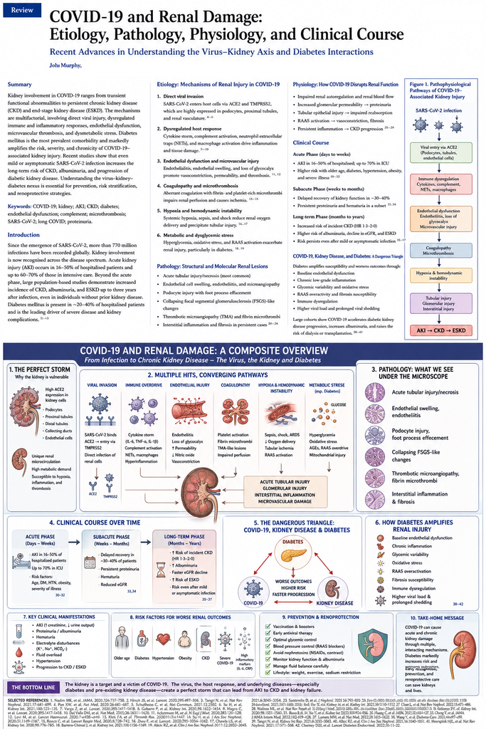
A Unified Pathophysiological Model
The evidence supports the following integrated model:
SARS-CoV-2 infection
↓
viral replication and systemic inflammatory activation
↓
cytokines + complement + platelet activation
↓
endothelial dysfunction
↓
microvascular dysfunction
↓
renal hypoxia
↓
tubular mitochondrial stress
↓
acute tubular injury
At the same time:
SARS-CoV-2
↓
RAAS perturbation
↓
vasoconstriction + oxidative stress + inflammation
↓
additional renal injury
And in genetically susceptible individuals:
SARS-CoV-2
↓
cytokine activation
APOL1 susceptibility
↓
podocyte injury
↓
collapsing glomerulopathy
↓
nephrotic proteinuria + AKI.
Finally:
AKI
↓
incomplete repair
↓
fibrosis
↓
nephron loss
↓
CKD
↓
kidney failure in susceptible patients.
The Metabolic Axis
COVID-19 simultaneously affects glucose metabolism:
SARS-CoV-2
↓
inflammation + insulin resistance + possible β-cell dysfunction
↓
hyperglycaemia
↓
endothelial dysfunction + oxidative stress
↓
renal injury
Meanwhile:
pre-existing diabetes
↓
endothelial and glomerular vulnerability
↓
more severe COVID-19
↓
greater AKI risk
Thus the biological system can be represented as:
SARS-CoV-2 ↔ diabetes ↔ kidney disease
rather than three independent diseases.
Outstanding Scientific Questions
Several fundamental questions remain unresolved.
1. How often does SARS-CoV-2 truly persist in renal tissue?
Detection of viral RNA or protein does not necessarily establish productive viral replication.
2. Which patients fail to recover from AKI?
The answer may involve:
- genetic susceptibility;
- baseline nephron reserve;
- APOL1;
- diabetes;
- endothelial phenotype;
- immune dysregulation.
3. Does persistent renal disease represent viral persistence or post-infectious pathology?
The answer may differ between patients.
4. Can renal Long COVID be identified before eGFR declines?
Urinary proteomics, metabolomics and tubular biomarkers may eventually provide the answer.
5. Is COVID-associated diabetes a distinct disease?
It may represent a heterogeneous combination of:
- unmasked pre-existing diabetes;
- stress hyperglycaemia;
- corticosteroid-induced diabetes;
- persistent insulin resistance;
- pancreatic injury.
6. Can early treatment prevent CKD?
This remains one of the most important unanswered therapeutic questions.
Conclusions
COVID-19 should no longer be regarded as exclusively a respiratory infection with occasional renal complications.
SARS-CoV-2 can produce a complex renal syndrome encompassing:
acute tubular injury
glomerular disease
endothelial dysfunction
microvascular injury
proteinuria
AKI
persistent renal dysfunction
and, in susceptible individuals,
CKD and kidney failure.
The predominant pathological lesion is acute tubular injury, while glomerular lesions such as collapsing glomerulopathy occur in specific clinical and genetic contexts. Evidence for direct renal infection exists, but competing biopsy and autopsy findings demonstrate that direct viral cytotoxicity is not sufficient to explain the renal phenotype. The current evidence instead supports an integrated model involving viral interaction with renal cells, systemic inflammation, endothelial dysfunction, complement and coagulation activation, hypoxaemia, haemodynamic stress, RAAS dysregulation and mitochondrial injury.
The APOL1-associated collapsing glomerulopathy phenotype further demonstrates that host genetics can determine the anatomical response to infection. SARS-CoV-2 may therefore function not as a solitary cause of renal pathology but as a biological trigger acting upon a susceptible host.
The relationship with diabetes is equally important.
Diabetes compromises renal microvascular and glomerular integrity before infection. COVID-19 can then superimpose inflammatory, endothelial, haemodynamic and metabolic stress. Conversely, epidemiological evidence increasingly supports an association between COVID-19 and subsequent diabetes. A 2025 meta-analysis involving more than 48 million people found a 41% increased relative risk of new-onset diabetes, while a 2026 prevalence meta-analysis found a substantial pooled prevalence but extreme heterogeneity among studies.
The most important recent renal evidence is longitudinal.
The 2025 OpenSAFELY study found nearly a doubling of kidney-failure risk following COVID-19 overall and a more than seven-fold hazard among patients requiring hospitalisation, with excess risk persisting beyond six months. The absence of increased kidney-failure risk among non-hospitalised individuals in that study argues against the proposition that every SARS-CoV-2 infection inevitably produces progressive renal disease. Instead, the evidence points toward a severity-dependent renal trajectory concentrated among individuals with substantial systemic disease.
The emerging paradigm can therefore be summarised as:
SARS-CoV-2 infection
↓
systemic inflammation + endothelial dysfunction + metabolic disturbance
↓
acute tubular and/or glomerular injury
↓
AKI
↓
complete recovery OR incomplete repair
↓
persistent nephron injury
↓
CKD
↓
accelerated kidney failure in a susceptible subset.
Diabetes may amplify each step.
The kidney may consequently represent one of the most important organs through which the long-term biological consequences of severe SARS-CoV-2 infection become measurable.
Future research should move beyond serum creatinine and conventional epidemiology toward longitudinal integration of renal histopathology, urinary proteomics, tubular biomarkers, metabolomics, endothelial biology, viral persistence, immune profiling and host genetics.
The decisive scientific objective is no longer merely to determine whether COVID-19 injures the kidney.
It is to determine which biological pathways convert an episode of SARS-CoV-2 infection into irreversible nephron loss—and which of those pathways can still be interrupted after the acute infection has disappeared.
Selected References
- Mahalingasivam V, Zheng B, Wing K, et al. Long-term kidney outcomes after COVID-19: a matched cohort study using the OpenSAFELY platform. Lancet Reg Health Eur. 2025;55:101338.
- Sharma P, Uppal NN, Wanchoo R, et al. Kidney biopsy findings in patients with COVID-19. J Am Soc Nephrol. 2020.
- Larsen CP, Bourne TD, Wilson JD, et al. Collapsing glomerulopathy in a patient with COVID-19. Kidney Int Rep. 2020.
- Wu H, Larsen CP, Hernandez-Arroyo CF, et al. AKI and collapsing glomerulopathy associated with COVID-19 and APOL1 high-risk genotype. J Am Soc Nephrol. 2020.
- Mahjani M, Parvin M, Ghobadi S, et al. Postmortem histopathologic findings and SARS-CoV-2 detection in autopsy kidneys of patients with COVID-19: a systematic review and meta-analysis. Am J Clin Pathol. 2023;159:429–436.
- Menez S, Robinson-Cohen C, Bansal N, et al. Overview of acute kidney manifestations and management of COVID-19. Kidney Int. 2021.
- Cocking E, Daher J, Alabbood M. New-onset diabetes mellitus post COVID-19 infection: a systematic review and meta-analysis. Diabetes Res Clin Pract. 2025;227:112417.
- Keels JN, LaPlante RD, Lee CS, Dwyer AA. Prevalence of new-onset diabetes following COVID-19 infection: a systematic review and meta-analysis. Diabetes Obes Metab. 2026;28:3182–3192.
- El-Naas A, Hamad O, Nair S, et al. New onset of type 1 and type 2 diabetes post-COVID-19 infection: a systematic review. Emerg Microbes Infect. 2025;14:2492211.
- Risk for newly diagnosed diabetes after COVID-19: a systematic review and meta-analysis. 2022.
- Chen IW, et al. Association between COVID-19 and the development of long-term renal dysfunction. 2025.
- Azim T, et al. Impact of COVID-19 on nephropathy in diabetes mellitus. 2024.
- Mehrotra-Varma S, et al. Patients with type 1 diabetes are at elevated risk of new hypertension, CKD and diabetic ketoacidosis following COVID-19. 2024.
- Post-acute metabolic changes and risk of new-onset diabetes following COVID-19: systematic review and meta-analysis. 2026.