John Murphy, CEO The COVID-19 Long-haul Foundation
Introduction
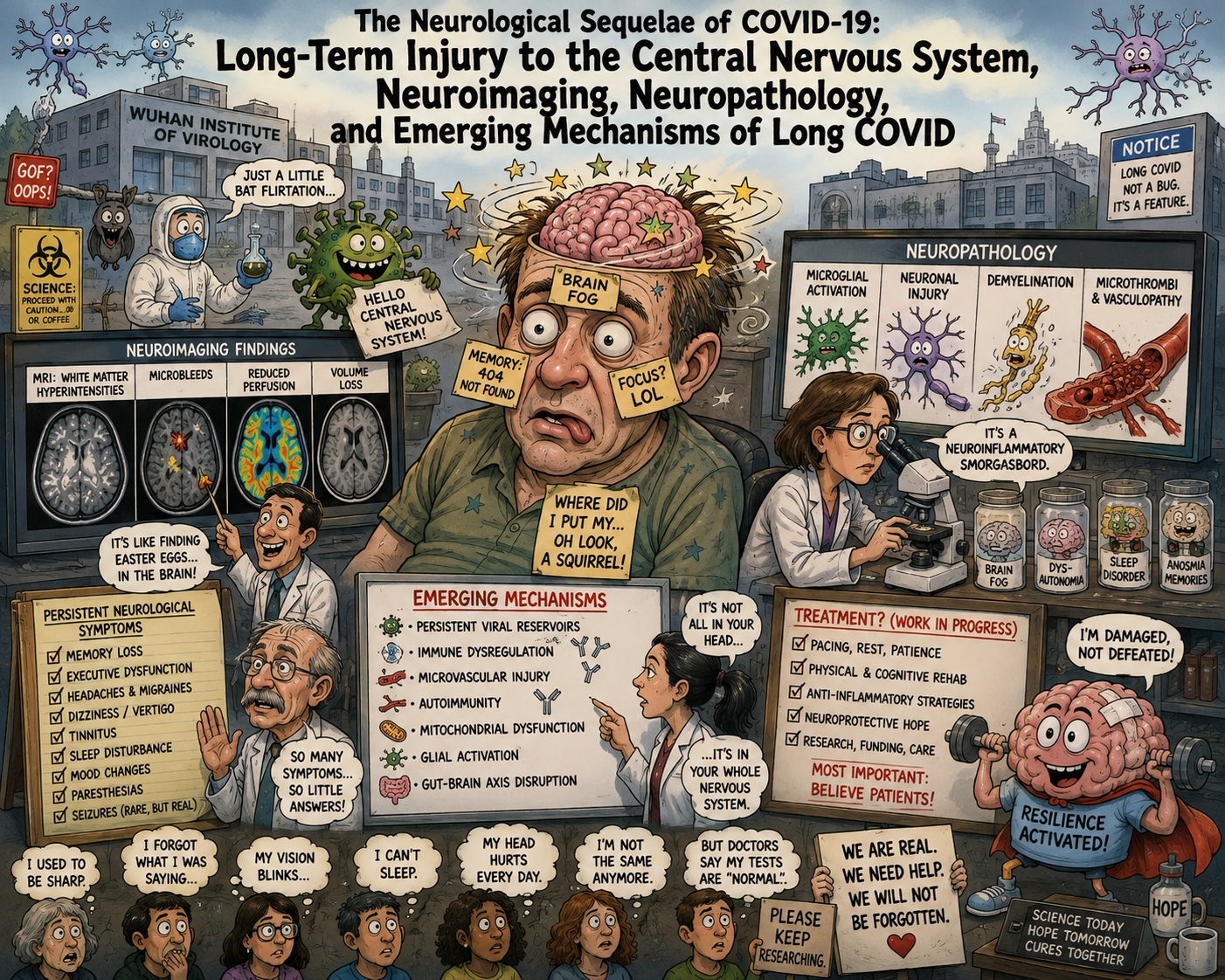
The neurological consequences of SARS-CoV-2 infection have evolved from an initially unexpected collection of clinical observations into a substantial body of neuropathological, neuroimaging, immunological and molecular evidence. What was first regarded largely as an acute respiratory disease with occasional neurological complications is now understood to have the capacity to produce persistent disturbances of cognition, autonomic regulation, sleep, sensory processing, motor function, pain perception and affect. The contemporary literature increasingly supports the proposition that, in at least a subset of patients, these manifestations reflect measurable alterations in the central nervous system (CNS), rather than merely nonspecific consequences of prolonged illness.
The terminology is important. Long COVID, or post-COVID-19 condition (PCC), is not synonymous with persistent viral encephalitis. Nor is it synonymous with structural brain destruction. Rather, it appears to represent a heterogeneous post-infectious syndrome in which several biological processes—immune dysregulation, neurovascular injury, blood-brain-barrier dysfunction, persistent antigen or viral RNA, glial activation, altered neuronal and synaptic function, metabolic dysfunction, autonomic disturbance and, in some patients, autoimmunity—may coexist in different proportions. The most comprehensive contemporary review, published in Nature Reviews Neurology in April 2026, explicitly identifies viral persistence, immune dysregulation, neuroinflammation, herpesvirus reactivation, microvascular injury, gut-brain-axis disturbance and structural and functional brain abnormalities among the principal candidate mechanisms.
The central neurological paradox of Long COVID is therefore this: patients may have profound neurological dysfunction despite relatively unimpressive conventional brain imaging. This apparent discrepancy disappears when the brain is examined with more sensitive techniques—volumetric MRI, diffusion imaging, susceptibility imaging, dynamic contrast-enhanced MRI, FDG-PET, magnetic-resonance spectroscopy, quantitative microstructural analysis and molecular biomarkers of neuronal and glial injury. At the same time, conventional imaging remains important because it can reveal the more severe consequences of vascular, inflammatory and hypoxic CNS injury.
The emerging picture is consequently not one of a single “COVID lesion,” but of a distributed disturbance involving gray matter, white matter, cerebral microvasculature, glia, limbic circuits, brainstem nuclei and neuroimmune interfaces.
1. The CNS as a Target of SARS-CoV-2
The earliest controversy concerned whether SARS-CoV-2 actually enters the brain. The answer is now more nuanced than either “yes” or “no.”
Autopsy investigations have demonstrated SARS-CoV-2 RNA and, in selected tissues, viral protein within the CNS. In a landmark Nature study of 44 autopsied patients, extensive CNS sampling was performed in 11 individuals. SARS-CoV-2 RNA was detected in CNS tissue in 10 of 11 cases, including in multiple brain regions in patients who died considerably later after the initial illness. In one patient, viral RNA remained detectable 230 days after symptom onset. Viral RNA and protein were identified in structures including the hypothalamus, cerebellum, basal ganglia and cervical spinal cord. Importantly, however, the investigators found relatively little overt inflammatory or cytopathic injury in relation to the extensive distribution of viral RNA. This observation is crucial: viral persistence does not necessarily imply widespread direct viral destruction of neurons.
Other neuropathological studies have identified viral material in the olfactory system, brainstem, vascular structures and, in some cases, neuronal and glial elements. Brainstem investigations have demonstrated SARS-CoV-2 nucleoprotein in neurons and glial cells, accompanied by increased Iba-1 expression, a marker of microglial activation. Vagus nerve involvement has also been reported, providing a plausible anatomical route connecting pulmonary and brainstem pathology.
Nevertheless, the proposition that Long COVID is primarily caused by persistent productive infection of the brain is not established. The evidence instead suggests that viral persistence, where present, may act as one component of a larger biological process.
2. The Blood-Brain Barrier: The Neurovascular Interface at the Center of the Disease
The blood-brain barrier (BBB) is formed principally by specialized endothelial cells joined by tight junctions, supported by pericytes, astrocytic endfeet, basement membrane and associated immune and glial cells. Its function is to maintain the biochemical environment required for neuronal signaling while excluding potentially harmful circulating molecules and inflammatory cells.
COVID-19 appears capable of disturbing this barrier.
The most important human evidence comes from a 2024 Nature Neuroscience investigation of patients with Long COVID-associated cognitive impairment. Dynamic contrast-enhanced MRI demonstrated increased BBB permeability in patients with brain fog, despite conventional MRI showing no clinically significant abnormalities. Leakage was particularly apparent in the frontal cortex and bilateral temporal lobes. The same patients exhibited persistent systemic inflammatory and coagulation abnormalities, increased adhesion of peripheral blood mononuclear cells to brain endothelial cells, and increased endothelial expression of inflammatory adhesion molecules.
This finding is particularly significant because it supplies a bridge between systemic inflammation and CNS dysfunction.
A compromised BBB may permit increased passage of cytokines, complement components, fibrinogen, immune cells and other circulating mediators into the CNS. It may also alter the ionic and metabolic environment surrounding neurons. Astrocytes and pericytes, which normally participate in BBB regulation, can themselves become reactive. The consequence is not necessarily an inflammatory lesion visible on conventional MRI; instead, it may be a distributed disturbance of neuronal homeostasis.
The BBB findings also provide a plausible explanation for why relatively subtle vascular abnormalities may produce disproportionately severe cognitive symptoms.
3. Cerebral Microvascular Disease
The cerebral microcirculation appears to be another major target.
Neuropathological investigations of severe COVID-19 have demonstrated endothelial injury, vascular congestion, fibrinogen leakage, platelet accumulation, activation of coagulation pathways and loss of small vessels. One mechanistic study demonstrated SARS-CoV-2-related injury to brain endothelial cells and formation of “string vessels”—empty basement-membrane tubes representing remnants of lost capillaries.
Microvascular dysfunction is important because neurons have extraordinary metabolic requirements and minimal tolerance for interruptions in oxygen and substrate delivery. Even without producing a macroscopic infarction, repeated or persistent microvascular dysfunction could cause:
- impaired regional cerebral perfusion;
- endothelial activation;
- impaired oxygen extraction;
- BBB leakage;
- microhemorrhages;
- altered neurovascular coupling;
- local tissue hypoxia;
- white-matter injury; and
- impaired cognitive processing.
This mechanism is particularly attractive in Long COVID because cognition depends upon the integrity of distributed neural networks rather than a single anatomical center. A modest reduction in perfusion across several interconnected regions can therefore produce substantial impairment without producing a classic stroke.
4. Neuropathological Findings at Autopsy
The neuropathology of COVID-19 has several recurring components.
Hypoxic-ischemic injury
In severe acute disease, hypoxic-ischemic injury is common. A classic New England Journal of Medicine autopsy study found acute hypoxic injury in the cerebrum and cerebellum of all examined patients, including neuronal loss in cerebral cortex, hippocampus and cerebellar Purkinje-cell layers. Importantly, thrombi and vasculitis were absent in that cohort, demonstrating that hypoxia alone can produce major neuropathological injury.
This distinction is important when interpreting later cognitive abnormalities: some survivors of severe COVID may have persistent consequences of hypoxic brain injury, prolonged ventilation, systemic hypotension, metabolic derangements or critical illness in addition to SARS-CoV-2-specific mechanisms.
Microglial activation
Microglial activation is one of the most reproducible findings.
A 2025 Nature Neuroscience autopsy investigation found regionally heterogeneous microglial abnormalities, including loss of the homeostatic microglial marker P2Y12R, disruption of the CX3CR1-CX3CL1 signaling axis and metabolic dysfunction at sites of vascular inflammation. Particularly striking abnormalities occurred in medullary autonomic nuclei, structures that regulate cardiovascular, respiratory and visceral functions.
These observations are potentially relevant to Long COVID manifestations such as:
- dysautonomia;
- abnormal heart-rate regulation;
- orthostatic intolerance;
- abnormal respiratory control;
- gastrointestinal autonomic dysfunction; and
- disordered sleep.
White-matter microglial pathology
An important Cell study demonstrated prominent white-matter-selective microglial reactivity after respiratory SARS-CoV-2 infection. The experimental model did not require direct brain infection. Instead, systemic respiratory infection was sufficient to produce persistent neuroinflammatory changes.
Human brain specimens from infected individuals exhibited similar white-matter microglial activation. In mice, this was accompanied by impaired hippocampal neurogenesis, loss of oligodendrocytes and myelin, with abnormalities persisting for at least seven weeks. Elevated CCL11 was implicated as one potential mediator.
The importance of this study is conceptual: the CNS can be injured without extensive direct neuronal infection.
5. Gray Matter: Cortical and Limbic Injury
The gray matter contains neuronal cell bodies, dendrites, synapses, astrocytes, microglia and other supporting structures. Several independent imaging studies now identify abnormalities in gray matter after SARS-CoV-2 infection.
The landmark longitudinal UK Biobank study is particularly persuasive because participants underwent MRI before and after infection. Among 401 infected individuals compared with 384 controls, SARS-CoV-2 infection was associated with greater reduction in gray-matter thickness and tissue contrast in the orbitofrontal cortex and parahippocampal gyrus, changes in tissue-damage measures in regions functionally connected with the primary olfactory cortex, and greater reduction in overall brain size. These effects remained detectable after excluding hospitalized patients.
The affected structures are anatomically meaningful.
Orbitofrontal cortex
The orbitofrontal cortex participates in:
- executive function;
- reward evaluation;
- decision-making;
- emotional regulation;
- sensory integration; and
- olfactory processing.
Parahippocampal and entorhinal regions
These structures are integral to:
- episodic memory;
- contextual memory;
- spatial processing; and
- communication between sensory and hippocampal systems.
Anterior cingulate cortex
The anterior cingulate participates in:
- attention;
- cognitive control;
- motivation;
- error monitoring;
- emotional processing;
- pain processing; and
- autonomic integration.
A 2025 Nature Medicine study of 351 previously hospitalized patients found persistent global cognitive deficits approximately one year after COVID-19 and identified reduced anterior cingulate cortex volume. The magnitude of cognitive impairment was associated with elevated serum neurofilament light chain (NfL) and glial fibrillary acidic protein (GFAP), biomarkers of neuronal/axonal and astroglial injury respectively.
6. The Hippocampus and Memory
The hippocampus deserves special consideration because memory impairment is among the most characteristic cognitive complaints of Long COVID.
The hippocampus is not simply a memory “storage center”; it is a highly plastic structure involved in memory consolidation, contextual processing, spatial representation and interaction with distributed cortical networks.
Evidence for hippocampal involvement comes from multiple levels:
- longitudinal MRI demonstrating abnormalities in parahippocampal and related structures;
- FDG-PET showing temporal and hippocampal hypometabolism;
- experimental evidence of impaired hippocampal neurogenesis;
- microglial activation in hippocampal circuitry;
- altered white-matter connectivity;
- increased biomarkers of neuronal injury; and
- emerging evidence of altered synaptic and immune signaling.
The precise mechanism remains unresolved. It may involve inflammation, altered neurogenesis, vascular dysfunction, metabolic stress, sleep disruption, autonomic dysfunction or autoimmunity rather than direct viral cytotoxicity alone.
7. The Thalamus, Brainstem and Autonomic Nuclei
The thalamus is a major relay and integration center connecting sensory, cortical, limbic and arousal systems. Abnormalities in thalamic structure or metabolism could therefore have wide-ranging consequences.
FDG-PET studies of Long COVID have identified hypometabolism involving the thalamus, temporal regions, brainstem and cerebellum. In one frequently cited cohort, hypometabolism was observed in the olfactory/orbital region, right temporal lobe including amygdala and hippocampus, right thalamus, bilateral pons/medulla and cerebellum. The brainstem and cerebellar abnormalities correlated with greater symptom burden.
The brainstem may be disproportionately important because it contains nuclei governing:
- respiration;
- cardiovascular regulation;
- arousal;
- sleep;
- swallowing;
- gastrointestinal function;
- vestibular function;
- cranial nerve activity; and
- autonomic control.
The 2025 autopsy work demonstrating microglial and neurovascular abnormalities in medullary autonomic nuclei therefore provides an intriguing pathological correlate for the clinical phenomenon of dysautonomia.
8. Cerebellar Involvement
The cerebellum is increasingly recognized as part of the neurological phenotype.
It is traditionally associated with coordination and balance, but modern neuroscience recognizes its involvement in cognition, language, affect and autonomic regulation.
PET studies have demonstrated cerebellar hypometabolism, while neuropathological studies have identified microglial activation and white-matter abnormalities within cerebellar structures. Autopsy series have also examined cerebellar nuclei and white matter as sites of inflammatory pathology.
This provides a possible anatomical substrate for:
- dizziness;
- impaired coordination;
- gait instability;
- altered balance;
- autonomic symptoms; and
- some forms of cognitive dysfunction.
9. White Matter: The Often-Overlooked Component
If gray matter represents the computational machinery of the brain, white matter represents its communication infrastructure.
White matter consists primarily of myelinated axons, oligodendrocytes, astrocytes, microglia, vascular structures and extracellular matrix. Damage here can disrupt communication between otherwise intact gray-matter regions.
Several imaging studies demonstrate abnormalities in white-matter microstructure following SARS-CoV-2 infection.
A 2024 Nature Communications investigation of Post-COVID Condition identified widespread microstructural abnormalities. The study found shifts in tissue volume from membrane-enclosed compartments toward free-water compartments, with abnormalities involving neocortical gray matter, thalamus, corpus callosum, internal capsule, cerebellum and brainstem. These alterations correlated with cognitive impairment, olfactory dysfunction and fatigue.
A 2025 review of neuroimaging literature similarly concluded that structural MRI studies have identified frontal and temporal gray-matter changes and white-matter hyperintensities, while diffusion imaging has revealed abnormalities in white-matter tracts including the sagittal stratum and thalamic radiations.
10. Oligodendrocytes and Myelin
One of the most intriguing areas of research concerns oligodendrocytes.
Oligodendrocytes generate myelin—the multilayered membrane surrounding axons that allows rapid and efficient saltatory conduction. Damage to oligodendrocytes therefore does not necessarily kill neurons; instead, it can degrade the efficiency of communication between neurons.
The 2022 Cell study demonstrated persistent depletion of mature oligodendrocytes and loss of myelin after respiratory SARS-CoV-2 infection in mice. The investigators also observed white-matter-selective microglial activation and impaired hippocampal neurogenesis. Human brain tissue demonstrated a similar pattern of white-matter microglial activation.
This is an important mechanistic observation because it provides a plausible biological bridge between systemic infection and the subjective experience of slowed information processing.
The concept is increasingly supported by advanced MRI. A 2025 multimodal MRI study used T1w/T2w imaging, diffusion imaging and magnetic-resonance spectroscopy and identified altered myelin-associated signal and tissue microstructure in Long COVID. The imaging abnormalities correlated with physical and cognitive function.
The human evidence does not yet establish widespread demyelinating disease analogous to multiple sclerosis. Rather, the emerging picture is one of subtle myelin and axonal dysregulation, which may be substantially more difficult to detect on routine clinical MRI.
11. Astrocytes: The Metabolic and Neuroimmune Interface
Astrocytes are no longer regarded as passive structural support cells. They regulate extracellular potassium, neurotransmitter concentrations, cerebral blood flow, BBB integrity, synaptic function and neuronal metabolism.
They are consequently a logical target for a systemic inflammatory disease.
GFAP, an intermediate filament expressed in astrocytes, is increasingly used as a biomarker of astroglial injury. The 2025 Nature Medicine study found persistently elevated GFAP approximately one year after hospitalization, together with elevated NfL.
A 2025 study using long-term human cortical organoids provided additional mechanistic evidence. SARS-CoV-2 infection occurred at low levels in astrocytes and several neuronal populations, with astrocytes showing the highest infection rate among infected cell types. Infection was accompanied by morphological changes and increased expression of astrogliosis-associated genes including SERPINA3, CD44 and S100A10, together with transcriptional changes involving inflammation and cellular metabolism.
This does not prove that the same degree of infection occurs in living human brains during Long COVID. But it demonstrates that human neural tissue can respond to SARS-CoV-2 with a characteristic astroglial inflammatory program.
12. Microglia: The Brain’s Persistent Immune Response
Microglia are the resident macrophage-like immune cells of the CNS. In their homeostatic state they survey the neural environment; when activated, they change morphology, metabolism, receptor expression and cytokine signaling.
The current literature increasingly suggests that persistent microglial dysfunction may be more important than persistent neuronal infection.
The 2025 Nature Neuroscience autopsy study identified loss of P2Y12R and dysfunction of the CX3CR1-CX3CL1 pathway—systems involved in maintaining normal neuron-microglia communication. The investigators also identified metabolic failure and focal neuropathology in association with vascular inflammation.
The implications are substantial.
Activated or dysfunctional microglia can:
- alter synaptic pruning;
- release inflammatory mediators;
- influence oligodendrocyte survival;
- affect hippocampal neurogenesis;
- alter neuronal excitability;
- disturb sleep regulation;
- modify pain processing; and
- impair network-level cognition.
Thus, the neurological consequences of COVID may persist after the acute inflammatory episode because the cellular state of the CNS immune system itself has been altered.
13. MRI: What the Films Actually Show
It is essential to distinguish routine clinical MRI from quantitative research MRI.
Routine MRI frequently appears normal in Long COVID. A prospective multicenter study of 140 patients with persistent neurological symptoms examined MRI at approximately six and twelve months. White-matter hyperintensities were not more prevalent than in healthy controls, and microbleeds occurred at frequencies comparable to population studies. Only 4% of patients had lesions suggestive of inflammatory or demyelinating disease. Cranial-nerve enhancement was detected in approximately 7%, predominantly involving the oculomotor nerve.
This is an important negative finding.
It means that a normal conventional MRI does not exclude CNS involvement.
T1-weighted MRI
T1 imaging can reveal:
- regional cortical thinning;
- gray-matter volume loss;
- hippocampal or parahippocampal alterations;
- ventricular enlargement;
- global volume changes.
The UK Biobank study demonstrated reductions in cortical thickness particularly involving the orbitofrontal cortex and parahippocampal gyrus.
T2/FLAIR
FLAIR is sensitive to increased water content and white-matter abnormalities.
Reported abnormalities include:
- periventricular hyperintensities;
- deep white-matter lesions;
- watershed injury;
- inflammatory lesions;
- ADEM-like lesions;
- corpus-callosum abnormalities; and
- lesions associated with vascular injury.
However, nonspecific white-matter hyperintensities are common with aging, hypertension, diabetes and cerebrovascular disease and therefore cannot automatically be attributed to COVID.
Diffusion-weighted imaging and DTI
Diffusion MRI is potentially more revealing.
Changes in:
- mean diffusivity;
- fractional anisotropy;
- orientation dispersion;
- extracellular water;
- axonal coherence; and
- tissue free-water fraction
can identify microstructural abnormalities invisible to conventional T1/T2 imaging.
The 2024 Nature Communications study demonstrated widespread microstructural changes involving cortical gray matter, thalamus, corpus callosum, internal capsule, cerebellum and brainstem.
Susceptibility-weighted imaging
SWI can identify:
- microhemorrhages;
- venous abnormalities;
- iron-related susceptibility;
- small vascular lesions.
These findings have been reported in acute COVID and in selected post-COVID cohorts, but the 2025 longitudinal cohort suggests that persistent microbleeds are not substantially more common than expected in the general population.
Dynamic contrast-enhanced MRI
This may ultimately be one of the most interesting techniques.
DCE-MRI can quantify BBB permeability rather than merely showing whether a gross lesion exists. The 2024 Nature Neuroscience study demonstrated abnormal permeability particularly in frontal and temporal regions in Long COVID patients with brain fog.
14. CT: Important, but Less Sensitive for Long COVID
CT is excellent for detecting:
- acute hemorrhage;
- large infarction;
- mass effect;
- cerebral edema;
- hydrocephalus;
- major vascular events;
- venous sinus thrombosis.
It is considerably less sensitive than MRI for subtle white-matter, limbic, cortical and microvascular abnormalities.
During acute COVID, CT and MRI studies documented ischemic infarction, hemorrhage, microhemorrhages, leukoencephalopathy, hypoxic injury, ADEM-like abnormalities and other inflammatory lesions.
For chronic Long COVID, however, a normal CT is not surprising and has little capacity to exclude microstructural or functional brain dysfunction.
The clinical interpretation should therefore be:
Normal CT does not mean normal brain physiology.
15. PET: Seeing the Brain When MRI Looks Normal
FDG-PET measures glucose metabolism and therefore provides a functional rather than purely anatomical picture.
One of the earliest Long COVID PET studies identified a characteristic pattern of hypometabolism involving:
- orbitofrontal and olfactory cortex;
- temporal cortex;
- amygdala;
- hippocampus;
- thalamus;
- pons;
- medulla; and
- cerebellum.
These abnormalities correlated with symptoms including anosmia, memory impairment, pain and insomnia.
Other studies have identified hypometabolism in the anterior and posterior cingulate cortex and precuneus even when conventional MRI was unremarkable.
This discrepancy between structural MRI and functional PET is biologically important. It suggests that a substantial component of Long COVID may involve neuronal network dysfunction preceding or occurring without gross anatomical destruction.
16. Biomarkers of Neuronal and Glial Injury
Two biomarkers have received particular attention:
Neurofilament light chain
NfL is released following axonal injury.
The 2025 Nature Medicine study found elevated NfL approximately one year after hospitalization, particularly among patients with neurological complications.
A 2024 Molecular Psychiatry study similarly found higher plasma NfL in Long COVID patients with neurocognitive symptoms after mild infection, with levels associated with cognitive impairment and fatigue.
However, not every study has reproduced persistent NfL elevation. A 2024 Scientific Reports study found normal NfL in a neurological Long COVID outpatient cohort, demonstrating that persistent axonal injury is not universal.
GFAP
GFAP is a marker of astroglial injury or activation.
Its elevation in Long COVID is arguably more consistent than persistent NfL elevation, although even GFAP findings vary among cohorts.
This heterogeneity is itself informative: Long COVID probably contains multiple neurological phenotypes rather than a single neuropathological disease.
17. Cerebrospinal Fluid and Neuroimmune Abnormalities
Routine CSF is often surprisingly normal.
A 2024 study of post-COVID cognitive impairment found that most patients had normal conventional CSF measurements. Nevertheless, single-cell analysis revealed molecular evidence of monocyte recruitment, chemokine signaling, cellular stress and suppressed interferon responses, particularly within myeloid cells. Longitudinal improvement was associated with changes in inflammatory genes and increased CXCL8, CCL3L1 and soluble TREM2.
Other studies have identified brain-reactive antibodies in serum and CSF. One cohort found anti-neuronal autoantibodies in 52% of patients with post-COVID cognitive complaints, including antibodies reacting with vascular endothelium, Purkinje cells, axon initial segments, astrocytic proteins and structures within basal ganglia and hippocampus. The presence of anti-neuronal antibodies in CSF was associated with poorer cognitive performance.
These findings suggest that some forms of Long COVID may represent a form of post-infectious neuroimmunological disease rather than persistent infection alone.
18. The 2026 Breakthrough: Evidence for Pathogenic Autoantibodies
The most provocative neurological development of 2026 is a Cell study by de Sá and colleagues.
Using tissue-based immunofluorescence, ELISA, a greater-than-21,000-protein human array and mass spectrometry, the investigators identified extensive autoantibody responses in Long COVID. Patients with neurocognitive symptoms demonstrated increased antibodies directed against CNS and peripheral nervous-system proteins.
Patient IgG reacted with human locus coeruleus and thalamic tissue and showed reactivity against peripheral nervous-system structures. Most importantly, passive transfer of patient IgG into mice produced phenotypes including fatigue-like behavior, impaired coordination, thermal hyperalgesia, small-fiber injury and increased pain-related neuronal activity.
This is a major conceptual advance because it moves the field beyond the observation that “autoantibodies are present” toward experimental evidence that some autoantibodies can actually transmit neurological phenotypes.
It does not mean that all Long COVID is autoimmune.
It does suggest, however, that a subset of patients may have a definable antibody-mediated neurological disorder.
19. The Choroid Plexus and Brain-CSF Interface
The choroid plexus has emerged as another structure of interest.
It produces cerebrospinal fluid and forms an important interface between blood, CSF and the CNS immune environment.
A 2025 Nature Communications study of cognitively impaired PASC patients found enlarged choroid plexus volume together with cortical thinning, increased hippocampal susceptibility and elevated astroglial injury-associated proteins. These abnormalities were reproduced in an independent validation cohort.
The finding is provocative because the choroid plexus is increasingly recognized as an immunological gateway. Alteration of this interface could contribute to sustained communication between systemic inflammation and the CNS.
20. The Neurodegenerative Question
One of the most consequential questions is whether Long COVID causes neurodegeneration.
The answer at present is not definitively established, but the question can no longer be dismissed.
The 2025 Nature Communications study identified cortical thinning in cingulate and insular cortices, increased hippocampal paramagnetic susceptibility, enlarged choroid plexus volume and altered proteins associated with oxidative stress, synaptic function and neurodegenerative pathways in cognitively impaired PASC patients. These findings were reproduced in a validation cohort.
Conversely, other studies have not found persistent neuronal or astrocytic biomarker abnormalities in all cognitively impaired patients.
Thus, the appropriate conclusion is not that Long COVID inevitably produces progressive neurodegenerative disease. Rather, a subset of patients exhibits biological signatures that overlap with processes involved in neurodegeneration, and longitudinal observation is required to determine whether these represent transient injury, maladaptive repair or progressive disease.
21. A Multisystem CNS Model
The most coherent interpretation of the current evidence is a model in which several processes interact:
SARS-CoV-2 infection
↓
Systemic immune activation
↓
Endothelial and microvascular dysfunction
↓
Blood-brain-barrier disruption
↓
Microglial and astrocytic activation
↓
Altered oligodendrocytes, myelin and synaptic function
↓
Neuronal metabolic stress and network dysfunction
↓
Limbic, cortical, thalamic, brainstem and cerebellar abnormalities
↓
Cognitive, sensory, autonomic, sleep and pain syndromes
But this pathway is not necessarily linear.
In another patient:
Persistent viral antigen or RNA
↓
Chronic immune stimulation
↓
Autoantibody production
↓
Neurovascular and neuronal targeting
↓
CNS dysfunction
And in another:
Severe acute disease
↓
Hypoxia + inflammation + thrombosis + critical illness
↓
Permanent structural brain injury
↓
Persistent neurological disability
These pathways can overlap.
This is why two patients with ostensibly identical Long COVID diagnoses may have completely different MRI scans, biomarker profiles and treatment responses.
22. What the Evidence Does—and Does Not—Demonstrate
Several conclusions can now be stated with considerable confidence.
Strongly supported
First, SARS-CoV-2 infection can produce persistent neurological symptoms lasting months or years. The 2026 Nature Reviews Neurology synthesis identifies cognitive impairment, fatigue, headache, sleep disturbance, dysautonomia, pain and psychiatric symptoms among the principal manifestations.
Second, objective brain abnormalities can occur after SARS-CoV-2 infection, including changes in cortical thickness, gray-matter volume, white-matter microstructure and cerebral metabolism.
Third, BBB dysfunction has been directly demonstrated in patients with Long COVID-associated cognitive impairment.
Fourth, neuronal and astroglial injury markers can remain elevated after COVID-19, although not consistently in every Long COVID cohort.
Fifth, microglial activation and neurovascular pathology are documented neuropathological features of COVID-19.
Sixth, oligodendrocyte and myelin abnormalities are biologically plausible and supported by experimental and imaging evidence.
Seventh, persistent viral RNA can occur in the CNS after acute infection, although its relationship to Long COVID symptoms remains uncertain.
Eighth, at least one neurological Long COVID phenotype now has experimental evidence implicating pathogenic autoantibodies.
Still uncertain
It remains uncertain:
- how frequently SARS-CoV-2 persists in the living human CNS of patients with Long COVID;
- whether persistent viral material is infectious or merely residual;
- whether microglial activation is causal or compensatory;
- whether MRI abnormalities progress or resolve;
- whether cortical thinning represents neuronal loss, altered dendritic architecture, inflammation, vascular change or measurement effects;
- whether Long COVID causes progressive neurodegenerative disease;
- which patients have primarily vascular, immune, autoimmune, metabolic or viral-persistence phenotypes; and
- which biomarkers can reliably identify each subtype.
23. The Most Important Diagnostic Implication
Perhaps the most important clinical lesson is that a normal routine MRI cannot be interpreted as evidence that the CNS is normal.
The contemporary hierarchy of neurological investigation is approximately:
CT
best for acute hemorrhage, large infarction, mass effect and major vascular pathology.
Conventional MRI
best for infarction, inflammatory lesions, demyelination, hemorrhage, encephalitis and structural abnormalities.
DWI/DTI/free-water MRI
better suited to microstructural white- and gray-matter abnormalities.
SWI
sensitive to microhemorrhage and vascular susceptibility abnormalities.
DCE-MRI
can identify BBB permeability abnormalities.
Volumetric MRI
can detect regional cortical and subcortical volume changes.
FDG-PET
can identify functional metabolic abnormalities even when conventional MRI is normal.
MRS
can identify altered neurochemical and metabolic states.
CSF and blood biomarkers
can reveal evidence of neuroaxonal injury, astroglial injury and neuroimmune activation.
The most informative future diagnostic approach may therefore be multimodal rather than dependent upon a single scan.
Conclusion
The neurological legacy of COVID-19 is increasingly difficult to reconcile with the proposition that Long COVID is merely a collection of subjective symptoms occurring after an otherwise resolved respiratory infection.
The evidence now spans several independent domains: longitudinal MRI demonstrates alterations in cortical thickness and limbic structures; diffusion imaging identifies abnormalities in gray- and white-matter microstructure; FDG-PET demonstrates persistent abnormalities of cerebral metabolism; DCE-MRI reveals disruption of the blood-brain barrier; blood biomarkers demonstrate evidence of neuronal and astroglial injury in subsets of patients; autopsy studies demonstrate microglial activation, neurovascular pathology and, in selected cases, persistent viral RNA; experimental studies demonstrate oligodendrocyte and myelin abnormalities; CSF studies identify persistent neuroimmune alterations; and emerging 2026 work provides experimental evidence that autoantibodies can directly reproduce neurological phenotypes in animals.
The most defensible interpretation is therefore not that SARS-CoV-2 produces one stereotyped neurological lesion. It is that SARS-CoV-2 can initiate a distributed CNS disease process involving the neurovascular unit, BBB, microglia, astrocytes, oligodendrocytes, myelin, neurons and synaptic networks, with different patients expressing different combinations of these abnormalities.
The brain regions most repeatedly implicated include the orbitofrontal cortex, anterior cingulate cortex, insula, parahippocampal and entorhinal cortices, hippocampus, amygdala, thalamus, corpus callosum, internal capsule, brainstem, medullary autonomic nuclei and cerebellum. These are not random anatomical observations: together they form interconnected systems governing memory, attention, executive function, emotion, olfaction, autonomic regulation, sleep, balance and sensory integration.
The most consequential development may ultimately be the recognition that the CNS consequences of Long COVID are neither exclusively structural nor exclusively functional. They appear to occupy the continuum between the two: inflammation and vascular dysfunction alter the cellular environment; glia change their phenotype; myelin and axonal integrity may deteriorate; neuronal metabolism and synaptic function become abnormal; and the resulting network dysfunction can precede or exceed what is visible on conventional imaging.
In this sense, the neurological component of Long COVID may be best understood as a post-viral systems disorder of the neurovascular, neuroimmune and neuronal networks, rather than as a single neurological disease.
The central scientific challenge for the next decade is consequently not simply to demonstrate that Long COVID affects the brain. That proposition is increasingly well supported. The challenge is to determine which biological mechanism dominates in which patient, which abnormalities are reversible, which represent permanent injury, and which molecular signatures can be converted into reliable diagnostic tests and mechanism-specific treatments.
That distinction will determine whether Long COVID neurology remains a descriptive field—or becomes a genuinely treatable branch of neuroimmunology.
Footnotes and Selected Peer-Reviewed References
1. Matthews R, Alam A, Bullmore E, et al. Understanding the long-term neurological effects of SARS-CoV-2 infection. Nature Reviews Neurology. 2026;22:351–365. Published April 13, 2026. This is the most recent comprehensive synthesis of neurological Long COVID available as of August 2026.
2. Douaud G, Lee S, Alfaro-Almagro F, et al. SARS-CoV-2 is associated with changes in brain structure in UK Biobank. Nature. 2022;604:697–707. Longitudinal pre-/post-infection MRI demonstrated gray-matter and limbic-system changes.
3. Wood GK, Sargent BF, Ahmad Z-U-A, et al. Posthospitalization COVID-19 cognitive deficits at 1 year are global and associated with elevated brain injury markers and gray matter volume reduction. Nature Medicine. 2025;31:245–257.
4. Greene C, Connolly R, Brennan D, et al. Blood-brain barrier disruption and sustained systemic inflammation in individuals with long COVID-associated cognitive impairment. Nature Neuroscience. 2024;27:421–432.
5. Rieg S, et al. Cerebral microstructural alterations in Post-COVID-condition are related to cognitive impairment, olfactory dysfunction and fatigue. Nature Communications. 2024;15:4256.
6. Fernandez-Castaneda A, et al. Mild respiratory COVID can cause multi-lineage neural cell and myelin dysregulation. Cell. 2022;185:2452–2468.e16.
7. Younger D. COVID-19 Postmortem Neuropathology: 2025 Update. Neurology. 2025. The updated autopsy literature identifies brainstem inflammation, perivascular T-cell infiltration and persistent viral RNA in a subset of cases.
8. Stein SR, et al. SARS-CoV-2 infection and persistence in the human body and brain at autopsy. Nature. 2022;612:758–763. Demonstrated persistent viral RNA in CNS tissue, including late after infection.
9. Microglia dysfunction, neurovascular inflammation and focal neuropathologies are linked to IL-1- and IL-6-related systemic inflammation in COVID-19. Nature Neuroscience. 2025. Demonstrated microglial P2Y12R loss, CX3CR1-CX3CL1 abnormalities and focal pathology, particularly involving medullary autonomic nuclei.
10. Altered brain tissue microstructure and neurochemical profiles in long COVID and recovered COVID-19 individuals: A multimodal MRI study. Brain, Behavior, & Immunity—Health. 2025;50:101142.
11. Brain MRI findings in patients with post COVID-19 condition: frequency and longitudinal changes in a nationwide cohort study. Frontiers in Neurology. 2025. Prospective 140-patient study demonstrating that routine MRI abnormalities are not sufficiently specific to diagnose Long COVID.
12. Neuroimaging biomarkers of post-acute sequelae of Coronavirus Disease 2019. British Journal of Radiology. 2025. Review of structural MRI, diffusion imaging and other neuroimaging biomarkers.
13. 18F-FDG brain PET hypometabolism in patients with long COVID. European Journal of Nuclear Medicine and Molecular Imaging. Demonstrated hypometabolism involving orbitofrontal/olfactory, temporal, thalamic, brainstem and cerebellar regions.
14. Needham EJ, et al. Brain injury in COVID-19 is associated with dysregulated innate and adaptive immune responses. Brain. 2022;145:4097–4107. Cited in the 2025 Nature Medicine synthesis as evidence linking brain injury with immune dysregulation.
15. de Sá KSG, Silva J, Bayarri-Olmos R, et al. A causal link between autoantibodies and neurological symptoms in long COVID. Cell. 2026;189:3214–3235.e37. The study provides experimental evidence that patient-derived IgG can reproduce neurological phenotypes in mice.
16. Bird L. Pathological potential of autoantibodies in long COVID. Nature Reviews Immunology. 2026;26:485. Commentary emphasizing neural, synaptic, neurogenesis and neuronal-apoptosis targets identified in the 2026 Cell study.
17. Wood H. Autoantibodies could trigger neurological symptoms of long COVID. Nature Reviews Neurology. 2026;22:480. Independent scientific commentary on the 2026 Cell findings.
18. Clinical and CSF single-cell profiling of post-COVID-19 cognitive impairment. Cell Reports Medicine. 2024. Identified CSF myeloid recruitment, chemokine signaling and altered interferon responses in patients with persistent cognitive impairment.
19. Association of cerebrospinal fluid brain-binding autoantibodies with cognitive impairment in post-COVID-19 syndrome. Demonstrated brain-reactive antibodies in serum/CSF and an association between CSF anti-neuronal antibodies and impaired cognitive performance.
20. SARS-CoV-2 neuropathology at autopsy studies demonstrate that the pathological spectrum includes hypoxic injury, microglial activation, white-matter lesions, vascular injury, brainstem abnormalities, hemorrhage and infarction, while the magnitude of direct neuronal infection varies considerably between studies.
21. The Spectrum of Neuroimaging Findings on CT and MRI in Adults With COVID-19. American Journal of Roentgenology. 2021;217:959–974. Provides the principal acute CT/MRI differential, including infarction, hemorrhage, leukoencephalopathy, hypoxic injury, ADEM, corpus-callosum lesions and cranial-nerve abnormalities.
22. A 2025 systematic review/meta-analysis of brain MRI findings in neurologically symptomatic COVID-19 patients reported acute/subacute infarction, olfactory abnormalities, white-matter abnormalities, microbleeds, gray-matter abnormalities, leptomeningeal enhancement, ADEM-like lesions, hemorrhage, cranial neuropathy and basal-ganglia abnormalities among reported findings. These figures primarily describe symptomatic COVID-19 rather than Long COVID and therefore should not be interpreted as prevalence estimates for PC