John Murphy, CEO The COVID-19 Long haul Foundation
Abstract
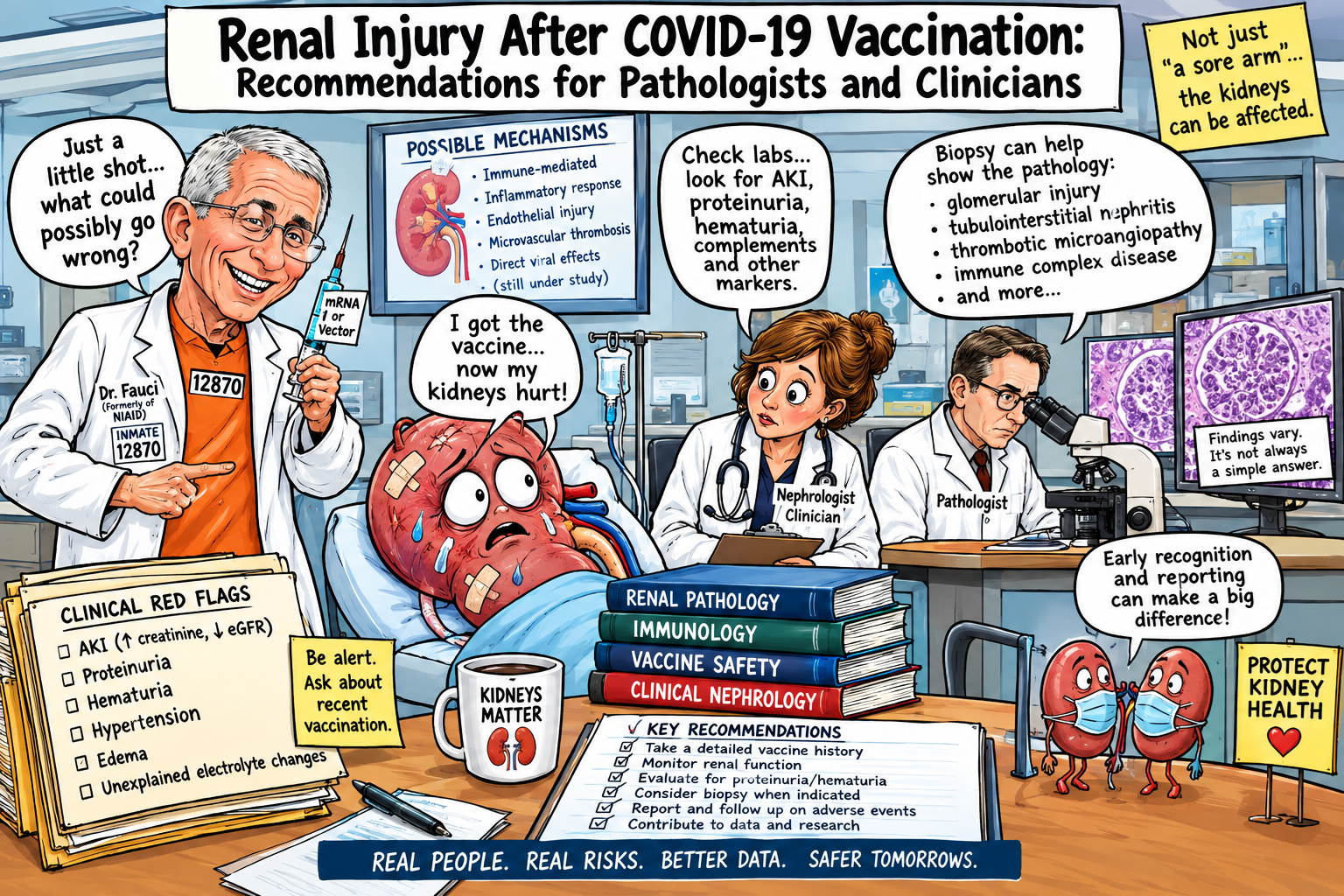
The global deployment of SARS-CoV-2 vaccines has been accompanied by reports of acute kidney injury (AKI), glomerular disease, interstitial nephritis, podocytopathy and thrombotic microangiopathy (TMA) occurring after vaccination. Most reported cases have been established by ante-mortem kidney biopsy rather than autopsy. Accordingly, the pathological literature presently supports a spectrum of renal lesions occurring in temporal association with vaccination, but does not establish that vaccination is the cause of most or all such lesions. This distinction is particularly important because SARS-CoV-2 infection itself is capable of producing acute tubular injury, endothelial injury, TMA, collapsing glomerulopathy and several immune-mediated glomerular diseases.
The principal pathological phenotypes reported after SARS-CoV-2 vaccination include minimal-change disease, focal segmental and collapsing glomerulosclerosis, IgA nephropathy, necrotising and crescentic glomerulonephritis, ANCA-associated vasculitis, anti-glomerular basement membrane disease, membranous nephropathy, acute tubulointerstitial nephritis and TMA. In biopsy series, glomerular disease has predominated over isolated tubulointerstitial disease. The potential mechanisms are heterogeneous and may include dysregulated adaptive immunity, autoimmunity, complement activation, endothelial injury, platelet activation and podocyte dysfunction rather than direct renal cytotoxicity by vaccine constituents. Demonstration of SARS-CoV-2 spike protein in renal tissue should not be interpreted in isolation as proof of vaccine-mediated injury, and several investigations have failed to demonstrate convincing renal deposition of spike or nucleoprotein.
For pathologists confronted with a death occurring after vaccination, the appropriate approach is therefore not to assign causation from temporal proximity or from nonspecific acute tubular injury. Rather, a comprehensive examination should integrate gross and histological examination, immunofluorescence, electron microscopy, immunohistochemistry, molecular virology, microbiology, complement studies, relevant serology, toxicology and clinical chronology. For clinicians, new haematuria, proteinuria, rapidly increasing serum creatinine, nephrotic syndrome, hypertension, thrombocytopenia or evidence of haemolysis after vaccination should prompt appropriate renal evaluation rather than attribution to an adverse vaccine event or dismissal as coincidental.
Introduction
The kidney occupies a particularly vulnerable position at the intersection of vascular biology, immunology and systemic inflammation. Its extensive capillary network, specialised glomerular filtration barrier, metabolically active tubular epithelium and dependence upon tightly regulated endothelial function make it susceptible to injury from systemic immune activation, complement dysregulation, thrombosis and haemodynamic disturbance.
During the COVID-19 pandemic, acute kidney injury emerged as an important complication of SARS-CoV-2 infection. Autopsy and biopsy studies identified acute tubular injury as the dominant lesion, with additional cases demonstrating thrombotic microangiopathy, endothelial injury, collapsing glomerulopathy and other glomerular abnormalities. Importantly, renal tissue from some COVID-19 autopsy series demonstrated substantial tubular injury without demonstrable SARS-CoV-2 nucleocapsid by immunohistochemistry or viral RNA by in-situ hybridisation, illustrating that renal dysfunction during systemic viral disease cannot automatically be equated with direct viral invasion of the kidney.
The subsequent introduction of SARS-CoV-2 vaccines generated reports of new-onset or recurrent renal disease occurring after immunisation. A systematic review of early reports identified minimal-change disease, IgA nephropathy and vasculitis among the most frequently described lesions, together with TMA and several other glomerular disorders.
The distinction between temporal association, biological plausibility and causation is central. A patient may develop renal disease after vaccination because vaccination triggered the disease; because vaccination coincided with an unrelated disease; because vaccination precipitated an already subclinical process; or because an alternative exposure, including SARS-CoV-2 infection itself, was responsible.
For the practising pathologist, therefore, the question is not simply whether a kidney is abnormal after vaccination. It is:
What lesion is present, what biological pathway produced it, and what evidence exists that vaccination rather than infection, medication, pre-existing disease or another systemic insult initiated that pathway?
The renal lesions of greatest pathological interest
A contemporary review of post-vaccination kidney biopsies found a predominance of glomerular lesions. In one series of 27 patients undergoing native-kidney biopsy after vaccination, 74% had glomerular disease; necrotising glomerulonephritis was the most common individual category. The reported spectrum included podocytopathies, IgA nephropathy, ANCA-associated disease, anti-GBM disease, membranous nephropathy, interstitial nephritis and TMA.
These findings should be understood as a phenotypic spectrum, not as a single vaccine-specific pathological entity.
Acute tubular injury
Acute tubular injury is characterised microscopically by varying degrees of:
- epithelial flattening;
- loss of proximal tubular brush borders;
- tubular dilatation;
- epithelial vacuolisation;
- luminal granular casts;
- epithelial cell detachment;
- focal tubular necrosis;
- regenerative epithelial changes.
At ultrastructural examination, mitochondrial swelling, loss of apical microvilli, disruption of epithelial tight junctions and cytoplasmic injury may be encountered.
ATI is important but nonspecific. It may result from hypotension, sepsis, haemodynamic compromise, nephrotoxic medications, pigment nephropathy, severe systemic inflammation or hypoxia. It is also the predominant renal lesion reported in COVID-19 itself.
Consequently, ATI alone should never be reported as evidence of vaccine-mediated nephrotoxicity.
In an autopsy, this caveat becomes even more important because postmortem autolysis can mimic or obscure acute tubular injury. Sampling interval, warm and cold ischaemia, resuscitation, shock and terminal haemodynamic events must be considered.
Glomerular endothelial injury and thrombotic microangiopathy
The glomerular endothelium is one of the most informative cellular targets when investigating suspected vaccine-associated renal injury.
TMA may demonstrate:
- endothelial swelling;
- narrowing or occlusion of glomerular capillary lumina;
- fibrin thrombi;
- platelet aggregates;
- mesangiolysis;
- glomerular basement-membrane duplication;
- endothelial detachment;
- arteriolar thrombi;
- arteriolar endothelial swelling;
- concentric intimal proliferation in chronic disease;
- cortical ischaemia and infarction.
A published post-vaccination renal biopsy demonstrated endothelial swelling, capillary obstruction, fibrin deposition and glomerular basement-membrane duplication, with platelet thrombi confirmed by CD61 immunostaining. Electron microscopy demonstrated endothelial hypertrophy, capillary occlusion, fibrin deposition and diffuse podocyte foot-process effacement.
Another reported case demonstrated severe endothelial injury by electron microscopy, including loss of endothelial fenestrations, endothelial swelling and capillary occlusion, interpreted as an early TMA pattern. The clinical circumstances, however, included concurrent SARS-CoV-2 infection, illustrating precisely why infection and vaccination cannot be separated merely by chronology.
Complement
Complement activation is a biologically plausible mechanism in endothelial injury. C3 activation can amplify itself through the alternative pathway and ultimately generate terminal complement complex C5b-9. In susceptible individuals, excessive complement activation can damage endothelial cells and promote platelet-rich microvascular thrombosis.
Therefore, suspected TMA should prompt consideration of:
- C3 and C4;
- soluble C5b-9 where clinically available;
- ADAMTS13 activity and inhibitor;
- haemolysis studies;
- peripheral blood smear;
- platelet count;
- LDH;
- haptoglobin;
- bilirubin;
- reticulocyte count;
- renal function;
- complement-regulatory genetic testing in selected cases.
A post-vaccination TMA phenotype is particularly compelling when accompanied by thrombocytopenia, microangiopathic haemolytic anaemia and renal failure. Nevertheless, alternative causes—including complement-mediated atypical HUS, malignant hypertension, pregnancy-associated disease, drugs, infection and autoimmune disease—must be excluded.
Podocyte injury and the filtration barrier
The podocyte is a highly differentiated epithelial cell responsible for maintaining the integrity of the glomerular filtration barrier. Its architecture depends upon an intricate cytoskeleton and slit-diaphragm complex involving proteins including nephrin, podocin and synaptopodin.
Immune perturbation can produce podocyte dysfunction without obvious glomerular structural destruction on routine light microscopy.
The classic manifestation is minimal-change disease, in which light microscopy may appear nearly normal while electron microscopy demonstrates diffuse podocyte foot-process effacement.
The early systematic literature on post-vaccination renal disease identified minimal-change disease as the most frequently reported pathological diagnosis.
The pathobiological hypothesis is not that vaccine material necessarily poisons podocytes directly. Rather, altered T-cell and cytokine signalling may perturb podocyte biology and the slit diaphragm, resulting in increased glomerular permeability.
In more severe podocyte disease, focal segmental glomerulosclerosis or collapsing glomerulopathy may develop.
Collapsing glomerulopathy deserves special attention because it is also strongly associated with SARS-CoV-2 infection and may interact with host susceptibility, particularly APOL1 genetic variation.
IgA nephropathy and immune-complex disease
IgA nephropathy can present after vaccination with:
- haematuria;
- proteinuria;
- acute kidney injury;
- nephritic syndrome;
- or recurrence of previously quiescent disease.
Histologically, mesangial IgA deposition is demonstrated by immunofluorescence, often accompanied by C3.
The pathologist should determine whether the lesion represents:
- genuinely new disease;
- an exacerbation of previously silent IgA nephropathy;
- an incidental background finding;
- or another immune-complex process.
The distinction is particularly important in older adults, in whom low-level IgA deposits can occasionally be found without clinically significant IgA nephropathy.
Necrotising and crescentic glomerulonephritis
Among reported post-vaccination biopsies, crescentic and necrotising GN represents one of the most clinically consequential phenotypes.
Histological examination may reveal:
- segmental fibrinoid necrosis;
- rupture of the glomerular basement membrane;
- cellular crescents;
- fibrocellular crescents;
- fibrous crescents;
- endocapillary proliferation;
- interstitial inflammation;
- tubular injury.
The differential diagnosis includes:
- ANCA-associated vasculitis;
- anti-GBM disease;
- immune-complex GN;
- infection-related GN;
- lupus nephritis;
- drug-induced disease.
Accordingly, renal pathology should always be integrated with:
- MPO-ANCA;
- PR3-ANCA;
- anti-GBM antibodies;
- ANA;
- anti-dsDNA where appropriate;
- C3/C4;
- infectious serologies;
- blood cultures when indicated.
The 2021 KDIGO glomerular disease framework remains an important reference for classification and clinical-pathological correlation.
Acute interstitial nephritis
Acute tubulointerstitial nephritis may demonstrate:
- interstitial oedema;
- lymphocytic infiltration;
- plasma cells;
- eosinophils;
- tubulitis;
- tubular epithelial injury.
A biopsy-confirmed case of acute interstitial nephritis following Pfizer-BioNTech vaccination has been reported, with AKI developing shortly after vaccination.
However, in this setting the medication history is indispensable.
Patients frequently receive multiple drugs around the time of vaccination, including antibiotics, proton-pump inhibitors, NSAIDs and other agents known to cause AIN. Consequently, AIN temporally following vaccination is not synonymous with vaccine-induced AIN.
The pathology establishes the lesion; causation requires the clinical investigation.
Electron microscopy: the underused arbiter
Electron microscopy can transform an apparently nonspecific biopsy into a mechanistically informative specimen.
In suspected post-vaccination disease, the ultrastructural examination should specifically assess:
Endothelium
- swelling;
- loss of fenestrations;
- subendothelial widening;
- capillary occlusion;
- endothelial detachment.
Podocytes
- diffuse or segmental foot-process effacement;
- podocyte hypertrophy;
- cytoskeletal abnormalities;
- detachment.
Glomerular basement membrane
- duplication;
- splitting;
- thickening;
- subendothelial or subepithelial deposits.
Intravascular material
- fibrin;
- platelet aggregates;
- electron-dense thrombotic material.
In one post-vaccination case, electron microscopy demonstrated endothelial swelling and loss of fenestrations that were not fully appreciated by light microscopy, leading to recognition of early TMA rather than minimal-change disease.
For the renal pathologist, this is an important practical lesson: when clinical circumstances suggest TMA or podocytopathy, electron microscopy should not be regarded as optional merely because light microscopy appears relatively bland.
Immunofluorescence
A complete renal immunofluorescence panel should ordinarily include:
- IgG;
- IgA;
- IgM;
- C3;
- C1q;
- fibrin/fibrinogen;
- kappa;
- lambda.
Additional stains should be selected according to the differential diagnosis.
Examples include:
IgA + C3: IgA nephropathy.
Granular IgG/C3 along capillary loops: membranous nephropathy or another immune-complex process.
Linear IgG: anti-GBM disease.
Pauci-immune pattern: ANCA-associated GN.
Fibrin/platelet-associated staining: supportive of thrombotic injury in appropriate morphological circumstances.
Immunohistochemistry and the question of spike protein
One of the most frequently proposed mechanisms is direct renal exposure to vaccine-associated spike antigen.
This hypothesis should be tested experimentally rather than assumed.
Potential targets include:
- SARS-CoV-2 spike;
- S1;
- nucleocapsid.
However, antibody specificity, tissue controls and assay validation are critical. Cross-reactivity and nonspecific staining can create misleading results.
Indeed, a detailed investigation of new-onset kidney disease after vaccination did not find definitive renal deposition of spike protein or nucleoprotein in the examined biopsy material. The investigators therefore considered an abnormal immune response more plausible than direct deposition as the principal mechanism.
This negative evidence is scientifically important.
Absence of renal spike does not prove absence of vaccine causality, because an immune-mediated mechanism would not require persistent renal antigen. Conversely, detection of spike staining alone would not prove causation.
Molecular pathology and genomics
A comprehensive investigation of unexplained severe renal injury could extend beyond conventional microscopy.
Host genomic analysis
Depending upon the phenotype, potentially informative analyses include:
- APOL1, particularly in collapsing glomerulopathy;
- complement-regulatory genes;
- alternative complement pathway genes;
- HLA genotype;
- selected autoimmune susceptibility loci;
- thrombosis and endothelial-disorder genes.
The purpose is not to find a “vaccine injury gene”, for which there is currently no established diagnostic test, but to identify pre-existing susceptibility that might explain an unusually severe response to an immune stimulus.
Transcriptomics
RNA sequencing of preserved tissue could assess:
- interferon signalling;
- JAK/STAT activation;
- NF-κB pathways;
- complement activation;
- endothelial activation;
- coagulation pathways;
- oxidative stress;
- tubular injury;
- podocyte stress.
In an ideal research design, renal tissue would be compared with:
- vaccinated individuals without renal disease;
- patients with fatal SARS-CoV-2 infection;
- patients with non-COVID AKI;
- age- and comorbidity-matched controls.
Such comparative biology would be considerably more informative than an isolated autopsy.
Microbiology and molecular virology
The exclusion of SARS-CoV-2 infection is essential.
SARS-CoV-2 infection can itself cause:
- acute tubular injury;
- endothelial injury;
- TMA;
- collapsing glomerulopathy;
- immune-mediated glomerular disease.
Autopsy work has demonstrated that severe COVID-associated renal injury can occur without detectable viral nucleocapsid or viral RNA in kidney tissue, supporting indirect systemic mechanisms in at least some cases.
In a suspected vaccine-associated death, testing should therefore include, where appropriate:
- SARS-CoV-2 RT-PCR;
- viral RNA in-situ hybridisation;
- nucleocapsid immunohistochemistry;
- viral sequencing;
- blood or respiratory specimens obtained before death;
- serological history distinguishing vaccination from infection where possible.
Broad molecular testing can also evaluate:
- CMV;
- EBV;
- BK virus;
- adenovirus;
- bacterial pathogens;
- fungi.
This is particularly important when necrotising or immune-complex lesions are present.
What constitutes an adequate renal autopsy examination?
When renal failure is unexplained and vaccination is part of the clinical history, the autopsy should not be restricted to representative cortex.
At minimum, the pathologist should obtain:
Both kidneys
- external examination;
- weight;
- cortical thickness;
- corticomedullary distinction;
- vascular examination;
- representative cortex;
- corticomedullary junction;
- medulla;
- any focal lesion.
Histology
Multiple paraffin blocks should be prepared to permit evaluation of:
- glomeruli;
- tubules;
- interstitium;
- arteries;
- arterioles;
- peritubular capillaries.
Special studies
- PAS;
- Jones methenamine silver;
- trichrome;
- Congo red where indicated;
- iron stain where indicated.
Immunofluorescence
Preferably performed on appropriately preserved tissue obtained at autopsy.
Electron microscopy
Particularly where TMA, podocytopathy, immune-complex disease or endothelial injury is suspected.
Immunohistochemistry
Including selected endothelial, platelet, complement and viral markers.
Molecular microbiology
SARS-CoV-2 and other relevant pathogens.
The pathologist should document the postmortem interval because autolysis can substantially compromise interpretation of tubular epithelial morphology.
Autopsy findings must be interpreted against the background kidney
The elderly patient frequently has pre-existing renal disease.
Diabetic nephropathy, hypertensive nephrosclerosis, atherosclerosis, chronic tubulointerstitial scarring, previous infection, obstructive disease and age-related glomerulosclerosis can all substantially reduce renal reserve.
An autopsy series of patients who died with COVID-19 demonstrated frequent chronic renal abnormalities, including diabetic nephropathy, tubulointerstitial scarring and vascular sclerosis. Acute tubular injury was sometimes difficult to evaluate because of postmortem autolysis.
Accordingly, the critical question is not simply:
“Is there renal disease?”
but:
“Is there an acute lesion superimposed on the individual’s pre-existing renal substrate, and is that acute lesion temporally and biologically compatible with the clinical event under investigation?”
Differential diagnosis in the post-vaccination patient
The following differential should be systematically considered:
| Pathological phenotype | Important alternative causes |
|---|---|
| Acute tubular injury | Sepsis, shock, dehydration, nephrotoxins, COVID-19 |
| TMA | Complement-mediated aHUS, malignant hypertension, drugs, infection, autoimmune disease |
| Minimal-change disease | Idiopathic disease, malignancy, medications, infection |
| FSGS/collapsing GN | APOL1-associated disease, infection, drugs, haemodynamic stress |
| IgA nephropathy | Coincidental disease, infection, systemic immune activation |
| Crescentic GN | ANCA vasculitis, anti-GBM disease, infection, lupus |
| AIN | Antibiotics, PPIs, NSAIDs, other drugs |
| Membranous nephropathy | Primary PLA2R-associated disease, malignancy, autoimmune disease |
| Immune-complex GN | Infection, lupus, autoimmune disease |
| Renal infarction | Atrial fibrillation, atherosclerosis, embolism, hypercoagulability |
This differential is essential because causal attribution becomes progressively weaker as competing explanations accumulate.
Recommendations for practicing physicians
The appropriate response to renal abnormalities following vaccination is neither reflexive attribution nor reflexive dismissal.
1. Establish a baseline
Where clinically appropriate, obtain or review:
- serum creatinine;
- eGFR;
- urinalysis;
- urine albumin/protein;
- blood pressure;
- medication history;
- prior renal diagnoses.
The value of a post-event creatinine is substantially increased when a reliable pre-event baseline exists.
2. Investigate hematuria and proteinuria
New persistent:
- haematuria;
- proteinuria;
- nephrotic-range proteinuria;
- RBC casts;
- rapidly rising creatinine
should trigger renal evaluation.
3. Look specifically for TMA
If AKI occurs with:
- thrombocytopenia;
- anaemia;
- elevated LDH;
- reduced haptoglobin;
- schistocytes;
- neurological abnormalities;
- severe hypertension,
TMA should be considered urgently.
ADAMTS13 testing, complement assessment and haematological evaluation may be lifesaving.
4. Exclude infection
SARS-CoV-2 infection should be assessed because infection itself can produce many of the renal phenotypes attributed temporally to vaccination.
5. Review every medication
The temporal association should not terminate the medication review.
NSAIDs, antibiotics, proton-pump inhibitors, diuretics and numerous other agents can cause renal injury.
6. Refer for nephrology evaluation when indicated
Rapidly progressive renal dysfunction, nephrotic syndrome, active urinary sediment, suspected vasculitis, TMA or unexplained AKI should prompt nephrology consultation.
7. Consider biopsy early when the result could change management
A kidney biopsy can distinguish among:
- ATI;
- AIN;
- minimal-change disease;
- FSGS;
- IgA nephropathy;
- membranous disease;
- crescentic GN;
- TMA;
- other glomerulopathies.
The published literature specifically supports early biopsy in selected patients with new renal disease after vaccination because clinical manifestations alone cannot reliably identify the underlying lesion.
Current KDIGO guidance provides the broader framework for evaluation and management of AKI and glomerular diseases; the 2026 AKI/AKD guideline is presently in public-review draft form.
Recommendations for pathologists
The pathology report should distinguish three separate statements:
1. Morphological diagnosis
For example:
“Acute thrombotic microangiopathy with glomerular and arteriolar fibrin thrombi.”
2. Clinicopathological interpretation
For example:
“The findings are compatible with severe renal microvascular endothelial injury.”
3. Causality
The report should generally avoid stating:
“This lesion was caused by vaccination.”
unless there is extraordinary evidence supporting that conclusion.
A more defensible formulation is:
“The lesion occurred in temporal association with SARS-CoV-2 vaccination. The histological findings are not specific for vaccine-associated injury. Correlation with clinical chronology, SARS-CoV-2 infection status, medication exposure, complement studies, haemolysis parameters and other potential causes of thrombotic microangiopathy is recommended.”
This distinction protects both scientific validity and the integrity of the autopsy report.
A proposed research framework for suspected fatal vaccine-associated renal injury
A particularly valuable research programme would establish a multicentre registry of post-vaccination renal deaths and severe AKI cases.
Each case should contain:
Clinical chronology
- vaccine manufacturer;
- formulation;
- dose number;
- vaccination date;
- onset of symptoms;
- creatinine trajectory;
- proteinuria;
- haematuria;
- blood pressure;
- thrombocytopenia;
- haemolysis.
Pathology
- light microscopy;
- immunofluorescence;
- electron microscopy;
- immunohistochemistry;
- digital pathology.
Molecular studies
- SARS-CoV-2 PCR;
- viral RNA localisation;
- spike/nucleocapsid studies;
- transcriptomics;
- host genomics.
Immunology
- complement;
- ANCA;
- anti-GBM;
- ANA;
- IgA;
- relevant autoantibodies.
Microbiology
- bacterial cultures;
- viral testing;
- fungal studies;
- broad metagenomic sequencing where appropriate.
Exposure data
- NSAIDs;
- antibiotics;
- PPIs;
- contrast;
- other nephrotoxins;
- anticoagulants;
- immunomodulatory drugs.
Controls
The most important scientific component would be appropriate control groups.
A useful design would compare:
vaccine-associated suspected renal injury
versus SARS-CoV-2 infection-associated renal injury
versus non-COVID AKI
versus vaccinated individuals without renal disease
versus age-, sex- and comorbidity-matched controls.
Only such comparative studies can begin to determine whether a particular molecular signature is genuinely associated with vaccination rather than simply representing severe systemic illness.
Conclusions
The renal pathology reported after SARS-CoV-2 vaccination is heterogeneous and encompasses lesions affecting virtually every major compartment of the kidney. The most important reported phenotypes include acute tubular injury, minimal-change disease and other podocytopathies, IgA nephropathy, necrotising and crescentic glomerulonephritis, ANCA-associated disease, acute interstitial nephritis and thrombotic microangiopathy.
The cellular biology is correspondingly heterogeneous. Tubular epithelial cells may undergo acute injury; podocytes may lose filtration-barrier integrity; glomerular endothelial cells may swell and lose fenestrations; complement activation may injure microvascular endothelium; platelets and fibrin may produce microvascular occlusion; and adaptive immune activation may generate immune-complex or pauci-immune glomerular disease.
Nevertheless, there is presently no single histological lesion that can be regarded as pathognomonic of COVID-19 vaccine injury.
This point is particularly important in autopsy practice. Most of the published evidence concerning post-vaccination renal pathology derives from kidney biopsies in living patients, not from rigorously characterised fatal autopsy cohorts. Conversely, substantial autopsy literature exists for COVID-19 infection itself and demonstrates acute tubular and vascular injury that can resemble some of the lesions reported after vaccination.
The appropriate scientific position is therefore neither to dismiss reported renal events nor to infer causation from chronology alone.
For pathologists, the imperative is phenotypic precision: identify the exact lesion, determine its cellular compartment, establish its ultrastructural and immunopathological characteristics, and investigate competing causes.
For physicians, the imperative is clinical vigilance: unexplained AKI, haematuria, proteinuria, nephrotic syndrome, rapidly progressive renal dysfunction, thrombocytopenia or microangiopathic haemolysis warrants investigation regardless of its temporal relationship to vaccination.
For researchers, the principal unmet need is comparative molecular pathology. Carefully controlled autopsy and biopsy studies integrating histology, electron microscopy, immunopathology, genomics, transcriptomics, complement biology and molecular microbiology could determine whether particular renal injury phenotypes occur more frequently after vaccination, identify susceptible individuals and distinguish immune-mediated adverse events from coincidental renal disease.
Such an approach would move the discussion from pharmacovigilance signals and individual case reports toward the standard demanded by modern clinical pathology: reproducible morphology, mechanistic evidence, appropriate controls and disciplined causal inference.
References
- Kanduri SR, et al. New-onset and relapsed kidney histopathology following COVID-19 vaccination: a systematic review. Kidney Int Rep. 2021. PMID: 34835183.
- Renal side effects of COVID-19 vaccination. Systematic review of 130 reported renal adverse events, including minimal-change disease, IgA nephropathy, ANCA-associated vasculitis and acute interstitial nephritis. [Journal details indexed in PubMed]. PMID: 36366292.
- Sharma P, et al. Histopathological findings and clinicopathologic correlation in COVID-19: a systematic review. Renal pathology review incorporating autopsy and biopsy studies.
- Pathophysiology of COVID-19-associated acute kidney injury. Review of renal autopsy and biopsy findings, including ATI, endothelial injury, TMA and collapsing glomerulopathy.
- Postmortem kidney pathology findings in patients with COVID-19. Autopsy study of 42 patients demonstrating predominantly acute tubular injury and focal renal fibrin thrombi.
- Autopsy findings in 32 patients with COVID-19: a single-institution experience. Demonstrated chronic renal disease, TMA and C5b-9-associated endothelial injury.
- Renal thrombotic microangiopathy following COVID-19 vaccination. Renal biopsy demonstrating endothelial injury, fibrin deposition and thrombotic lesions.
- Endothelial injury and thrombotic microangiopathy following SARS-CoV-2 vaccination. Renal biopsy/electron-microscopic investigation of endothelial injury.
- Tanaka F, Katayama K, Joh K, et al. Minimal change disease with thrombotic microangiopathy following the Pfizer-BioNTech COVID-19 vaccine. Clin Kidney J. 2022;15:567–568. doi:10.1093/ckj/sfab234. PMID: 35198160.
- Minimal change disease following COVID-19 vaccination: a systematic review. PLoS One. 2024. PMID: 38442131; PMCID: PMC10914286.
- Minimal change disease and COVID-19 vaccination: four cases and review of literature. [Nephrology literature]. PMID: 35923765.
- Collapsing glomerulopathy in patients with COVID-19. Reports linking SARS-CoV-2-associated collapsing glomerulopathy with APOL1 risk variation and interferon-associated podocyte injury.
- Lim J-H, et al. New-onset kidney diseases after COVID-19 vaccination: a case series. Vaccines. 2022;10:302. doi:10.3390/vaccines10020302. PMID: 35214760.
- A systematic review of perinuclear antineutrophil cytoplasmic antibody-associated glomerulonephritis following coronavirus disease 2019 vaccination: a 2024 update. Thirty-five reported patients; predominantly mRNA vaccines and MPO-ANCA disease. PMID: 38817489.
- Thammathiwat T, Banjongjit A, Iampenkhae K, Townamchai N, Kanjanabuch T. ANCA-associated glomerulonephritis following SARS-CoV-2 vaccination: a case series and systematic review. Vaccines (Basel). 2023;11:983. doi:10.3390/vaccines11050983. PMID: 37243087.
- Acute interstitial nephritis following COVID-19 vaccination. Biopsy-confirmed reports of acute tubulointerstitial nephritis following SARS-CoV-2 vaccination.
- New insights into kidney disease after COVID-19 infection and vaccination: histopathological and clinical findings. Study examining renal pathology and viral antigen in post-vaccination kidney disease; no definitive spike or nucleoprotein deposition was demonstrated in examined renal biopsies. PMID: 37402613.
- New-onset and relapsed kidney histopathology following COVID-19 vaccination. The original systematic review identified 48 reported cases, with MCD, IgA nephropathy and vasculitis predominating.
- New-onset biopsy-proven glomerular disease following COVID-19 vaccination. IgA nephropathy and ANCA-associated RPGN were the most frequent phenotypes in the reported cohort; the authors emphasised that further studies are necessary to establish causality. PMID: 39124761.
- Renal complications following COVID-19 vaccination: a narrative literature review. Review of proposed mechanisms including dysregulated T-cell responses, cytokine activation, molecular mimicry, delayed hypersensitivity and NET-associated pathways. PMID: 37323744.
Footnotes
Footnote 1 — Terminology. The term “post-vaccination renal injury” is used here to denote renal disease occurring after vaccination. It does not imply that vaccination caused the disease.
Footnote 2 — Autopsy versus biopsy. Published evidence concerning renal lesions after COVID-19 vaccination is predominantly derived from renal biopsies obtained during life. Evidence from complete autopsy series specifically demonstrating fatal vaccine-caused renal injury remains comparatively limited.
Footnote 3 — Acute tubular injury. ATI is a morphological diagnosis rather than an aetiological diagnosis. It should not be interpreted as evidence of vaccine nephrotoxicity without supporting clinical and mechanistic evidence.
Footnote 4 — Spike antigen. Immunohistochemical detection of spike protein is not, in isolation, proof that vaccine-derived antigen caused renal injury. Assay validation, appropriate negative and positive controls, localisation and correlation with nucleocapsid testing are essential.
Footnote 5 — SARS-CoV-2 infection as a confounder. COVID-19 itself produces several renal lesions that overlap with those reported after vaccination, making exclusion or documentation of infection essential in causality assessment.
Footnote 6 — Genetic susceptibility. APOL1 and complement-pathway variants may identify susceptibility to particular renal phenotypes but cannot establish that vaccination caused the initiating injury.
Footnote 7 — Causality. A robust attribution requires temporal plausibility, biological plausibility, pathological compatibility, exclusion of competing causes, reproducibility and, ideally, controlled epidemiological evidence.
Footnote 8 — Clinical management. The presence of renal disease following vaccination does not imply that vaccination should be discontinued universally. Decisions regarding subsequent vaccination should be individualised according to the specific renal diagnosis, severity of the event, evidence for causality, infection risk and specialist assessment.