
John Murphy, CEO The COVID-19 Long haul Foundation
Abstract
The neurological consequences of infection with severe acute respiratory syndrome coronavirus 2 (SARS-CoV-2) extend beyond the central nervous system and include a heterogeneous spectrum of peripheral nervous-system disorders. These manifestations range from acute inflammatory polyradiculoneuropathies, including Guillain–Barré syndrome (GBS), to cranial neuropathies, critical-illness–associated axonal injury, and persistent sensory, autonomic, and small-fiber neuropathies associated with post-acute sequelae of COVID-19 (PASC), commonly termed Long COVID. Among the latter, small-fiber neuropathy (SFN) has emerged as a particularly important phenotype because conventional nerve-conduction studies may remain normal despite substantial sensory and autonomic dysfunction.
The evidence accumulated since 2020 supports several biologically plausible relationships between SARS-CoV-2 infection and peripheral nerve dysfunction, including post-infectious immune dysregulation, neuroinflammation, microvascular injury, endothelial dysfunction, autonomic disturbance, and potentially direct or indirect effects on peripheral neural tissues. The evidence is strongest for an association between COVID-19 and GBS in the acute or early post-infectious period and for an association between Long COVID and clinically suspected or biopsy-confirmed SFN. However, the magnitude of risk, causal mechanisms, and optimal treatment of PASC-associated neuropathy remain incompletely established.
Recent cohort and case-control investigations have provided objective evidence of reduced intraepidermal nerve-fiber density, autonomic abnormalities, altered quantitative sensory testing, and neurovascular dysfunction in subsets of patients with Long COVID. These findings are important because they move the discussion of post-COVID neuropathic symptoms beyond subjective symptom inventories toward demonstrable abnormalities of peripheral nerve structure and function. Nevertheless, selection bias, heterogeneous diagnostic criteria, absence of appropriate controls in many studies, pre-existing metabolic risk factors, and the referral nature of several cohorts limit generalizability.
A rational clinical approach therefore requires careful phenotyping, exclusion of common causes of neuropathy, appropriate use of electrodiagnostic testing and skin biopsy, assessment for autonomic dysfunction when clinically indicated, and recognition that normal nerve-conduction studies do not exclude SFN. Treatment should presently remain phenotype-directed and evidence-based, with symptomatic management constituting the foundation of care. Immunotherapies such as intravenous immunoglobulin remain investigational for Long COVID-associated SFN despite encouraging preliminary observations.
Keywords: COVID-19; SARS-CoV-2; Long COVID; post-acute sequelae of SARS-CoV-2 infection; peripheral neuropathy; small-fiber neuropathy; autonomic neuropathy; Guillain–Barré syndrome; neuroinflammation; dysautonomia.
Introduction
The neurological phenotype of COVID-19 is considerably broader than the encephalopathy, anosmia, dysgeusia, and cerebrovascular complications initially recognized during the pandemic. SARS-CoV-2 infection may affect the peripheral nervous system as well as the brain and spinal cord, producing disorders involving sensory, motor, autonomic, cranial, and peripheral nerve structures. A systematic review of peripheral nervous-system involvement identified 1,320 neurological events among 1,004 patients reported in 225 studies; 350 events were classified as peripheral nervous-system manifestations and another 165 involved both peripheral and cranial nerves. Guillain–Barré syndrome and related disorders represented the dominant peripheral neuropathic phenotype in that early literature.
The clinical problem subsequently changed as increasing numbers of patients reported persistent neurological symptoms after the acute infection had resolved. The World Health Organization recognizes post-COVID-19 condition as a syndrome occurring following probable or confirmed infection, generally beginning approximately three months after the initial illness and persisting for at least two months when another diagnosis does not adequately explain the symptoms. Neurological manifestations are among the recognized sequelae.
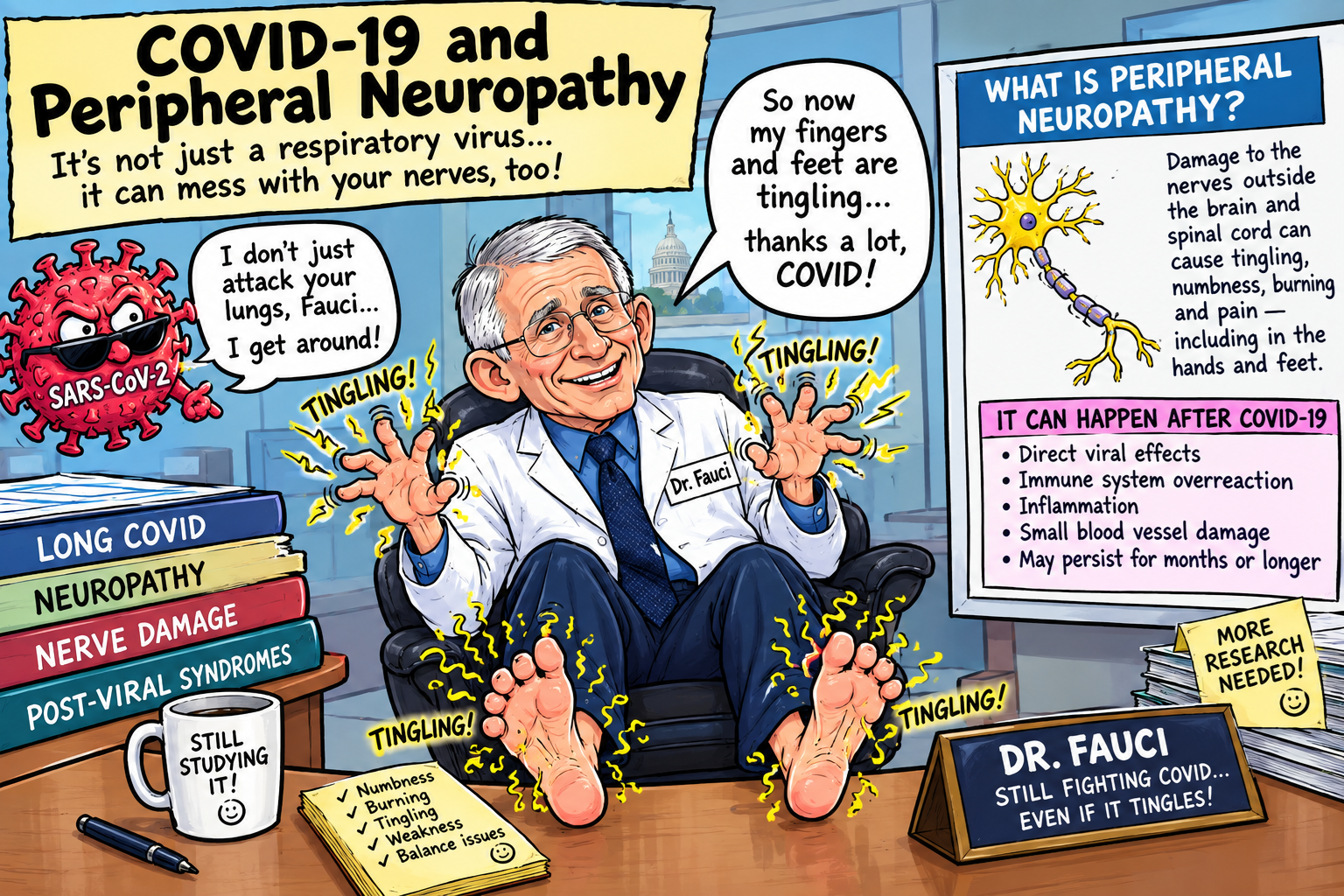
Among patients with PASC, neuropathic symptoms—including burning pain, electric or stabbing sensations, paresthesia, numbness, dysesthesia, altered temperature sensation, and autonomic dysfunction—have attracted increasing scientific attention. Small-fiber neuropathy is particularly relevant because the unmyelinated C fibers and thinly myelinated Aδ fibers subserve pain, temperature, sudomotor, vasomotor, and other autonomic functions and therefore can produce profound disability without abnormalities on routine nerve-conduction studies.
The emerging literature consequently suggests that “COVID-related neuropathy” should not be regarded as a single disease. It encompasses several mechanistically and clinically distinct disorders.
1. The Peripheral Nervous System in COVID-19
Peripheral neuropathy may involve sensory fibers, motor fibers, autonomic fibers, roots, plexuses, cranial nerves, or combinations thereof. COVID-19-associated peripheral neurological disease can broadly be divided into at least five phenotypes:
- Acute inflammatory neuropathies, particularly GBS and its variants;
- Small-fiber neuropathy, frequently accompanied by autonomic dysfunction;
- Large-fiber polyneuropathy, including axonal neuropathies;
- Critical-illness–associated neuromuscular injury, particularly in severely ill or mechanically ventilated patients;
- Focal or multifocal neuropathies, including cranial neuropathies, mononeuropathies, plexopathies, and compressive injuries occurring during prolonged hospitalization.
These categories can overlap.
The distinction is clinically consequential because their diagnostic tests, mechanisms, prognosis, and treatments differ substantially.
2. Guillain–Barré Syndrome
GBS is the best-characterized acute peripheral neuropathic disorder reported in association with COVID-19.
The syndrome is an acute immune-mediated polyradiculoneuropathy characterized by progressive weakness, reduced or absent reflexes, sensory symptoms, and, frequently, autonomic dysfunction. Severe cases can involve respiratory muscle weakness and require mechanical ventilation.
Early reports consisted predominantly of case reports and small case series. Subsequent systematic reviews substantially enlarged the evidence base. A 2023 systematic review encompassing 156 studies and 436 reported patients found that neurological symptoms of GBS began, on average, approximately 19 days after COVID-19. The most frequent manifestations were generalized weakness, reduced reflexes, facial paresis or paralysis, and hypoesthesia. Acute inflammatory demyelinating polyneuropathy was the predominant electrophysiological subtype, with smaller numbers of acute motor axonal, acute motor-sensory axonal, pharyngeal-cervical-brachial, and Miller Fisher presentations.
An earlier systematic review of 73 cases found that the clinical and electrophysiological characteristics generally resembled conventional post-infectious GBS. Cerebrospinal-fluid albuminocytologic dissociation was present in approximately 71% of reported cases, whereas SARS-CoV-2 RNA was absent from CSF in the patients in whom it was tested. More than 70% had a favorable outcome, particularly following standard GBS treatment.
These observations favor an immune-mediated or post-infectious mechanism rather than direct viral invasion of peripheral nerves.
Importantly, association does not establish that SARS-CoV-2 caused every reported case. GBS occurs spontaneously and following numerous infections, and the early pandemic generated extraordinary reporting intensity. Robust epidemiological comparisons are therefore essential when estimating excess risk attributable specifically to SARS-CoV-2.
3. Small-Fiber Neuropathy: An Emerging Long-COVID Phenotype
The more persistent and clinically challenging peripheral neurological phenotype is small-fiber neuropathy.
Small fibers include unmyelinated C fibers and thinly myelinated Aδ fibers. They participate in nociception, thermal sensation, sudomotor regulation, vasomotor control, and visceral/autonomic signaling. Damage can consequently produce symptoms that appear simultaneously neurological, vascular, and autonomic.
Patients may describe:
- burning feet or hands;
- electric-shock sensations;
- pins and needles;
- painful hypersensitivity;
- allodynia;
- numbness;
- altered temperature perception;
- unexplained itching or dysesthesia;
- sweating abnormalities;
- color or temperature changes in extremities;
- orthostatic intolerance;
- palpitations;
- gastrointestinal dysmotility;
- bladder dysfunction;
- sexual dysfunction;
- abnormal heart-rate responses; and
- fatigue or exercise intolerance.
The clinical phenotype is important because conventional electromyography and nerve-conduction studies predominantly evaluate large myelinated fibers. A patient can therefore have substantial small-fiber pathology while having a “normal EMG.”
A 2021 study of 13 patients who developed new paresthesias during or after SARS-CoV-2 infection found that all developed symptoms within two months of infection. Six had skin-biopsy-confirmed SFN, while several others demonstrated autonomic abnormalities despite normal biopsy findings. The authors concluded that SFN could underlie at least some of the persistent paresthesias observed in Long COVID.
Subsequent evidence has strengthened that observation.
A 2022 evaluation of patients with prolonged Long COVID found that 59% had at least one test confirming neuropathy. Skin biopsy was abnormal in 63% of tested participants, whereas only 17% of those undergoing electrodiagnostic testing had abnormal results. At least ten participants received a diagnosis of SFN. Longitudinal improvement averaged approximately 52%, although complete resolution was not reported.
These findings illustrate a fundamental diagnostic principle:
A normal nerve-conduction study does not exclude clinically important post-COVID small-fiber neuropathy.
4. Objective Evidence From Skin Biopsy
Skin biopsy provides an objective method of evaluating small-fiber integrity by quantifying intraepidermal nerve-fiber density (IENFD), typically using immunohistochemical staining of distal-leg and sometimes proximal specimens.
In conventional SFN, reduced IENFD supports a structural small-fiber abnormality. The technique is particularly useful in patients whose symptoms strongly suggest SFN but whose conventional electrophysiological studies are normal.
A 2025 prospective-style cohort of patients with Long COVID and persistent neuropathic pain provides important contemporary evidence. Eighteen patients met initial criteria and 17 underwent analysis. Fourteen of the 17 patients—82%—had abnormal skin-biopsy results. Seventy percent reported autonomic symptoms, and every patient with autonomic complaints had an abnormal biopsy. At six months, ten of 17 reported improvement in pain and/or dysautonomia.
The study is small and highly selected; it therefore cannot establish the prevalence of SFN among all patients with Long COVID. Nevertheless, its multimodal design—incorporating skin biopsy, quantitative sensory testing, laser-evoked potentials, and electrochemical skin conductance—provides stronger physiological evidence than symptom reporting alone.
5. Small-Fiber Neuropathy and Dysautonomia
The intersection between SFN and autonomic dysfunction may be particularly important in PASC.
Small autonomic fibers regulate vascular tone, heart rate, sweating, gastrointestinal motility, bladder function, and other involuntary physiological processes. Their dysfunction can therefore produce an apparently disparate collection of symptoms.
A study comparing 87 patients with post-COVID condition, 50 patients with ME/CFS, and 50 healthy controls found evidence of autonomic abnormalities and small-fiber dysfunction in the post-COVID group. POTS was identified in 13.8% of patients with post-COVID condition, while abnormal Sudoscan findings occurred in 19.5%. The investigators also found evidence compatible with non-length-dependent SFN.
Earlier Mayo Clinic experience similarly documented autonomic dysfunction after COVID-19. Among 27 patients evaluated for suspected post-COVID autonomic dysfunction, symptoms developed from the time of infection to 122 days afterward. Lightheadedness was reported by 93%, while objective abnormalities involved sudomotor, cardiovagal, and cardiovascular adrenergic testing. Twenty-two percent fulfilled POTS criteria.
These observations provide a plausible physiological bridge between neuropathic sensory symptoms and systemic manifestations such as orthostatic intolerance, tachycardia, abnormal sweating, gastrointestinal disturbance, and exercise intolerance.
6. Proposed Mechanisms
6.1 Immune Dysregulation
The leading mechanistic hypothesis for post-COVID SFN is immune-mediated injury.
SARS-CoV-2 infection generates a complex innate and adaptive immune response. In susceptible individuals, persistent or aberrantly regulated immune activation could damage peripheral axons either directly or indirectly through inflammatory mediators, vascular injury, autoantibodies, or altered neuroimmune signaling.
The hypothesis is consistent with the post-infectious timing of many neuropathic syndromes and with the established immune pathogenesis of disorders such as GBS.
It should nevertheless be emphasized that the existence of immune abnormalities in Long COVID does not establish that immune-mediated nerve destruction is the universal mechanism. Long COVID is biologically heterogeneous, and more than one mechanism may operate simultaneously.
6.2 Microvascular and Endothelial Dysfunction
Peripheral nerves are highly metabolically active structures dependent upon an extensive microvascular supply. Endothelial dysfunction, impaired perfusion, inflammation, and microvascular abnormalities could therefore compromise nerve function even in the absence of gross vascular occlusion.
This mechanism is particularly attractive for explaining combinations of neuropathy, dysautonomia, exercise intolerance, and other manifestations of PASC.
The hypothesis remains biologically plausible but should not be converted into the categorical claim that “microclots cause Long COVID neuropathy.” Direct causal evidence remains incomplete.
6.3 Neuroinflammation
Inflammatory signaling involving cytokines, chemokines, immune cells, and neurovascular interfaces may alter peripheral nerve function. Neuroinflammatory processes could produce pain and sensory dysfunction without necessarily causing immediately demonstrable axonal degeneration.
This may help explain why some patients experience prominent neuropathic symptoms while conventional structural testing remains relatively unrevealing.
6.4 Autonomic Dysfunction
Autonomic dysfunction may be both a consequence and a perpetuating component of small-fiber disease.
Damage to autonomic fibers can impair vascular constriction during standing, alter sweating, affect gastrointestinal motility, and disturb cardiovascular reflexes. Conversely, chronic autonomic dysregulation may itself alter tissue perfusion and physiological homeostasis.
The relationship is therefore likely bidirectional rather than linear.
6.5 Direct Viral Neurotropism
SARS-CoV-2 has demonstrated effects on neural tissues, but direct infection of peripheral nerves is not required to explain most post-COVID neuropathies.
The available evidence for GBS, for example, favors an immune-mediated process: early systematic reviews found CSF SARS-CoV-2 RNA absent in tested cases and clinical timing frequently compatible with a post-infectious process.
Accordingly, claims that persistent peripheral neuropathy is simply caused by ongoing direct viral infection of peripheral nerves remain unproven.
7. Large-Fiber and Critical-Illness Neuropathy
Not every post-COVID neuropathy is small-fiber disease.
Patients who experience severe COVID-19 requiring prolonged intensive-care treatment may develop critical-illness polyneuropathy or critical-illness myopathy. These conditions can produce profound weakness, impaired ambulation, respiratory muscle weakness, and prolonged rehabilitation requirements.
COVID-19 may also coexist with conventional causes of neuropathy, including:
- diabetes mellitus;
- renal dysfunction;
- vitamin B12 deficiency;
- alcohol exposure;
- thyroid disease;
- chemotherapy;
- paraproteinemia;
- nutritional deficiency;
- autoimmune disease;
- medication toxicity; and
- mechanical nerve compression.
Consequently, temporal association with SARS-CoV-2 infection should not automatically be interpreted as proof of causation.
The appropriate clinical question is not simply:
“Did the patient have COVID?”
but rather:
“Does the patient’s neuropathic phenotype, timing, objective testing, and exclusion of competing causes support a causal relationship to SARS-CoV-2 infection?”
8. Clinical Diagnosis
Diagnosis begins with careful phenotyping.
A detailed neurological history should establish:
- onset relative to SARS-CoV-2 infection;
- distribution of symptoms;
- distal versus proximal involvement;
- sensory versus motor manifestations;
- autonomic symptoms;
- progression versus fluctuation;
- orthostatic symptoms;
- bowel and bladder involvement;
- sexual dysfunction;
- sleep disturbance;
- prior neuropathy;
- diabetes and metabolic risk;
- alcohol exposure;
- medications;
- nutritional status; and
- family history.
The examination should include strength, reflexes, vibration, proprioception, pinprick, temperature, gait, coordination, cranial nerves, and autonomic findings where appropriate.
Conventional electrodiagnostic testing
Nerve-conduction studies and electromyography are indispensable when large-fiber neuropathy, radiculopathy, motor disease, or GBS is suspected.
They are, however, insensitive to isolated SFN.
Consequently, a normal study should not terminate evaluation when a convincing small-fiber phenotype remains.
Skin biopsy
IENFD measurement remains one of the principal objective tests for SFN.
Quantitative sensory testing
QST can evaluate thermal and sensory thresholds and may provide complementary evidence. In the 2025 Long COVID cohort, QST showed greater sensitivity than several other noninvasive measures, although it remained imperfect.
Autonomic testing
Depending upon the phenotype, evaluation may include:
- tilt-table testing;
- autonomic reflex testing;
- quantitative sudomotor testing;
- heart-rate variability;
- Valsalva testing;
- deep-breathing responses; and
- other specialized autonomic assessments.
No single test establishes PASC-associated SFN in every patient.
9. Differential Diagnosis
Because SFN has numerous causes, the diagnostic evaluation should ordinarily address common alternative etiologies.
Depending upon clinical circumstances, laboratory evaluation may include:
- fasting glucose and HbA1c;
- vitamin B12 and related metabolites when indicated;
- thyroid function;
- serum protein electrophoresis with immunofixation;
- folate;
- complete blood count;
- metabolic profile;
- inflammatory markers;
- selected autoimmune serologies; and
- targeted infectious or toxicological investigations.
The work-up should be individualized rather than indiscriminately extensive.
This issue is particularly important because diabetes, prediabetes, renal disease, nutritional abnormalities, and other metabolic disorders are themselves major causes of peripheral neuropathy. Failure to evaluate them can lead to erroneous attribution of neuropathy to COVID-19.
10. Treatment
There is currently no universally accepted disease-modifying treatment specifically approved for PASC-associated peripheral neuropathy.
Treatment should therefore be individualized according to the phenotype.
Symptomatic treatment
Neuropathic pain may be treated using established approaches that can include:
- gabapentinoids;
- serotonin-norepinephrine reuptake inhibitors;
- selected tricyclic antidepressants;
- topical lidocaine;
- topical capsaicin; and
- nonpharmacological pain-management strategies.
Drug selection must account for renal function, age, drug interactions, falls risk, cognitive effects, and autonomic symptoms.
Rehabilitation
Physical and occupational therapy can be useful for weakness, gait impairment, balance disturbance, and functional limitation. However, patients with post-exertional malaise require individualized pacing rather than indiscriminate graded exercise escalation.
Autonomic management
Management of orthostatic intolerance may include fluid and salt strategies where medically appropriate, compression garments, positional measures, medication review, and selected pharmacological interventions under specialist supervision.
Immunotherapy
Immunomodulatory therapy is one of the most controversial areas.
A 2024 Yale case-control study identified 16 patients with biopsy-confirmed SFN after COVID-19. Nine received IVIG and all nine reported clinical improvement in neuropathic symptoms, compared with three of seven untreated patients; the difference was statistically significant. The investigators appropriately classified the evidence as Class III and called for larger clinical trials.
This result is intriguing but does not establish IVIG as standard therapy.
The study was retrospective, nonrandomized, small, and subject to treatment-selection and other biases. Consequently, IVIG should presently be regarded as an investigational or specialist-directed therapy for selected patients rather than routine treatment for Long COVID neuropathy.
11. Prognosis
The natural history of post-COVID peripheral neuropathy remains incompletely defined.
Some patients improve substantially over months, whereas others experience persistent symptoms for years. In the earlier prolonged-Long-COVID neuropathy cohort, average longitudinal improvement was approximately 52%, but no participant reported complete resolution.
The 2025 multimodal SFN cohort similarly found that approximately 59% of participants reported improvement in pain and/or dysautonomia at six months.
These observations suggest that post-COVID SFN is not necessarily permanent, but neither should it be characterized as uniformly self-limited.
Prognosis probably depends upon the underlying phenotype, severity, autonomic involvement, metabolic comorbidities, duration before diagnosis, and potentially the mechanisms responsible for the neuropathy.
12. COVID-19 Vaccination and the Differential Diagnosis
Reports of neuropathic syndromes following COVID-19 vaccination should be distinguished carefully from neuropathy following SARS-CoV-2 infection.
A 2025 case series described 16 patients with suspected SFN after COVID-19 vaccination, nine of whom fulfilled diagnostic criteria for SFN. The authors emphasized that causality was difficult to establish and that additional surveillance and investigation were required.
Accordingly, temporal proximity alone should not establish causation. Infection itself is a major potential confounder, and neuropathies have multiple spontaneous and immune-mediated causes.
The appropriate scientific position is therefore neither to dismiss such reports nor to treat temporal association as proof. Carefully designed epidemiological studies, objective neurological phenotyping, appropriate control groups, and mechanistic investigation are required.
13. The Larger Significance of Peripheral Neuropathy in Long COVID
Peripheral neuropathy may represent more than an isolated neurological complication.
The convergence of neuropathic pain, autonomic dysfunction, orthostatic intolerance, impaired exercise physiology, fatigue, and cognitive symptoms raises the possibility that peripheral neural dysfunction participates in a larger systems-level disorder.
The 2024 Yale study is notable in this regard because patients with biopsy-confirmed SFN also demonstrated neurovascular dysregulation on invasive cardiopulmonary exercise testing. Ninety-two percent reported post-exertional malaise.
Such findings raise an important hypothesis: small-fiber pathology may constitute one component of a broader neurovascular-autonomic syndrome in a subset of patients with PASC.
This hypothesis remains to be tested prospectively.
It would be particularly valuable for future studies to integrate:
- skin biopsy;
- autonomic testing;
- quantitative sensory testing;
- nerve-conduction studies;
- immunological profiling;
- vascular/endothelial biomarkers;
- metabolic measurements;
- cardiopulmonary exercise testing; and
- longitudinal clinical outcomes.
Such multimodal investigation could determine whether clinically defined SFN identifies a biologically coherent subgroup of Long COVID.
14. Research Priorities
Several major questions remain unanswered.
First, the true prevalence of SFN after SARS-CoV-2 infection is unknown. Referral cohorts cannot provide population prevalence.
Second, diagnostic heterogeneity remains substantial. Studies use different combinations of symptoms, skin biopsy, QST, autonomic testing, and electrodiagnostic criteria.
Third, the causal mechanisms remain unresolved. Immune dysregulation, microvascular injury, autonomic dysfunction, neuroinflammation, metabolic disturbances, and other mechanisms may coexist.
Fourth, biomarkers capable of identifying patients with active peripheral nerve injury are lacking.
Fifth, the natural history requires prospective longitudinal studies beginning immediately after infection and extending for several years.
Finally, treatment trials are urgently needed. The preliminary IVIG findings are sufficiently interesting to justify randomized controlled trials, but not sufficient to establish efficacy.
Conclusion
The neurological consequences of SARS-CoV-2 infection extend substantially beyond the acute respiratory syndrome. Peripheral nervous-system involvement encompasses acute immune-mediated polyradiculoneuropathies such as Guillain–Barré syndrome, critical-illness–associated neuropathy, cranial and focal neuropathies, and a growing body of evidence concerning small-fiber and autonomic dysfunction in Long COVID.
The most important emerging observation is that neuropathic symptoms in Long COVID can correspond to objectively demonstrable abnormalities of peripheral nerve structure and function. Skin-biopsy abnormalities, altered sensory testing, autonomic dysfunction, and neurovascular abnormalities have now been documented in several independent clinical cohorts.
Small-fiber neuropathy deserves particular clinical attention because conventional nerve-conduction studies can remain normal despite substantial disease. Patients presenting with burning pain, paresthesia, numbness, dysesthesia, altered thermal sensation, autonomic symptoms, or orthostatic intolerance after SARS-CoV-2 infection therefore warrant consideration of SFN when more common explanations have been excluded.
At the same time, scientific rigor requires restraint. The existing evidence does not establish that SARS-CoV-2 causes peripheral neuropathy in every patient who develops neuropathic symptoms after COVID-19, nor does it establish a single pathogenic mechanism or a universally effective disease-modifying treatment.
The most defensible conclusion is that SARS-CoV-2 infection can be followed by clinically significant peripheral neuropathic syndromes, including objectively demonstrable small-fiber neuropathy in a subset of patients, and that this phenomenon warrants systematic neurological investigation rather than dismissal as nonspecific post-viral symptomatology.
The field has progressed from anecdotal reports toward objective physiological and pathological evidence. The next step must be adequately powered prospective studies that establish prevalence, causal mechanisms, biological subtypes, prognosis, and effective treatment.
Footnotes and References
1. World Health Organization. A clinical case definition of post COVID-19 condition by a Delphi consensus. WHO; 2021. The WHO definition describes post-COVID condition as symptoms occurring after probable or confirmed infection, usually approximately three months after onset, lasting at least two months, and not otherwise explained.
2. World Health Organization. Neurology and COVID-19: scientific brief. Updated 2024. The WHO review recognizes both acute neurological manifestations and neurological sequelae associated with post-COVID condition.
3. Hernández-Fernández F, et al. Peripheral nervous system involvement associated with COVID-19: a systematic review of the literature. This review identified 225 eligible publications involving 1,004 patients and 1,320 neurological events.
4. Abu-Rumeileh S, et al. Guillain–Barré syndrome spectrum associated with COVID-19: an up-to-date systematic review of 73 cases. J Neurol. 2021;268:1133–1170. The review found clinical and electrophysiological patterns broadly resembling conventional GBS and evidence favoring an immune-mediated/post-infectious process.
5. Aladawi M, et al. Guillain-Barré syndrome as a complication of COVID-19: a systematic review. The review identified 109 cases with confirmed infection and found a mean interval of approximately 12 days between COVID-19 and neurological manifestations.
6. Post-COVID-19 small-fiber neuropathy was initially documented in small clinical series. Patients developed new paresthesias during or after SARS-CoV-2 infection, with some demonstrating reduced intraepidermal nerve-fiber density despite normal large-fiber nerve-conduction studies.
7. Oaklander AL. Increasing associations of long-COVID with small-fiber neuropathy. Pain. 2024;165(9)–e95. The review/editorial literature highlights the accumulating association between Long COVID and SFN.
8. Novak P, et al. Peripheral neuropathy evaluations of patients with prolonged Long COVID. Neurol Neuroimmunol Neuroinflamm. The cohort demonstrated objective neuropathy in a substantial subset, with SFN predominating and incomplete recovery over longitudinal follow-up.
9. McAlpine L, Zubair AS, Joseph P, Spudich S. Case-control study of individuals with small fiber neuropathy after COVID-19. Neurol Neuroimmunol Neuroinflamm. 2024;11(3). This retrospective study found biopsy-confirmed SFN and reported an association between IVIG treatment and symptomatic improvement, but the authors classified the evidence as Class III and called for larger trials.
10. Drobinska N, et al. Small Fiber Neuropathy in Long COVID: A Cohort Study with Multimodal Assessment and Follow-Up. Eur Neurol. 2025;88(2):52–63. Among 17 analyzed patients with post-COVID neuropathic pain, 82% had abnormal skin-biopsy findings and 70% had autonomic complaints.
11. Dysautonomia and small fiber neuropathy in post-COVID condition and chronic fatigue syndrome. The comparative study identified autonomic and small-fiber abnormalities among patients with post-COVID condition and demonstrated overlap with ME/CFS phenotypes.
12. Autonomic dysfunction following COVID-19 infection: an early experience. The Mayo Clinic series documented objective autonomic abnormalities and POTS/orthostatic intolerance in patients evaluated after COVID-19.
13. Post-COVID-19 Small Fiber Neuropathy as a New Emerging Quality of Life-Threatening Disease: A Systematic Review. The systematic review describes the emerging phenotype of post-COVID SFN, including distal burning pain, numbness, dysautonomia, and multisystem manifestations, while emphasizing the continuing limitations of the evidence base.
14. Small fiber neuropathy following COVID-19 vaccination: a case series. J Neurol Sci. 2025;474:123536. The authors reported nine diagnostically confirmed SFN cases among 16 evaluated patients but explicitly emphasized the difficulty of establishing causality.
15. The distinction between infection-associated and vaccination-associated neuropathy is essential in epidemiological studies because temporal association alone cannot establish causation. The infection itself is a potential confounder, and peripheral neuropathy has numerous established causes.
16. The present review should not be interpreted as establishing IVIG, corticosteroids, plasma exchange, antivirals, anticoagulants, or other immunomodulatory therapies as standard treatment for PASC-associated SFN. Controlled clinical trials remain necessary to determine efficacy, patient selection, dosing, duration, and risk-benefit relationships.