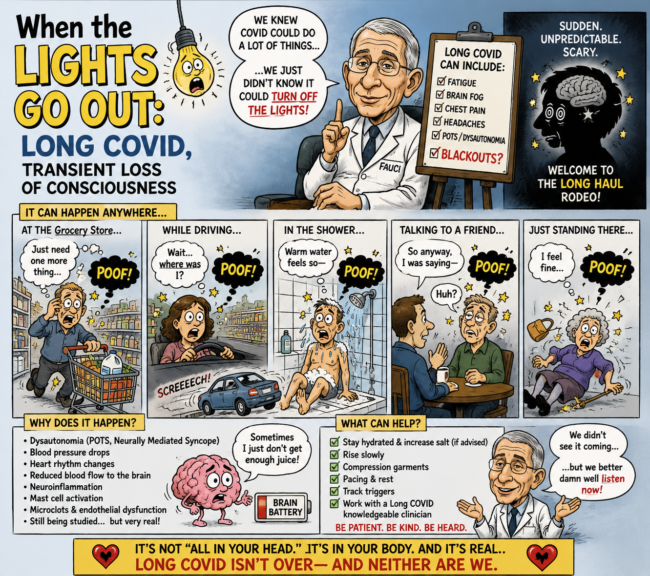
John Murphy, CEO The COVID-19 Long haul Foundation
Abstract
Long COVID, or post-acute sequelae of SARS-CoV-2 infection (PASC), has increasingly revealed itself as a disorder extending well beyond pulmonary injury. Among its most consequential neurological manifestations are dizziness, orthostatic intolerance, autonomic dysfunction, cognitive impairment, altered consciousness, and episodes described by patients as “blackouts.” The word is deceptively simple. A blackout may represent true syncope caused by transient cerebral hypoperfusion; an arrhythmia; orthostatic hypotension or postural tachycardia syndrome (POTS); seizure; impaired cerebral autoregulation; sleep intrusion; or a transient period of profound cognitive disconnection in which the patient remains technically conscious but loses effective awareness of the surrounding world.
The emerging literature provides biological reasons to take these reports seriously. Studies of Long COVID have identified abnormalities in cerebral blood-flow regulation, autonomic function, brain metabolism, white matter, functional connectivity, and neuroinflammation. A 2025 systematic review encompassing 78 neuroimaging and neurophysiological studies and more than 5,900 participants found abnormalities involving cognition, functional connectivity, electrophysiology and cerebral blood flow, although findings were heterogeneous.
The most compelling mechanistic possibility is that, in at least a subset of patients, SARS-CoV-2 leaves behind a disorder of the neurovascular-autonomic system: the machinery responsible for maintaining adequate cerebral perfusion when a person stands, exercises, becomes dehydrated, experiences stress, or undergoes changes in blood pressure and carbon dioxide. When that compensatory system fails, the brain can briefly receive inadequate blood flow. The clinical result can be presyncope, visual dimming, cognitive interruption, collapse—and, in extreme circumstances, loss of consciousness.
This phenomenon deserves greater clinical recognition because a blackout occurring while driving, climbing stairs, bathing, working with machinery, or crossing a road can transform an otherwise invisible neurological disorder into a potentially fatal event.
The Patient’s Description: “Everything Went Black”
There is a particular vocabulary shared by patients with post-COVID neurological illness.
“I suddenly wasn’t there.”
“Everything went black.”
“I was awake, but I couldn’t process anything.”
“I don’t remember what happened for several seconds.”
“I stood up and the room disappeared.”
“I was driving and suddenly I realized I didn’t know where I was.”
These statements are clinically important precisely because they do not map neatly onto a single neurological diagnosis.
The brain does not possess a single “blackout switch.”
Consciousness is produced through the coordinated operation of the cerebral cortex, thalamus, brainstem arousal systems, cerebral circulation, autonomic nervous system, glucose and oxygen delivery, and innumerable supporting metabolic processes. A temporary disturbance anywhere along this network can produce an alteration of consciousness.
Consequently, the first question when a patient reports a Long-COVID blackout should not be “Is this Long COVID?”
It should be:
What physiological system temporarily failed?
1. The Cerebral Circulation: The Most Important Piece of the Puzzle
The brain represents only a small fraction of body mass but consumes approximately one-fifth of resting oxygen and energy.
It therefore cannot tolerate prolonged interruption of blood flow.
Fortunately, the brain possesses an extraordinarily sophisticated mechanism called cerebral autoregulation. Cerebral arterioles continually adjust their diameter to maintain relatively stable cerebral perfusion despite changes in systemic blood pressure.
When a healthy person stands upright, gravity causes blood to pool in the legs and abdomen.
The autonomic nervous system responds almost immediately:
- sympathetic activity increases;
- peripheral blood vessels constrict;
- heart rate rises;
- cardiac output is preserved;
- cerebral perfusion is maintained.
In some patients after COVID-19, this system appears to malfunction.
Studies of Long-COVID patients undergoing tilt testing have demonstrated abnormal reductions in cerebral blood flow during orthostatic stress. Importantly, abnormalities in cerebral blood flow can persist even when conventional measures of heart rate and blood pressure become less abnormal over time.
This observation may be extraordinarily important.
A patient can therefore have a blood pressure reading that looks acceptable in a conventional office examination while experiencing abnormal cerebral perfusion during standing.
The blood pressure cuff may say:
“Normal.”
The brain may effectively be saying:
“Not enough.”
2. POTS and the Long-COVID Brain
POTS has emerged as one of the most recognizable autonomic phenotypes associated with Long COVID.
The characteristic physiology is excessive tachycardia upon standing, accompanied by symptoms such as:
- lightheadedness;
- dizziness;
- palpitations;
- weakness;
- exercise intolerance;
- cognitive dysfunction;
- visual disturbances;
- presyncope;
- and sometimes fainting.
But the deeper issue may not be the heart rate itself.
It may be where the blood is going.
A patient can develop substantial peripheral pooling while cerebral circulation fails to compensate adequately. The result is a mismatch between cardiovascular compensation and cerebral demand.
The patient may therefore experience:
standing → autonomic stress → cerebral hypoperfusion → visual disturbance → cognitive interruption → presyncope → syncope.
A 2025 deep-phenotyping study of Long-COVID POTS reported widespread autonomic abnormalities together with abnormal cerebral blood-flow measures.
This provides a plausible physiological bridge between the patient’s apparently cardiovascular complaint—“I almost faint”—and a neurological complaint—“my brain stopped working.”
3. The Brainstem: The Possible Autonomic Command Center
The brainstem deserves particular attention.
It contains critical networks controlling:
- respiration;
- blood pressure;
- heart rate;
- arousal;
- sleep-wake transitions;
- swallowing;
- sympathetic and parasympathetic activity.
Long-COVID neuroimaging studies have identified abnormalities involving the brainstem and cerebellum, while reviews of the neurological literature have proposed brainstem dysfunction as one contributor to persistent autonomic abnormalities.
This does not establish that SARS-CoV-2 has physically destroyed the brainstem in every Long-COVID patient.
That distinction matters.
The more defensible interpretation is that infection may disrupt several interacting systems—vascular, inflammatory, autonomic, metabolic and neural—which can collectively disturb brainstem-mediated regulation.
The result can resemble a malfunctioning control system rather than a conventional focal brain lesion.
4. The Vagus Nerve and the Autonomic Highway
The vagus nerve represents one of the principal communication pathways between the brain and the internal organs.
It participates in regulation of:
- heart rate;
- gastrointestinal activity;
- respiratory control;
- inflammation;
- blood pressure;
- and visceral sensory information.
Post-COVID neurological reviews have described abnormalities involving autonomic regulation and proposed dysfunction of vagal pathways among possible mechanisms.
If autonomic signaling becomes unreliable, the cardiovascular system may fail to make sufficiently rapid adjustments to posture or physiological stress.
That creates an important conceptual model:
Long COVID → autonomic dysregulation → impaired cardiovascular compensation → cerebral hypoperfusion → transient neurological dysfunction.
It is a hypothesis supported by several converging lines of evidence, rather than a single proven mechanism.
5. The Neurovascular Unit
Perhaps the most intriguing explanation lies not inside neurons themselves but at the interface between neurons and blood vessels.
The brain depends upon the neurovascular unit, consisting broadly of:
- neurons;
- endothelial cells;
- pericytes;
- astrocytes;
- vascular smooth muscle;
- and the blood-brain barrier.
These structures communicate continuously to match local blood flow to neural activity.
Long-COVID research has implicated endothelial dysfunction, altered cerebral perfusion, blood-brain-barrier disturbance and inflammatory processes. Reviews of neurological Long COVID specifically identify vascular dysfunction and hypoperfusion as potentially important mechanisms.
This suggests a provocative possibility:
The Long-COVID blackout may sometimes be less a disease of neurons than a disease of the relationship between neurons and their blood supply.
That distinction could eventually change treatment.
6. Neuroinflammation and Microglial Activation
Another component is inflammation.
The brain contains resident immune cells called microglia. Under appropriate circumstances they protect neural tissue. When persistently activated, however, they can alter neuronal signaling and contribute to a state of chronic neuroinflammation.
PET studies in Long COVID have identified patterns consistent with altered brain metabolism and microglial activity. Hypometabolic abnormalities have been reported in regions including the olfactory system, thalamus, brainstem and cerebellum.
These findings are important because they suggest that a patient can have profound neurological dysfunction without having a conventional large stroke visible on routine MRI.
7. MRI Can Be “Normal” While the Brain Is Not
This is one of the most misunderstood aspects of Long COVID.
A routine MRI is excellent at identifying many structural abnormalities.
It is not a direct measurement of:
- moment-to-moment cerebral perfusion;
- autonomic regulation;
- synaptic efficiency;
- neurovascular coupling;
- metabolic efficiency;
- or transient electrical dysfunction.
Recent reviews report structural abnormalities in some Long-COVID cohorts, including changes in gray matter and white matter, while functional imaging has demonstrated altered connectivity and metabolism.
A normal conventional MRI therefore cannot be interpreted as:
“Nothing is wrong with the brain.”
It means something considerably narrower:
“No detectable structural abnormality was identified by this examination.”
That is an important distinction.
8. The Frontal Lobes and Executive Failure
The frontal lobes are essential for:
- attention;
- planning;
- working memory;
- decision-making;
- behavioral inhibition;
- multitasking;
- and situational awareness.
Long-COVID neuroimaging reviews have reported frontal and temporal gray-matter changes and abnormalities in white-matter pathways, with associations between some imaging findings and cognitive deficits.
A transient reduction in effective cerebral perfusion could therefore produce a particularly dangerous phenomenon:
The patient may remain physically awake but lose the cognitive capacity necessary to respond appropriately.
This may be experienced as:
“I was conscious, but my brain disappeared.”
That is not necessarily syncope.
It may represent transient impairment of higher-order cortical processing.
9. The Thalamus: The Brain’s Relay Station
The thalamus participates in:
- consciousness;
- sensory processing;
- attention;
- cortical communication;
- and sleep-wake regulation.
Long-COVID PET studies have identified abnormalities involving the thalamus among other regions.
The thalamus is therefore particularly interesting when patients describe episodes of altered awareness.
The symptom may not be simply “fainting.”
It may be a temporary disruption of the enormous network through which the brain maintains conscious engagement with the environment.
10. The Hippocampus and Memory Gaps
Another characteristic of blackout stories is the missing interval.
A patient may say:
“I remember driving down the road. Then suddenly I was three miles farther along.”
This is profoundly different from ordinary forgetfulness.
The hippocampus is central to the formation of new episodic memories. Long-COVID research has reported abnormalities in regions involved in memory, including the hippocampal formation, although causality remains uncertain.
A memory gap, however, does not prove a hippocampal lesion.
It may result from:
- syncope;
- seizure;
- hypoperfusion;
- hypoglycemia;
- sleep intrusion;
- medication effects;
- arrhythmia;
- or impaired attention.
This is why the word “blackout” must never be treated as a diagnosis.
11. Seizure Must Be Considered
An episode of loss of awareness after COVID should not automatically be attributed to dysautonomia.
Seizures can produce:
- sudden behavioral arrest;
- staring;
- impaired awareness;
- automatisms;
- memory loss;
- confusion;
- collapse;
- or generalized convulsions.
Consequently, unexplained episodes may warrant neurological evaluation and, when clinically appropriate, EEG.
The distinction is particularly important because seizure-related impairment can occur without dramatic convulsions.
12. Cardiac Blackouts Are Equally Dangerous
COVID has also been associated with cardiac complications and arrhythmias.
A published case illustrates the danger particularly well.
A 62-year-old man developed sudden loss of consciousness while driving and subsequently crashed his vehicle. Investigation identified asystole, a prolonged absence of effective cardiac electrical activity. The patient had not experienced warning symptoms such as chest pain, palpitations or lightheadedness before losing consciousness.
This case is not proof that Long COVID caused the accident.
Nor does it establish that every COVID-associated blackout is neurological.
It demonstrates something more fundamental:
Sudden loss of consciousness while driving is a medical emergency regardless of the presumed cause.
13. A True Patient Story: The Fire Outside the Window
A particularly illuminating firsthand account was published by UCSF.
Ali Zahir described his experience following COVID. While attempting to sleep after an overnight shift, he developed nausea, dizziness, confusion and a severe headache. He subsequently stumbled out of bed and fell to the ground. When he recovered sufficiently to look outside, he discovered a fire across the street.
This is not a laboratory experiment.
It is one person’s documented account.
Its value lies in demonstrating how neurological symptoms can become dangerous in ordinary life. Confusion, dizziness, impaired balance and altered awareness do not need to produce classic syncope to create a life-threatening situation.
14. The Accident Problem
The societal significance of Long-COVID blackouts is potentially much larger than the number of people who actually faint.
Imagine a patient whose symptoms consist of:
- sudden visual dimming;
- cognitive slowing;
- several seconds of impaired awareness;
- dizziness;
- transient confusion;
- or inability to process information.
Now put that person behind the wheel of an automobile.
The difference between syncope and impaired awareness becomes almost academic.
A car traveling 55 mph covers approximately 80 feet every second.
A five-second episode therefore means approximately 400 feet traveled while the driver’s neurological performance may be severely compromised.
The same principle applies to:
- motorcycles;
- tractors;
- ATVs;
- ladders;
- industrial machinery;
- swimming pools;
- firearms;
- and construction equipment.
The risk is not simply that the patient might fall.
It is that the patient may continue moving while the brain temporarily fails to perform.
15. Why Patients May Call It a “Blackout”
Patients frequently lack the vocabulary to distinguish neurological states.
Consider five possibilities.
Syncope
Temporary loss of consciousness caused by transient global cerebral hypoperfusion, usually followed by relatively rapid recovery.
Presyncope
The patient remains conscious but experiences:
- dimming vision;
- weakness;
- dizziness;
- nausea;
- auditory changes;
- and impending collapse.
Seizure
Abnormal electrical activity produces impaired awareness or loss of consciousness.
Cognitive blackout
The patient remains technically awake but temporarily loses effective attention, processing or memory.
Microsleep
The brain briefly enters sleep, sometimes for only seconds.
These conditions can feel remarkably similar to the person experiencing them.
16. The “Gray Zone” May Be the Most Important
The most clinically interesting patients may be those who never completely lose consciousness.
They describe:
“I was there, but I wasn’t there.”
This could represent transient cerebral hypoperfusion without complete syncope.
A person might continue walking.
Continue driving.
Continue speaking.
Yet subsequently have no memory of several seconds.
This phenomenon deserves systematic study because conventional definitions of syncope may exclude precisely the patients who are experiencing clinically meaningful cerebral dysfunction.
17. Carbon Dioxide and Cerebral Blood Flow
Carbon dioxide is another underappreciated regulator of cerebral circulation.
Hyperventilation lowers arterial CO₂.
Lower CO₂ causes cerebral vasoconstriction.
Consequently:
hyperventilation → hypocapnia → cerebral vasoconstriction → reduced cerebral blood flow.
Some Long-COVID autonomic studies have identified abnormal breathing patterns and hypocapnia during orthostatic testing. One deep-phenotyping study reported abnormal cerebral blood flow alongside autonomic abnormalities.
This creates another possible pathway toward lightheadedness and cognitive dysfunction.
The brain may therefore be receiving less blood because of a combination of:
- inadequate autonomic compensation;
- altered vascular tone;
- abnormal respiration;
- reduced CO₂;
- impaired autoregulation;
- and inflammatory vascular dysfunction.
18. The Mitochondrial Dimension
There is yet another layer.
Neurons require enormous quantities of ATP.
If mitochondrial function is impaired, the neuron may be able to survive but operate inefficiently.
This provides a possible explanation for the paradox of Long COVID:
The brain can appear structurally intact while functioning abnormally.
Neuroimaging studies showing altered metabolism provide support for the possibility of persistent cerebral metabolic dysfunction in at least some patients.
The blackout, in this model, would not necessarily represent destruction of brain tissue.
It could represent a temporary failure to meet energetic demand.
19. The Emerging Concept: A Neurovascular-Autonomic Syndrome
Taken together, these observations suggest a broader model.
Rather than viewing Long COVID as a collection of unrelated symptoms, one can conceptualize some neurological phenotypes as a failure of a tightly coupled system:
autonomic nervous system
↓
cardiovascular regulation
↓
cerebral autoregulation
↓
neurovascular coupling
↓
cerebral blood flow
↓
neuronal metabolism
↓
consciousness and cognition
An abnormality anywhere along this chain can theoretically destabilize the system.
An abnormality at several points simultaneously could make the patient dramatically more vulnerable.
20. Why the Evidence Must Still Be Interpreted Carefully
The scientific literature does not currently establish that Long COVID routinely causes sudden blackouts.
That claim would go beyond the evidence.
Instead, the evidence establishes several facts:
- neurological symptoms are common in Long COVID;
- autonomic dysfunction occurs in a subset of patients;
- POTS and orthostatic intolerance occur after COVID;
- cerebral blood-flow abnormalities have been documented;
- neuroimaging studies have identified structural and functional abnormalities in some cohorts;
- altered brain metabolism has been demonstrated;
- neuroinflammatory and vascular mechanisms remain plausible;
- individual patients have reported episodes of severe dizziness, altered awareness and collapse;
- and COVID-associated cardiac events can produce genuine loss of consciousness.
The unanswered question is how frequently these mechanisms actually produce true syncope or transient loss of consciousness in the Long-COVID population.
That question deserves prospective study.
21. What a Definitive Long-COVID Blackout Study Should Measure
A serious investigation should not rely solely upon questionnaires.
Patients reporting blackouts should ideally undergo a structured protocol incorporating, where clinically appropriate:
Cardiovascular
- continuous ECG;
- ambulatory rhythm monitoring;
- orthostatic blood pressure;
- heart-rate response;
- echocardiography where indicated.
Autonomic
- tilt-table testing;
- beat-to-beat blood pressure;
- heart-rate variability;
- Valsalva testing;
- deep-breathing testing;
- sudomotor evaluation.
Cerebrovascular
- transcranial Doppler;
- cerebral blood-flow velocity;
- CO₂ monitoring;
- cerebral oxygenation/NIRS where available.
Neurological
- EEG when seizure is suspected;
- brain MRI;
- neuropsychological testing;
- vestibular assessment when indicated.
Metabolic
- glucose;
- electrolytes;
- oxygenation;
- hemoglobin;
- renal and hepatic function;
- medication review.
This is especially important because hypoglycemia, anemia, arrhythmia, dehydration, medication effects and metabolic disturbances can mimic Long-COVID neurological phenomena.
22. A Particularly Important Differential Diagnosis: Hypoglycemia
A person with diabetes can experience:
- confusion;
- sweating;
- tremor;
- visual disturbance;
- abnormal behavior;
- impaired consciousness;
- seizure;
- and ultimately coma.
Thus a glucose measurement during or immediately after an episode can be extraordinarily informative.
A “Long-COVID blackout” should never be diagnosed without considering ordinary but potentially dangerous causes of transient loss of consciousness.
23. The Central Mystery
The most fascinating question is not simply:
Does Long COVID cause blackouts?
It is:
Why does SARS-CoV-2 appear capable of disturbing the machinery by which the brain maintains consciousness and cerebral perfusion in some individuals months or years after the initial infection?
Several hypotheses converge:
persistent antigen or viral remnants
↓
immune dysregulation
↓
endothelial and microvascular dysfunction
↓
autonomic dysfunction
↓
abnormal cerebral perfusion
↓
neuroinflammation and altered metabolism
↓
impaired neural network function
↓
cognitive dysfunction / presyncope / altered awareness
The hypothesis remains incomplete.
But it is increasingly biologically coherent.
24. The Patient Is Not Imagining the Darkness
Perhaps the most important conclusion is also the simplest.
When a patient says:
“Sometimes my brain just shuts off.”
the correct response is not automatically:
“Your MRI is normal.”
Nor is it:
“That’s anxiety.”
The correct response is:
“Let’s determine exactly what happens when your consciousness disappears.”
Modern medicine possesses the technology to begin answering that question.
Tilt-table testing can interrogate autonomic physiology.
Continuous ECG can reveal arrhythmia.
EEG can investigate seizures.
Transcranial Doppler can measure cerebral blood-flow responses.
MRI can examine structure.
PET can examine metabolism.
Neuropsychological testing can quantify cognitive dysfunction.
Together, these technologies could turn the patient’s vague description of a “blackout” into a measurable physiological event.
Conclusion
Long COVID has forced neurology to confront an uncomfortable possibility: a brain can be profoundly dysfunctional without necessarily being grossly structurally destroyed.
The emerging evidence points toward a complex interaction among the autonomic nervous system, cerebral circulation, neurovascular coupling, inflammation, brain metabolism and neural networks. Cerebral hypoperfusion during orthostatic stress has been documented in Long-COVID cohorts; autonomic abnormalities are well described; and neuroimaging studies have demonstrated alterations in brain structure, connectivity and metabolism.
For some patients, the clinical manifestation may be little more than dizziness.
For others, it may be cognitive failure.
For a smaller but critically important group, it may be an episode of transient loss of awareness or consciousness.
And for the patient who experiences such an event while driving, operating machinery, bathing alone, climbing stairs or walking near traffic, a neurological symptom can suddenly become an accident.
The darkness itself is therefore not the diagnosis.
It is the final visible moment of a physiological cascade whose origins may lie in the heart, autonomic nervous system, cerebral circulation, brainstem, neurovascular unit, immune system, neuronal metabolism—or several of these systems simultaneously.
The scientific challenge now is to determine which pathway is failing in which patient.
And the clinical imperative is more immediate:
A person reporting an unexplained blackout should be investigated for its cause rather than having the episode simply attributed to Long COVID.
Selected scientific sources
- Pettemeridou E, et al. Cognitive and Psychological Symptoms in Post-COVID-19 Condition: A Systematic Review of Structural and Functional Neuroimaging, Neurophysiology, and Intervention Studies. 2025.
- Neuroimaging biomarkers of post-acute sequelae of Coronavirus Disease 2019. British Journal of Radiology, 2025.
- Talkington GM, et al. Neurological sequelae of long COVID: a comprehensive review of diagnostic imaging, underlying mechanisms, and potential therapeutics. Frontiers in Neurology, 2025.
- Current update on the neurological manifestations of long COVID: more questions than answers. 2025 review.
- Long-Haul COVID Patients: Prevalence of POTS Are Reduced but Cerebral Blood Flow Abnormalities Remain Abnormal with Longer Disease Duration.
- Asystole in a COVID-19 patient without systemic illness: a case report.
- Zahir A. My Long COVID Experience. UCSF Synapse.