John Murphy, CEO The COVID-19 Long haul Foundation
Abstract
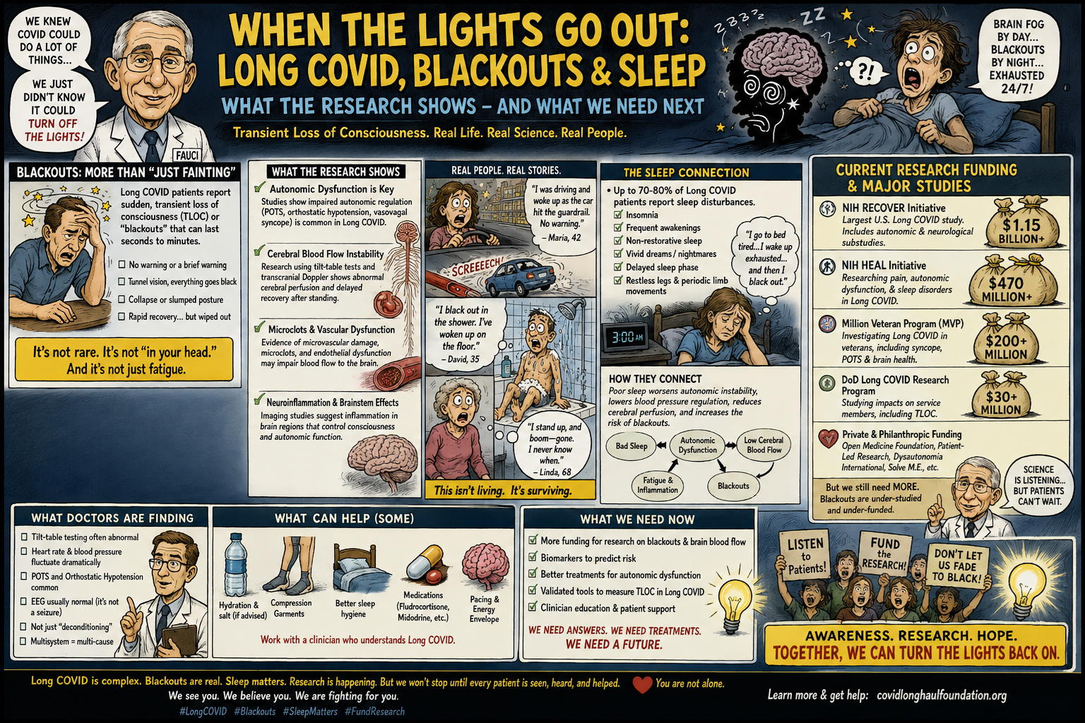
Long COVID, or post-acute sequelae of SARS-CoV-2 infection (PASC), has increasingly been recognized as a multisystem disorder in which neurological, cardiovascular, autonomic, vascular, metabolic, and sleep abnormalities may intersect. Among its most alarming manifestations is transient loss of consciousness (TLOC)—the sudden, temporary interruption of awareness and postural control commonly described by patients as a “blackout.” Although syncope has multiple potential causes and cannot automatically be attributed to Long COVID, accumulating evidence identifies orthostatic intolerance, postural orthostatic tachycardia syndrome (POTS), impaired cerebral blood-flow regulation, autonomic dysfunction, cardiac rhythm abnormalities, and neurovascular dysregulation as plausible contributors in a subset of patients.[1–7]
Sleep represents a second, increasingly important dimension of this problem. Systematic reviews indicate that sleep disturbance affects a substantial proportion of people with Long COVID, while dedicated clinical investigations have identified insomnia, hypersomnolence, sleep apnea, circadian disruption, REM-behavior disorder, and other sleep syndromes. Poor sleep may itself aggravate autonomic instability, fatigue, cognitive dysfunction, and orthostatic intolerance, potentially creating a self-reinforcing physiological cycle.[8–12] The National Institutes of Health (NIH) has consequently made both autonomic dysfunction and sleep disturbance explicit targets of the RECOVER clinical-trial program.
The scientific challenge is now to determine whether blackouts in Long COVID represent merely an occasional consequence of dysautonomia or a recognizable neurological-cardiovascular phenotype involving impaired cerebral perfusion, abnormal autonomic control, sleep-related physiological stress, and—in selected patients—cardiac or neurovascular pathology. The answer has implications not only for diagnosis and treatment but also for public safety, particularly when TLOC occurs while driving, bathing, climbing stairs, or operating machinery.
Keywords: Long COVID; PASC; transient loss of consciousness; syncope; dysautonomia; POTS; orthostatic intolerance; cerebral hypoperfusion; sleep disturbance; autonomic nervous system; neurovascular dysfunction; RECOVER.
1. The Moment When the Brain Goes Dark
There is a deceptively simple phrase used by patients with Long COVID: “The lights went out.”
Sometimes it means a few seconds of dimming vision followed by recovery. Sometimes it means awakening on the floor with no memory of the preceding event. Sometimes there is a warning—lightheadedness, nausea, sweating, tunnel vision, palpitations, weakness or a sensation that the world is receding. In other patients, consciousness appears to disappear almost without warning.
Medically, these experiences belong to a broader spectrum of transient loss of consciousness. Syncope is traditionally defined as a transient loss of consciousness caused by transient global cerebral hypoperfusion, characterized by rapid onset, short duration and spontaneous complete recovery. But not every “blackout” is syncope. Seizures, cardiac arrhythmias, hypoglycemia, medication effects, sleep attacks, concussion, metabolic disturbances and other neurological disorders can produce superficially similar episodes.
That distinction is critical.
The appropriate scientific question is therefore not:
“Does Long COVID cause blackouts?”
The more defensible question is:
“In which patients with Long COVID does transient loss of consciousness occur, what physiological pathway produces it, and how can those patients be identified before injury occurs?”
Evidence accumulated since 2020 suggests that a subset of patients develops objective abnormalities involving autonomic regulation, orthostatic cardiovascular responses and cerebral blood flow.[1–7]
2. The Autonomic Nervous System: The Body’s Hidden Control Room
The autonomic nervous system (ANS) regulates functions that ordinarily occur without conscious effort: heart rate, vascular tone, blood pressure, sweating, gastrointestinal activity, respiration and numerous other homeostatic processes.
Standing upright represents an enormous physiological challenge.
Approximately 500–800 mL of blood can transiently shift toward the lower extremities and abdomen. Normally, arterial pressure sensors detect the change and rapidly activate sympathetic pathways. Heart rate rises, peripheral vessels constrict, venous return is supported and cerebral perfusion is preserved.
A simplified model is:
Standing → venous pooling → reduced venous return → baroreceptor response → sympathetic activation → vasoconstriction + increased cardiac output → preservation of cerebral perfusion
In autonomic dysfunction, some portion of this compensatory system may fail.
Figure 1. Proposed Long COVID blackout pathway
                         SARS-CoV-2 INFECTION
                                  │
                                  ▼
                ┌─────────────────────────────────┐
                │ Persistent / dysregulated        │
                │ immune, vascular & neural state  │
                └─────────────────────────────────┘
                     │          │          │
                     ▼          ▼          ▼
                Autonomic    Vascular    Sleep/circadian
                dysfunction  dysfunction  disruption
                     │          │          │
                     └─────┬────┴─────┬────┘
                           ▼          ▼
                     Orthostatic   Fatigue /
                      instability  impaired recovery
                           │          │
                           ▼          ▼
                   ↓ cerebral blood-flow regulation
                           │
                           ▼
                 PRESYNCOPE / "BRAIN FOG"
                           │
                    ┌──────┴──────┐
                    ▼             ▼
              recovery       TLOC / SYNCOPE
                                    │
                         ┌──────────┼──────────┐
                         ▼          ▼          ▼
                       fall      injury      accident
This diagram represents a proposed mechanistic framework, not a proven single pathway for every patient.
3. Cerebral Blood Flow May Be the Missing Variable
One of the most important developments in this field is the growing recognition that blood pressure and heart rate do not necessarily tell the whole story.
A patient can stand up, maintain a seemingly acceptable blood pressure, and yet experience a substantial reduction in cerebral blood flow.
A review of orthostatic intolerance emphasizes that Long COVID and related disorders can exhibit impaired cerebral blood-flow regulation even when conventional blood-pressure and heart-rate measurements appear relatively normal.[4]
This observation changes the conceptual model.
For years, clinical reasoning often implicitly followed:
Normal blood pressure + normal heart rate = adequate cerebral perfusion.
That equation is not necessarily true.
Transcranial Doppler, extracranial Doppler, near-infrared spectroscopy and other techniques can provide information about cerebral circulation that conventional vital signs cannot.
A study of Long COVID patients undergoing autonomic evaluation found reduced orthostatic cerebral blood-flow velocity, while other investigations have identified abnormalities in autonomic function, small-fiber function and vascular regulation.[5–7]
The result is a plausible physiological sequence:
upright posture → impaired vascular compensation → reduced cerebral perfusion → dizziness/visual disturbance → presyncope → possible TLOC.
The distinction is important because the patient may not necessarily have dramatic hypotension immediately before losing consciousness.
4. What the Clinical Studies Actually Show
Early autonomic studies following COVID-19 infection documented a spectrum of abnormalities rather than a single syndrome.
In one Mayo Clinic series of 27 patients evaluated for post-COVID autonomic dysfunction, 93% reported lightheadedness and 11% reported syncope; 22% fulfilled criteria for POTS.[1]
Figure 2. Selected autonomic manifestations in an early post-COVID autonomic cohort
These numbers should not be interpreted as the prevalence of these conditions among all people with Long COVID. The patients were a referred clinical population, enriched for autonomic symptoms.[1]
More recent work has strengthened the physiological argument.
A 2026 study of 169 patients with Long COVID referred for orthostatic intolerance found evidence of autonomic abnormalities and measured reductions in orthostatic cerebral blood-flow velocity using transcranial Doppler. Patients with greater central-sensitization symptoms demonstrated somewhat greater cerebral blood-flow reductions and higher IL-6 levels, although the study does not establish that inflammation caused the cerebral hypoperfusion.[7]
Another study of 94 people with Long COVID examined orthostatic hemodynamics and cardiac autonomic function, finding distinct groups with initial orthostatic hypotension, POTS and apparently normal orthostatic hemodynamics.[13]
The emerging picture is therefore heterogeneous:
Long COVID ≠ one autonomic disorder.
Rather, Long COVID may contain several overlapping physiological phenotypes.
5. The Blackout Is Sometimes the End of a Physiological Cascade
The classical faint is not necessarily instantaneous.
A patient may first experience:
- blurred or narrowing vision;
- “gray-out” or tunnel vision;
- ringing in the ears;
- lightheadedness;
- nausea;
- sweating;
- palpitations;
- weakness;
- cognitive slowing;
- inability to remain upright.
If cerebral perfusion falls sufficiently, consciousness can disappear.
This explains why some patients describe the event as if someone had switched off a light.
The brain does not need to be structurally destroyed for consciousness to disappear. Conscious awareness requires adequate and continuously regulated cerebral perfusion and metabolic support.
That distinction is central to understanding TLOC.
6. Real-World Clinical Cases
Case 1: Repeated syncope after COVID-19
A published report described a 74-year-old woman who developed repeated syncope following COVID-19. Investigation identified sinus arrest and cardiovascular autonomic dysfunction. Even after pacemaker implantation, she experienced another syncopal episode; subsequent tilt-table testing demonstrated a vasodepressor form of reflex syncope.[14]
Importantly, the case eventually evolved favorably: follow-up testing approximately six months later was normal and the patient became asymptomatic.
This case illustrates two principles.
First, post-COVID syncope cannot simply be assumed to be POTS.
Second, cardiac rhythm disease must be excluded before attributing loss of consciousness to Long COVID dysautonomia.
Case 2: A patient who could not stand without fainting
Another published case involved a 20-year-old man with poorly controlled type 1 diabetes who developed severe autonomic dysfunction following COVID-19. He exhibited features of both POTS and orthostatic hypotension, with symptoms severe enough that he could not stand without experiencing syncope.[15]
This is a particularly instructive example because diabetes itself can produce autonomic neuropathy.
The case therefore demonstrates an essential methodological problem:
Long COVID may interact with pre-existing physiological vulnerabilities rather than operating in isolation.
Diabetes, dehydration, medications, anemia, cardiac disease, autonomic neuropathy and other conditions can all influence susceptibility to syncope.
Case 3: The post-COVID “blackout”
A published report described a 24-year-old woman who experienced loss of consciousness after recovering from COVID-19. Before falling, she experienced generalized weakness, tremor, blurred vision and an ascending sensation. The episode occurred approximately 18 days after quarantine.[16]
Such cases cannot establish a universal Long COVID mechanism, but they demonstrate that transient loss of consciousness has been reported in temporal association with SARS-CoV-2 infection.
Case 4: The severe cardioinhibitory response
Another case report documented severe cardioinhibitory reflex syncope associated with COVID-19, including prolonged pauses in cardiac activity. Subsequent tilt-table testing remained abnormal at three months but normalized by approximately six months.[17]
The authors proposed autonomic imbalance with excessive vagal activation as a possible contributor.
Again, the lesson is not that every Long COVID blackout is caused by vagal overactivity.
It is that autonomic physiology can profoundly alter cardiac output and consciousness.
7. Sleep: The Other Half of the Equation
Perhaps the most underappreciated component of the blackout problem is sleep.
Sleep disturbance is extraordinarily common in Long COVID.
A systematic review and meta-analysis encompassing 29 studies and 13,935 Long COVID patients estimated a pooled prevalence of:
- 46% for sleep disturbance;
- 56% for poor sleep quality;
- 38% for insomnia;
- 14% for excessive daytime sleepiness.[8]
Figure 3. Reported sleep burden in Long COVID
The sleep story has become more sophisticated than simply saying that patients are “tired.”
A specialized sleep clinic study of 42 patients with PASC identified multiple formal sleep disorders, including obstructive sleep apnea, chronic insomnia, idiopathic hypersomnia, narcolepsy type 2, REM behavior disorder and new circadian-phase delay.[9]
A separate prospective observational study using actigraphy found persistent sleep disturbances after COVID-19, with many participants reporting that the problem began after recovery from the acute infection.[10]
Thus Long COVID can affect both:
the quantity of sleep
and
the architecture, timing and physiological quality of sleep.
8. How Could Poor Sleep Promote Blackouts?
The sleep-blackout relationship remains an important hypothesis rather than a fully proven causal pathway.
But several connections are physiologically plausible.
Pathway 1: Sleep deprivation → autonomic instability
Insufficient or fragmented sleep can alter sympathetic and parasympathetic balance.
That matters because blood pressure regulation during standing is itself an autonomic process.
Pathway 2: Sleep fragmentation → impaired vascular regulation
Repeated awakenings, abnormal respiratory events and altered autonomic activity can interfere with normal cardiovascular regulation.
Pathway 3: Sleep loss → greater fatigue and reduced physiological reserve
A patient already operating close to the threshold of orthostatic intolerance may have less reserve after a night of severely disrupted sleep.
Pathway 4: Sleep disturbance → cognitive vulnerability
Sleep loss can worsen attention, reaction time, executive function and memory. A patient experiencing orthostatic cerebral hypoperfusion may therefore experience more severe cognitive impairment when sleep-deprived.
Pathway 5: Sleep apnea → intermittent physiological stress
Obstructive sleep apnea can produce repeated episodes of airway obstruction, oxygen desaturation and sympathetic activation. In a patient already experiencing autonomic dysfunction, this may add another physiological burden.
These mechanisms should be regarded as potential interactions, not proof that insomnia causes syncope in Long COVID.
9. The Proposed “Blackout–Sleep Spiral”
The emerging hypothesis can be represented as a feedback system:
             LONG COVID
                 │
        ┌────────┴─────────┐
        ▼                  ▼
  Dysautonomia        Sleep disruption
        │                  │
        ▼                  ▼
 Orthostatic          Poor recovery
 instability              │
        │                  ▼
        │             ↑ fatigue
        │             ↑ cognitive dysfunction
        │             ↑ autonomic stress
        └──────────┬───────┘
                   ▼
          Reduced physiological
                 reserve
                   │
                   ▼
        Cerebral hypoperfusion
                   │
          ┌────────┴────────┐
          ▼                 ▼
      Presyncope          TLOC
          │                 │
          ▼                 ▼
     fear of sleep       injury /
     or activity       activity restriction
          │                 │
          └────────┬────────┘
                   ▼
             worse sleep
This is a research model, not an established disease pathway.
Its importance lies in the possibility that Long COVID is not simply a collection of independent symptoms. Dysautonomia, sleep disruption, vascular dysfunction, fatigue and cognitive dysfunction may interact as components of a larger physiological network.
10. Why Conventional Testing Can Miss the Problem
A patient may enter an examination room and have:
- normal oxygen saturation;
- normal resting blood pressure;
- normal resting heart rate;
- normal neurological examination;
- normal conventional brain MRI.
And yet experience profound symptoms when standing.
That is because many relevant abnormalities are dynamic rather than static.
The physiology changes when the patient:
- stands;
- walks;
- exercises;
- becomes dehydrated;
- eats;
- becomes overheated;
- sleeps poorly;
- experiences emotional stress.
Consequently, evaluation may require more than a single set of resting vital signs.
Potentially useful investigations—selected according to the clinical scenario—include:
Autonomic
- orthostatic blood pressure and heart rate;
- active stand testing;
- tilt-table testing;
- beat-to-beat blood-pressure monitoring;
- autonomic reflex testing;
- sudomotor testing.
Cardiac
- ECG;
- ambulatory rhythm monitoring;
- echocardiography when indicated;
- evaluation for arrhythmia or structural heart disease.
Neurological
- neurological examination;
- EEG when seizure is suspected;
- brain imaging when clinically indicated;
- assessment of peripheral/autonomic small-fiber dysfunction in appropriate patients.
Cerebrovascular
- transcranial Doppler;
- cerebral perfusion studies in selected research or specialist settings.
Sleep
- sleep history;
- validated sleep questionnaires;
- polysomnography;
- home sleep apnea testing when appropriate;
- actigraphy;
- evaluation for hypersomnia, narcolepsy or circadian disorders.
11. What the NIH Is Now Studying
The research landscape has changed substantially.
The NIH established the RECOVER Initiative in 2021 with an initial $1.15 billion congressional appropriation to investigate Long COVID.[18]
In 2024, NIH announced another $515 million investment, while an additional $147 million was subsequently made available through reallocation from the Public Health and Social Services Emergency Fund. RECOVER reports these as a combined additional $662 million, allocated across fiscal years 2025–2029.[18,19]
Figure 4. RECOVER funding milestones
This represents an extraordinary investment in Long COVID research, but funding magnitude should not be confused with scientific certainty. The existence of substantial research funding means that the scientific questions are being investigated—not that every proposed mechanism has been validated.
12. RECOVER-AUTONOMIC: From Observation to Randomized Trials
The most important change in the field may be the transition from descriptive reports to randomized clinical trials.
RECOVER-AUTONOMIC was designed to study Long COVID-associated POTS and autonomic dysfunction. The program evaluates pharmacological and coordinated non-pharmacological approaches, including IVIG, ivabradine and structured supportive interventions.[20,21]
The trial enrolled approximately 380 participants across its autonomic program, with different studies evaluating severe and moderate POTS.[20]
The preliminary 2026 results are instructive.
In the ivabradine trial, the drug lowered heart rate but did not significantly improve the primary patient-reported measure of orthostatic intolerance compared with placebo.[22]
That is a valuable scientific result.
It suggests that simply correcting one physiological abnormality—heart rate—may not correct the entire disease.
The implication is profound:
The tachycardia may be a marker of a deeper physiological disturbance rather than the entire disturbance itself.
13. RECOVER-SLEEP: Finally Treating Sleep as a Primary Target
The NIH has also created a dedicated Long COVID sleep program.
RECOVER-SLEEP encompasses two trials involving approximately 1,000 participants:
- a hypersomnia trial evaluating modafinil or solriamfetol;
- a complex sleep-disturbance trial evaluating melatonin and light therapy.[23]
Participants undergo sleep assessments, activity tracking, sleep diaries and symptom measurements.
The existence of a dedicated randomized clinical-trial program is important because sleep had historically been treated as a secondary symptom of chronic illness.
RECOVER is treating it as a target worthy of direct intervention.
14. The Next Scientific Question: Are Sleep and Dysautonomia One Network?
The most interesting research question may not be whether Long COVID causes insomnia or POTS separately.
It may be whether they are physiologically connected.
Consider the following network:
                 LONG COVID
                     │
        ┌────────────┼────────────┐
        ▼            ▼            ▼
     Immune       Autonomic     Vascular
   dysregulation  dysfunction   dysfunction
        │            │            │
        │            ▼            ▼
        │       BP/HR instability
        │            │
        ▼            ▼
      Sleep ←──────→ Cerebral
    disruption       perfusion
        │              │
        └──────┬───────┘
               ▼
        fatigue / brain fog
               │
               ▼
       reduced resilience
               │
               ▼
       presyncope / TLOC
This model generates testable hypotheses.
For example:
Do patients with severe Long COVID sleep disturbance have greater orthostatic cerebral hypoperfusion?
Does treating sleep apnea reduce orthostatic symptoms?
Does improving circadian regularity improve autonomic stability?
Can cerebral blood-flow monitoring identify patients at risk of TLOC before blood pressure falls substantially?
Do specific inflammatory or autonomic biomarkers identify the blackout phenotype?
These are precisely the kinds of questions that next-generation pathobiology research should address.
15. The Emerging Importance of Biomarkers
One of the largest deficiencies in Long COVID medicine remains the absence of a universally accepted diagnostic biomarker.
RECOVER’s newest pathobiology funding opportunity is explicitly designed to use existing observational and clinical-trial data and biospecimens to investigate mechanisms, identify biomarkers, discover therapeutic targets and develop bioassays.[24]
The current opportunity permits proposed budgets of up to $800,000 in total costs per year for as much as two years, subject to the program’s requirements.[24]
For the blackout phenotype, a high-value biomarker program might integrate:
- autonomic testing;
- cerebral blood-flow measurements;
- inflammatory markers;
- endothelial biomarkers;
- cardiac rhythm data;
- sleep architecture;
- continuous glucose monitoring when relevant;
- wearable physiological measurements;
- small-fiber testing;
- neurocognitive measurements.
The objective should not merely be to prove that Long COVID exists.
It should be to determine which physiological phenotype a particular patient has.
16. A Dangerous Blind Spot: The Patient Who Faints While Driving
The public-health implications of TLOC extend beyond symptom burden.
A blackout while sitting in a chair may produce little physical damage.
A blackout while:
- driving;
- crossing a road;
- climbing stairs;
- showering;
- swimming;
- working at height;
- operating machinery;
can be catastrophic.
A patient who has unexplained loss of consciousness should therefore not assume that an episode is simply “brain fog” or Long COVID.
Cardiac syncope can be life-threatening.
Seizures require a different evaluation.
Hypoglycemia can cause loss of consciousness.
Medication-related hypotension can produce falls.
Anemia, dehydration and metabolic abnormalities can reduce physiological reserve.
Long COVID may ultimately prove to be one contributor within this differential—not a substitute for it.
17. What Clinicians Should Look For
A useful clinical history should ask questions that conventional Long COVID screening may overlook.
Before the episode
- Were you standing?
- Had you just gotten out of bed?
- Had you eaten?
- Were you dehydrated?
- Was the environment hot?
- Had you exercised?
- Had you slept poorly?
- Did your heart race?
- Did your vision narrow?
During the episode
- Did anyone observe the event?
- Was there complete loss of consciousness?
- How long did it last?
- Was there abnormal movement?
- Was there tongue biting?
- Was there urinary incontinence?
- Was there injury?
After the episode
- How rapidly did awareness return?
- Was there confusion?
- Was there prolonged fatigue?
- Did you remember the event?
- Was there chest pain or palpitations?
Across weeks and months
- How many episodes?
- Are they becoming more frequent?
- Do they occur only upright?
- Do they correlate with poor sleep?
- Do they correlate with meals?
- Do they occur during illness or dehydration?
Such longitudinal information may prove more informative than a single normal office examination.
18. What We Know—and What We Do Not
Reasonably well supported
1. Long COVID can involve autonomic dysfunction.
Multiple cohorts and reviews have documented orthostatic intolerance, POTS and other autonomic abnormalities.[1–7]
2. Syncope has been documented after COVID-19.
Published case reports and clinical cohorts describe syncope and TLOC in post-COVID populations.[1,14–17]
3. Cerebral blood-flow abnormalities occur in some patients with orthostatic intolerance and Long COVID.
These abnormalities may occur even when conventional cardiovascular measurements do not fully explain symptoms.[4–7]
4. Sleep disturbances are common.
Systematic reviews and clinical sleep evaluations demonstrate substantial rates of insomnia, poor sleep, hypersomnolence and other sleep disorders.[8–10]
5. NIH is treating autonomic dysfunction and sleep as legitimate research targets.
RECOVER has dedicated randomized trials for both.[20–23]
Still uncertain
1. How frequently does true TLOC occur in Long COVID?
There is no definitive population-level estimate.
2. Does cerebral hypoperfusion directly cause blackouts in Long COVID?
The physiology is compelling, but direct causal evidence remains incomplete.
3. Does poor sleep increase blackout risk?
The interaction is biologically plausible, but prospective studies specifically connecting sleep disturbance to TLOC are needed.
4. Are there distinct Long COVID blackout phenotypes?
This is increasingly plausible but not yet established.
5. Which treatments prevent TLOC?
This remains a major unanswered question.
19. The Research Agenda Should Change
Future Long COVID research should stop treating “blackouts” as merely another symptom on a checklist.
Instead, TLOC should be studied as a potentially measurable physiological phenotype.
A high-quality study would simultaneously measure:
Brain
→ cerebral blood flow
→ cognition
→ EEG where indicated
Heart
→ continuous ECG
→ heart-rate variability
→ rhythm abnormalities
Circulation
→ beat-to-beat blood pressure
→ stroke volume
→ venous return
→ orthostatic hemodynamics
Autonomic system
→ sympathetic function
→ parasympathetic function
→ sudomotor function
→ baroreflex function
Sleep
→ polysomnography
→ sleep architecture
→ oxygenation
→ actigraphy
→ circadian timing
Immune/vascular biology
→ inflammatory biomarkers
→ endothelial markers
→ coagulation/microvascular measures where scientifically justified
Patient experience
→ episode diaries
→ wearable data
→ falls
→ near-falls
→ sleep quality
→ functional status.
The ultimate goal would be a predictive model:
Who is at risk of losing consciousness—and under what physiological circumstances?
That is a clinically actionable question.
20. The Larger Meaning of “The Lights Go Out”
The phrase “brain fog” can sound trivial.
It is not.
For some patients, Long COVID produces something considerably more dramatic: a transient failure of the systems that maintain alertness, upright posture, cognition and cerebral perfusion.
The science does not yet justify declaring that SARS-CoV-2 universally causes syncope or that every blackout in a Long COVID patient is neurological.
But the evidence has moved far beyond anecdote.
Autonomic dysfunction has been repeatedly demonstrated.
Orthostatic cerebral hypoperfusion has been measured.
Sleep abnormalities have been objectively documented.
Syncope has appeared in clinical cohorts and case reports.
And the NIH has moved from simply cataloguing these symptoms toward randomized trials designed to test interventions.
The next step should be integration.
The brain, autonomic nervous system, cardiovascular system, cerebral circulation and sleep system should not be studied as isolated compartments.
They are parts of one physiological network.
When that network fails to maintain cerebral perfusion, the patient’s description may be remarkably simple:
“The lights went out.”
The scientific challenge is to understand exactly why—and to know how to keep them on.
Footnotes and References
1. Novak P, et al. Autonomic dysfunction following COVID-19 infection: an early experience. Mayo Clinic experience demonstrating lightheadedness, syncope and POTS among patients referred for autonomic evaluation.
2. Goldstein DS. Post-COVID dysautonomias: what we know and (mainly) what we don’t know. Nature Reviews Neurology. 2024;20:99–113. The review emphasizes the heterogeneous and incompletely understood nature of post-COVID autonomic dysfunction.
3. Raj SR and colleagues. Studies of POTS and autonomic dysfunction following SARS-CoV-2 infection have documented orthostatic cardiovascular abnormalities and impaired autonomic regulation.
4. Novak P, et al. Cerebral Blood Flow in Orthostatic Intolerance. The review emphasizes that cerebral blood-flow reductions can occur despite relatively normal heart rate and blood pressure.
5. Van Campen CLMC, et al. Orthostatic Symptoms and Reductions in Cerebral Blood Flow in Long-Haul COVID-19 Patients. The study examined cerebral blood-flow responses to orthostatic stress in Long COVID and ME/CFS.
6. Long COVID/POTS case-control and autonomic studies have reported reduced orthostatic cerebral blood-flow velocity, autonomic dysfunction, small-fiber abnormalities and inflammatory abnormalities in subsets of patients.
7. Novak P, et al. Central sensitization in Long COVID: associations with autonomic symptom burden, cerebral hypoperfusion, and neuroinflammation. 2026. In 169 patients referred for orthostatic intolerance, central sensitization was associated with autonomic symptom burden, cerebral blood-flow changes and IL-6.
8. Goh DY, et al. Prevalence of sleep disturbances in patients with long COVID assessed by standardised questionnaires and diagnostic criteria: a systematic review and meta-analysis. The meta-analysis included 29 studies and 13,935 patients and reported pooled prevalence estimates for sleep disturbance, poor sleep quality, insomnia and daytime sleepiness.
9. Coelho FMS, et al. Sleep disorder syndromes of post-acute sequelae of SARS-CoV-2 (PASC)/Long Covid. A specialized sleep clinic cohort identified obstructive sleep apnea, chronic insomnia, hypersomnia, REM-behavior disorder and circadian abnormalities.
10. Tański W, et al. Sleep Disturbances as a Consequence of Long COVID-19: Insights from Actigraphy and Clinimetric Examinations. J Clin Med. 2024.
11. Goh DY, et al. A 2025 systematic review and meta-analysis examined randomized trials of interventions for Long COVID-related sleep disturbance, illustrating the transition from descriptive research toward treatment trials.
12. A 2025 statewide representative cohort study found prospective associations between Long COVID and unhealthy sleep outcomes nearly three years after SARS-CoV-2 infection.
13. Recent physiological work has characterized distinct Long COVID orthostatic phenotypes and attenuated cardiac autonomic function in patients with abnormal orthostatic hemodynamics.
14. Repeated Syncope Following COVID-19 Infection. Published case report of a 74-year-old woman with sinus arrest and cardiovascular autonomic dysfunction followed by recurrent syncope and abnormal tilt testing.
15. A case of dysautonomia after COVID-19 infection in a patient with poorly controlled type I diabetes. The patient developed severe POTS/orthostatic hypotension and syncope.
16. An Unusual Case of Blackout in a COVID-19 Patient: COVID-19 Brain Fog. Published report describing a 24-year-old woman with post-COVID loss of consciousness preceded by weakness, tremor and blurred vision.
17. A case report of severe cardioinhibitory reflex syncope associated with coronavirus disease 2019. Follow-up demonstrated eventual normalization of tilt-table findings, illustrating the potential dynamic nature of post-COVID autonomic abnormalities.
18. NIH RECOVER funding records report the original $1.15 billion congressional appropriation for RECOVER.
19. RECOVER reports an additional $515 million NIH investment and $147 million PHSSEF reallocation, totaling $662 million in subsequent funding allocated over FY2025–FY2029.
20. RECOVER-AUTONOMIC is a randomized platform program evaluating interventions for Long COVID-related POTS, including IVIG, ivabradine and coordinated non-pharmacological care.
21. The published RECOVER-AUTONOMIC design describes a multicenter, randomized, double-blind, placebo-controlled adaptive platform trial.
22. Preliminary 2026 RECOVER-AUTONOMIC results indicated that ivabradine reduced heart rate but did not significantly improve patient-reported orthostatic intolerance compared with placebo.
23. RECOVER-SLEEP encompasses trials of modafinil/solriamfetol for hypersomnia and melatonin/light therapy for complex sleep disturbances, with approximately 1,000 participants planned across the program.
24. In August 2026, RECOVER opened a new pathobiology Research Opportunity Announcement allowing investigators to use RECOVER clinical-trial data and biospecimens; proposed budgets may reach $800,000 in total costs per year for up to two years.