
John Murphy, CEO The COVID-19 Long haul Foundation
Abstract
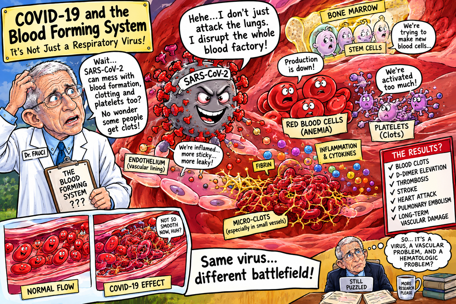
Coronavirus disease 2019 (COVID-19), caused by severe acute respiratory syndrome coronavirus 2 (SARS-CoV-2), is not exclusively a respiratory disorder. From the earliest clinical observations, abnormalities involving erythrocytes, leukocytes, lymphocytes, platelets, coagulation, and inflammatory biomarkers demonstrated that the disease substantially perturbs the hematopoietic and immune systems. Subsequent pathological investigations have established that COVID-19 can profoundly alter the bone marrow, spleen, lymph nodes, circulating blood cells, and the regulatory microenvironment governing hematopoiesis.
The hematological phenotype is heterogeneous and varies with disease severity and stage. Acute infection commonly produces lymphopenia, neutrophilia, thrombocytopenia, anemia, increased immature myeloid forms, altered platelet biology, and a transition from physiologic to stress hematopoiesis. Bone-marrow examinations have demonstrated myeloid left shift, histiocytosis, hemophagocytosis, altered erythropoiesis, megakaryocytic abnormalities, and, in some reports, reduced cellularity. Importantly, the question of direct SARS-CoV-2 infection of hematopoietic cells remains unsettled. Some experimental and pathological studies have identified viral proteins or RNA in selected hematopoietic populations, including megakaryocytes, whereas a carefully performed 2023 autopsy series failed to detect SARS-CoV-2 RNA in bone marrow and concluded that the hematopoietic abnormalities were predominantly indirect.[1–3]
COVID-19 also profoundly affects secondary hematopoietic and lymphoid organs. Splenic white-pulp depletion, lymphocyte loss, impaired germinal-center formation, altered T- and B-cell compartments, plasmablast expansion, macrophage activation, and hemophagocytosis have been documented. These changes provide a pathological substrate for the profound lymphopenia and immune dysregulation observed clinically.
The hematopoietic consequences may extend beyond the acute infection. Studies of Long COVID have reported persistent alterations in erythrocyte characteristics, anemia, lymphocyte populations, inflammatory markers, and coagulation-related biomarkers. Whether these abnormalities represent persistent marrow dysfunction, altered immune regulation, endothelial disease, chronic inflammation, nutritional deficiency, medication effects, or combinations thereof remains incompletely resolved.
This review examines the relationship between SARS-CoV-2 and the hematopoietic system, with particular emphasis on bone marrow pathology, hematopoietic stem and progenitor cells, erythropoiesis, leukopoiesis, megakaryopoiesis, platelet biology, hemophagocytosis, spleen and lymph-node pathology, immune memory, persistent hematological abnormalities, and the clinical implications for diagnosis and treatment.
Keywords: COVID-19; SARS-CoV-2; bone marrow; hematopoiesis; hematopoietic stem cells; erythropoiesis; anemia; lymphopenia; thrombocytopenia; megakaryocytes; platelets; spleen; lymph nodes; hemophagocytosis; HLH; Long COVID.
Introduction
Hematopoiesis is among the body’s most dynamic biological systems. Every day, the bone marrow generates enormous numbers of erythrocytes, granulocytes, monocytes, lymphocytes, and platelets. This production is ordinarily tightly regulated by interactions among hematopoietic stem cells (HSCs), progenitor cells, stromal cells, endothelial cells, macrophages, cytokines, chemokines, growth factors, and metabolic signals.
Severe infection fundamentally changes this equilibrium.
Instead of maintaining steady-state hematopoiesis, the marrow enters a state of stress or emergency hematopoiesis, redirecting production toward cell populations required to confront the infectious threat. SARS-CoV-2 infection is particularly consequential because the virus produces simultaneous pulmonary, vascular, inflammatory, metabolic, and immunological disturbances. The hematopoietic system consequently becomes both a responder to systemic infection and a potential contributor to disease pathology.[1]
The clinical consequences are readily visible in the complete blood count. Lymphopenia, neutrophilia, thrombocytopenia, anemia, altered neutrophil morphology, immature myeloid cells, and changes in platelet activation occur with variable frequency. Severe abnormalities frequently correlate with disease severity.
Yet the peripheral blood represents only the visible end product of a much larger biological system.
Understanding COVID-19 hematopathology therefore requires examination of the bone marrow and its microenvironment, the spleen, the lymph nodes, circulating hematopoietic cells, and the regulatory networks connecting these organs.
The Normal Hematopoietic System
Adult hematopoiesis is centered principally in the bone marrow.
Hematopoietic stem cells possess two defining properties:
- long-term self-renewal; and
- multipotent differentiation.
Through successive progenitor stages, HSCs generate the principal blood-cell lineages:
- erythrocytes;
- neutrophils;
- eosinophils;
- basophils;
- monocytes;
- lymphocytes;
- megakaryocytes; and
- platelets.
The marrow is therefore not merely a factory of blood cells. It is a highly organized biological niche in which hematopoietic cells interact continuously with vascular endothelial cells, stromal cells, macrophages, osteoblast-lineage cells, adipocytes, extracellular matrix, and soluble signaling molecules.
Infection disrupts this equilibrium.
The resulting response is commonly described as emergency hematopoiesis, in which production is preferentially redirected toward myeloid and innate immune populations while lymphoid production and maturation may be impaired.[1]
COVID-19 provides an unusually dramatic example of this phenomenon.
COVID-19 as a Systemic Hematopoietic Disease
Although SARS-CoV-2 initially infects the respiratory tract, severe COVID-19 produces systemic inflammatory and vascular effects capable of profoundly altering hematopoiesis.
The characteristic hematologic pattern includes:
- lymphopenia;
- neutrophilia;
- thrombocytopenia;
- anemia;
- elevated immature granulocytes;
- abnormal monocyte populations;
- altered platelet activation;
- increased D-dimer;
- hyperferritinemia;
- elevated inflammatory cytokines; and
- evidence of stress erythropoiesis.
A comprehensive review of SARS-CoV-2 hematopoietic responses concluded that the disease shifts normal hematopoietic equilibrium toward emergency hematopoiesis, characterized by increased myeloid production at the expense of lymphoid populations.[1]
This phenomenon is clinically significant because it provides a mechanistic bridge between systemic inflammation and the abnormalities observed in the peripheral blood.
Bone Marrow Pathology
Cellularity
Bone-marrow findings in COVID-19 are heterogeneous.
Autopsy and biopsy studies have reported:
- normocellular marrow;
- hypercellularity;
- variable decreases in cellularity;
- myeloid hyperplasia;
- left-shifted granulopoiesis;
- altered erythropoiesis;
- megakaryocytic abnormalities;
- histiocytosis; and
- hemophagocytosis.
An early pathological study examining 20 autopsies and two living patients found predominantly normocellular-to-hypercellular marrow with maturing trilineage hematopoiesis and frequent myeloid left shift.[4]
These findings argue against the concept that COVID-19 ordinarily causes a simple generalized marrow aplasia.
Instead, the marrow often remains active but becomes dysregulated.
Myeloid Left Shift and Emergency Granulopoiesis
One of the most consistent marrow abnormalities is a left shift of myelopoiesis.
A left shift signifies the release of increasingly immature granulocytic precursors into the peripheral circulation.
Clinically, this may manifest as:
- increased band forms;
- immature granulocytes;
- neutrophilia;
- toxic neutrophil changes; and
- increased neutrophil-to-lymphocyte ratio.
The phenomenon reflects emergency granulopoiesis.
The marrow is responding to inflammatory signals demanding rapid production of innate immune cells.
The problem is that quantity does not necessarily equal functional competence.
COVID-19 has been associated with populations of immature or dysfunctional neutrophils, suggesting that accelerated emergency production can produce cells that differ substantially from the normally generated neutrophil compartment.[1]
Erythropoiesis and Anemia
Anemia is common during severe COVID-19, but its origin is multifactorial.
Potential mechanisms include:
- inflammation-mediated suppression of erythropoiesis;
- altered iron metabolism;
- reduced erythrocyte lifespan;
- hemolysis;
- blood loss from repeated phlebotomy;
- renal dysfunction and reduced erythropoietin;
- nutritional deficiency;
- marrow stress;
- macrophage-mediated erythrophagocytosis; and
- direct or indirect disruption of erythroid precursor biology.
Inflammation induces hepcidin-mediated sequestration of iron, thereby restricting iron availability for erythropoiesis.
At the same time, systemic inflammatory signaling can alter erythroid maturation and promote the production of immature erythroid cells.
A major review of COVID-19 hematopoiesis described stress erythropoiesis as a characteristic component of the disease and discussed evidence for substantial effects on erythroid progenitors.[1]
Erythrophagocytosis
One of the more striking pathological findings is erythrophagocytosis, in which macrophages engulf erythrocytes or erythroid material.
A 2023 study of 28 COVID-19 autopsy cases found erythrophagocytosis in 15 of 28 cases (54%), with siderophages present in 11 of those 15 cases. Erythrophagocytosis correlated with lower hemoglobin concentrations.[2]
This finding provides a plausible pathological mechanism for anemia in severe COVID-19.
It also demonstrates that anemia cannot always be understood merely as “anemia of inflammation.”
Macrophage-mediated destruction or sequestration of erythroid material may make a substantive contribution in some critically ill patients.
Is SARS-CoV-2 Directly Infecting the Bone Marrow?
This remains one of the most controversial questions.
Several experimental studies have suggested that SARS-CoV-2-related proteins may interact with hematopoietic cells and that certain progenitor populations express molecular machinery potentially relevant to viral entry.[1]
Some studies have further reported viral material within bone-marrow-associated cells.
However, evidence is inconsistent.
The 2023 Histopathology study by Marques-Maggio and colleagues examined bone marrow from 28 COVID-19 autopsy cases and five controls. Although severe abnormalities in hematopoiesis were observed, highly sensitive PCR did not detect SARS-CoV-2 in bone marrow, leading the authors to conclude that the hematopoietic effects were predominantly indirect rather than the consequence of widespread viral replication within the marrow microenvironment.[2]
This is an important distinction.
Evidence supports:
- profound marrow dysfunction during severe COVID-19;
- altered hematopoietic differentiation;
- macrophage activation;
- erythrophagocytosis;
- emergency myelopoiesis;
- megakaryocytic abnormalities.
Evidence does not yet establish:
- widespread productive SARS-CoV-2 infection of normal human HSCs;
- persistent viral replication throughout the marrow;
- a universal direct cytopathic effect of SARS-CoV-2 on hematopoietic stem cells.
Thus, claims that COVID-19 universally “infects the bone marrow” would exceed the current evidence.
Megakaryocytes and Platelet Production
Megakaryocytes are the enormous marrow cells responsible for generating circulating platelets.
COVID-19 profoundly alters platelet biology.
Thrombocytopenia occurs frequently in severe disease, while paradoxically many patients also exhibit platelet hyperreactivity and a strongly prothrombotic state.
This apparent contradiction illustrates that platelet number and platelet function are separate biological variables.
A patient may have fewer platelets but a population of unusually activated platelets.
Recent pathological evidence has made the marrow component particularly intriguing.
A 2025 study of 11 critically ill COVID-19 patients with thrombocytopenia reported reduced marrow cellularity and decreased megakaryocytic lineage, together with increased emperipolesis and vacuolization. The investigators also reported SARS-CoV-2-associated proteins and double-stranded RNA within marrow megakaryocytes, raising the possibility of direct viral involvement in this cell population.[5]
Because the study was small, these observations should be considered provocative rather than definitive.
Nevertheless, they provide a potential biological explanation for at least part of COVID-associated thrombocytopenia.
Platelets as Immunological Cells
Platelets are not merely fragments involved in hemostasis.
They participate in:
- innate immunity;
- leukocyte recruitment;
- endothelial interactions;
- inflammatory signaling;
- complement activation;
- neutrophil extracellular trap formation; and
- immunothrombosis.
COVID-19 therefore creates a pathological intersection between hematopoiesis and vascular disease.
The marrow generates platelets; platelets interact with activated endothelium; endothelial activation promotes thrombosis; thrombosis generates additional inflammatory signals.
The result is a self-reinforcing immunothrombotic circuit.
This helps explain why hematological abnormalities and vascular complications are so tightly associated in severe COVID-19.
Lymphopenia
Lymphopenia became one of the characteristic laboratory abnormalities of COVID-19.
Both CD4+ and CD8+ T-cell populations may decline, particularly in severe disease.
Potential mechanisms include:
- cytokine-mediated apoptosis;
- lymphocyte redistribution;
- tissue recruitment;
- metabolic exhaustion;
- direct or indirect cellular injury;
- glucocorticoid effects;
- impaired lymphopoiesis; and
- destruction or dysfunction of secondary lymphoid organs.
The reduction is therefore not necessarily caused by failure of marrow production alone.
It may reflect simultaneous abnormalities in production, trafficking, survival, activation, and tissue organization.
The Spleen as a Major Hematopoietic and Immunological Target
The spleen occupies a unique position at the intersection of hematology and immunity.
It removes aged erythrocytes, participates in platelet sequestration, filters blood-borne pathogens, and supports adaptive immune responses.
COVID-19 can substantially alter splenic architecture.
Pathological studies have described:
- white-pulp depletion;
- reduced lymphocyte numbers;
- red-pulp congestion;
- hemorrhage;
- necrosis;
- macrophage activation;
- hemophagocytosis;
- altered B-cell compartments; and
- impaired germinal-center architecture.
A systematic pathological review found white-pulp depletion among the recurring splenic abnormalities in COVID-19 autopsy material.[6]
The consequence is potentially profound: an organ designed to coordinate immune surveillance becomes structurally and functionally altered during severe infection.
Lymph Nodes and Germinal Centers
Lymph nodes provide the anatomical environment in which antigen-presenting cells, T cells, and B cells coordinate adaptive immunity.
One of the most important pathological discoveries in early COVID-19 was the alteration or loss of germinal centers.
Kaneko and colleagues reported striking depletion of Bcl-6-expressing germinal-center B cells and T follicular helper cells in lymph nodes and spleens from patients dying with acute COVID-19.[7]
This finding was important because germinal centers are critical for:
- affinity maturation;
- somatic hypermutation;
- class switching;
- durable B-cell memory; and
- generation of high-quality long-lived plasma cells.
Disruption of germinal-center architecture could therefore influence the quality and durability of humoral immunity.
SARS-CoV-2 in Secondary Lymphoid Organs
Several autopsy studies have detected SARS-CoV-2 antigens in spleen and lymph-node tissue.
One study found viral nucleocapsid and spike antigen primarily in macrophage and dendritic-cell populations within the spleen and lymph nodes, rather than demonstrating widespread infection of T or B lymphocytes.[8]
This observation again emphasizes an important distinction.
The presence of viral antigen within an immune organ does not automatically mean that the virus is productively replicating throughout that organ.
Nevertheless, antigen exposure within macrophages and dendritic cells could substantially modify antigen presentation, cytokine production, and immune-cell trafficking.
Hemophagocytosis and Hyperinflammatory Disease
Hemophagocytosis is the engulfment of blood cells by activated macrophages.
It may occur in severe systemic inflammation and is a characteristic pathological feature of hemophagocytic lymphohistiocytosis (HLH).
COVID-19 has been associated with secondary HLH-like inflammatory syndromes.
Bone-marrow examinations have frequently demonstrated histiocytosis and hemophagocytosis. In one early pathological series, hemophagocytic histiocytes were identified in all 19 evaluable bone-marrow specimens.[4]
However, hemophagocytosis alone does not establish clinical HLH.
The diagnosis requires integration of clinical and laboratory criteria, which may include:
- persistent fever;
- cytopenias;
- hyperferritinemia;
- hypertriglyceridemia;
- hypofibrinogenemia;
- splenomegaly;
- elevated soluble IL-2 receptor;
- reduced NK-cell activity; and
- pathological evidence of hemophagocytosis.
This distinction is important because critically ill COVID-19 patients can exhibit several HLH-like features without fulfilling the full diagnostic syndrome.
Cytokines and the Marrow Microenvironment
The bone marrow responds to cytokines released throughout the body.
Among the important mediators are:
- IL-6;
- IL-1β;
- TNF-α;
- GM-CSF;
- interferons;
- CXCL10;
- G-CSF; and
- other chemokines and growth factors.
The resulting environment favors emergency myelopoiesis and changes the balance among hematopoietic lineages.
This is one reason the hematologic manifestations of COVID-19 should not be interpreted as isolated abnormalities.
The marrow is effectively receiving a continuous stream of systemic instructions.
The peripheral CBC is therefore a readout of systemic inflammatory physiology.
Neutrophils
Neutrophils are among the most strongly affected circulating populations.
Severe COVID-19 may produce:
- neutrophilia;
- immature granulocytes;
- abnormal segmentation;
- toxic granulation;
- altered metabolic programming; and
- excessive neutrophil extracellular trap formation.
Neutrophil extracellular traps (NETs) can capture pathogens but can also injure endothelium and promote thrombosis.
Thus, emergency granulopoiesis may become part of the mechanism linking hematopoiesis to immunothrombosis.
Monocytes and Macrophages
Monocytes and macrophages occupy another critical position.
COVID-19 has been associated with changes in:
- monocyte subsets;
- antigen presentation;
- macrophage activation;
- inflammatory cytokine production;
- tissue recruitment; and
- hemophagocytic activity.
Bone marrow macrophages may participate directly in erythrophagocytosis, while tissue macrophages contribute to inflammatory injury in the lung, spleen, and lymphoid organs.
The macrophage therefore becomes a central cellular link between inflammation, hematopoiesis, and tissue injury.
Long COVID and Persistent Hematological Abnormalities
The hematological story does not necessarily end when acute infection resolves.
A review of hematological findings in Long COVID identified persistent abnormalities involving:
- erythrocyte characteristics;
- anemia;
- lymphopenia;
- ferritin;
- D-dimer;
- IL-6; and
- other inflammatory markers.[9]
These findings are intriguing but require careful interpretation.
Persistent abnormalities may reflect:
- residual inflammation;
- altered iron metabolism;
- persistent immune activation;
- endothelial dysfunction;
- renal disease;
- nutritional deficiency;
- occult blood loss;
- medication effects;
- autoimmune phenomena;
- persistent coagulation activation; or
- altered marrow signaling.
Consequently, persistent anemia or thrombocytopenia after COVID-19 should not automatically be attributed to “Long COVID.”
It requires conventional hematological investigation.
Bone-Marrow Memory and Long-Term Immunity
The bone marrow has another role that is highly relevant to COVID-19: long-term immune memory.
Long-lived plasma cells reside within specialized bone-marrow niches and continuously produce antibodies.
A landmark Nature study demonstrated SARS-CoV-2 spike-specific bone-marrow plasma cells in individuals recovering from mild COVID-19, including 18 individuals examined seven to eight months after infection. These cells were quiescent and consistent with a stable long-lived plasma-cell compartment.[10]
This finding fundamentally changes how the marrow should be conceptualized.
The bone marrow is not merely the site at which acute blood cells are produced.
It is also a repository of immunological memory.
COVID-19 therefore affects the marrow in two very different ways:
- acute infection can disrupt hematopoietic homeostasis; and
- recovery can establish long-lived antigen-specific plasma-cell populations.
These are complementary rather than contradictory phenomena.
Bone Marrow, Immunity, and Vaccination
The marrow’s role in long-lived plasma-cell formation is also relevant to vaccination.
A successful vaccine response involves:
antigen exposure → B-cell activation → germinal-center maturation → plasma-cell differentiation → migration to marrow niches → long-lived antibody production.
The quality and durability of this process depends upon intact lymphoid architecture and appropriate germinal-center responses.
The observation that acute severe COVID-19 can disrupt germinal centers while convalescent infection can nevertheless generate long-lived bone-marrow plasma cells illustrates the complexity of SARS-CoV-2 immunity.
The immune system can sustain durable memory despite profound transient disruption of lymphoid architecture.
Clinical Evaluation of Persistent Hematological Abnormalities
Patients with persistent blood abnormalities after COVID-19 should undergo a conventional hematological assessment rather than having abnormalities attributed reflexively to post-viral disease.
A reasonable initial evaluation may include:
- complete blood count with differential;
- peripheral blood smear;
- reticulocyte count;
- iron;
- ferritin;
- transferrin saturation;
- vitamin B12;
- folate;
- LDH;
- haptoglobin;
- bilirubin;
- creatinine and renal function;
- liver enzymes;
- CRP and/or ESR;
- coagulation studies when clinically indicated.
Depending upon the abnormality, additional testing may include:
- serum protein electrophoresis;
- immunoglobulins;
- direct antiglobulin testing;
- erythropoietin;
- copper;
- zinc;
- autoimmune testing;
- viral studies;
- JAK2 or other myeloproliferative testing;
- flow cytometry; and
- bone-marrow aspiration and biopsy.
The purpose is to distinguish post-infectious hematological dysfunction from diseases such as:
- myelodysplastic syndrome;
- leukemia;
- lymphoma;
- aplastic anemia;
- immune thrombocytopenia;
- autoimmune hemolytic anemia;
- myeloproliferative neoplasms;
- nutritional deficiency;
- renal anemia; and
- occult blood loss.
When Is Bone-Marrow Biopsy Appropriate?
Bone-marrow biopsy is not routinely indicated for uncomplicated COVID-19.
It becomes appropriate when persistent or unexplained abnormalities raise concern for an underlying marrow disorder.
Potential indications include:
- unexplained pancytopenia;
- persistent severe thrombocytopenia;
- unexplained macrocytic anemia;
- abnormal circulating blasts;
- unexplained leukocytosis;
- suspected leukemia or lymphoma;
- suspected myelodysplastic syndrome;
- suspected HLH;
- unexplained severe reticulocytopenia;
- persistent cytopenias without an alternative explanation; or
- abnormal peripheral-smear findings.
COVID-19 should therefore be viewed as a possible precipitant or modifier of hematological disease rather than an explanation that automatically supersedes all other diagnoses.
Therapeutic Considerations
Treatment depends on the specific hematological abnormality.
There is no single “marrow treatment” for COVID-19.
Anemia
Treatment should address the mechanism:
- iron deficiency should receive iron replacement;
- vitamin deficiency should be corrected;
- renal anemia may require appropriate management of kidney disease;
- active blood loss must be identified;
- hemolysis requires cause-specific treatment;
- severe symptomatic anemia may require transfusion.
Thrombocytopenia
The differential diagnosis includes:
- immune thrombocytopenia;
- consumption;
- disseminated intravascular coagulation;
- marrow suppression;
- megakaryocyte dysfunction;
- drug-induced thrombocytopenia;
- sepsis; and
- thrombotic microangiopathy.
Treatment must therefore be mechanism-specific.
Leukopenia or lymphopenia
Persistent lymphopenia requires evaluation for:
- medication effects;
- immunodeficiency;
- chronic infection;
- nutritional abnormalities;
- autoimmune disease; and
- hematological malignancy.
Anticoagulation and the Hematopoietic System
COVID-19 creates a particularly difficult therapeutic balance between thrombosis and bleeding.
Platelet activation, endothelial dysfunction, NET formation, coagulation activation, and inflammation promote thrombosis.
At the same time, thrombocytopenia, endothelial injury, invasive procedures, gastrointestinal bleeding, and other complications can increase hemorrhagic risk.
Anticoagulation should therefore be individualized according to contemporary clinical guidelines and the patient’s thrombotic and bleeding risks.
The existence of COVID-associated thrombocytopenia does not itself mean that anticoagulation is contraindicated, nor does the existence of elevated D-dimer alone establish an indication for therapeutic anticoagulation.
Prognosis
The prognosis of COVID-associated hematological abnormalities depends heavily upon severity and mechanism.
Many acute abnormalities resolve with recovery.
Neutrophilia and lymphopenia frequently improve as systemic inflammation subsides.
Platelet counts may recover as inflammation and consumption decrease.
Anemia may improve as inflammation resolves, although patients with renal disease, nutritional deficiency, blood loss, or chronic inflammation may remain anemic.
The more consequential question concerns patients with persistent abnormalities.
Persistent cytopenias should not simply be assumed to represent a benign residue of SARS-CoV-2 infection.
They warrant evaluation because COVID-19 may:
- unmask a pre-existing hematological disorder;
- precipitate an autoimmune cytopenia;
- exacerbate renal or inflammatory anemia;
- alter immune-cell homeostasis; or
- potentially leave prolonged disturbances in hematopoietic regulation.
A Model of COVID-19 Hematopoietic Dysfunction
The available evidence supports a multidimensional model:
SARS-CoV-2 infection
↓
Pulmonary and systemic inflammation
↓
IL-6 / TNF-α / interferon / GM-CSF and other signaling changes
↓
Emergency hematopoiesis
↓
Myeloid expansion + lymphoid suppression
↓
Neutrophilia + lymphopenia
↓
Altered megakaryopoiesis + platelet activation
↓
Thrombocytopenia + immunothrombosis
At the same time:
Inflammation + hypoxia + altered iron metabolism + erythrophagocytosis
↓
Stress erythropoiesis
↓
Anemia
And:
Secondary lymphoid-organ injury
↓
White-pulp depletion + germinal-center disruption + altered B/T-cell organization
↓
Immune dysregulation
This model explains why the hematological manifestations of COVID-19 are so diverse.
Conclusions
COVID-19 profoundly affects the hematopoietic and blood-forming systems. The evidence demonstrates that SARS-CoV-2 infection can alter peripheral blood counts, bone-marrow architecture, hematopoietic differentiation, megakaryocyte biology, platelet function, splenic organization, lymph-node architecture, and immune-cell development.
The most reproducible abnormalities include lymphopenia, neutrophilia, thrombocytopenia, anemia, myeloid left shift, stress erythropoiesis, macrophage activation, and hemophagocytosis. Bone-marrow studies demonstrate that severe COVID-19 is associated with active but dysregulated hematopoiesis rather than a uniform marrow aplastic state.[1–4]
The question of direct marrow infection remains unresolved. Evidence for SARS-CoV-2-associated effects on hematopoietic progenitors and megakaryocytes is biologically provocative, and recent pathological work has reported viral material within megakaryocytes.[5] Conversely, a larger autopsy analysis using sensitive PCR failed to detect viral RNA in bone marrow, supporting the interpretation that much of the hematopoietic pathology is mediated indirectly through systemic inflammation, altered cytokine signaling, hypoxia, endothelial dysfunction, and immune activation.[2] The scientifically appropriate conclusion is therefore that COVID-19 clearly disrupts hematopoiesis, while the extent to which direct viral infection of marrow cells contributes remains uncertain.
The spleen and lymph nodes are similarly affected. White-pulp depletion, lymphocyte loss, germinal-center disruption, macrophage activation, and hemophagocytosis indicate that COVID-19 alters not only blood-cell production but also the anatomical infrastructure required for adaptive immunity.[6–8]
At the same time, the marrow has a paradoxical restorative function. It becomes a reservoir for long-lived SARS-CoV-2-specific plasma cells capable of sustaining antibody production long after acute infection.[10]
The emerging picture is therefore not one of simple marrow destruction. Rather, COVID-19 produces a dynamic remodeling of the hematopoietic system involving emergency hematopoiesis, altered lineage allocation, immune-cell depletion, platelet dysregulation, macrophage activation, and remodeling of secondary lymphoid organs. In some patients these abnormalities resolve with recovery; in others, persistent hematological abnormalities may remain.
For patients with prolonged anemia, thrombocytopenia, leukopenia, lymphopenia, or unexplained abnormalities of the peripheral smear after COVID-19, the appropriate clinical response is not diagnostic attribution by exclusion to “Long COVID.” Such abnormalities deserve conventional hematological investigation, including consideration of marrow examination when clinically indicated.
The hematopoietic system may ultimately prove to be one of the most informative windows into the systemic biology of SARS-CoV-2 infection. The blood is not merely a passive reflection of COVID-19. It is a dynamic record of the body’s attempt to reorganize hematopoiesis, immunity, inflammation, coagulation, and tissue repair in response to a systemic viral insult.
References and Footnotes
1. Elahi S. Hematopoietic responses to SARS-CoV-2 infection. Cellular and Molecular Life Sciences. 2022;79:187. This comprehensive review describes the transition from steady-state to stress hematopoiesis, including lymphopenia, neutrophilia, thrombocytopenia, stress erythropoiesis, and altered hematopoietic progenitor biology.
2. Marques-Maggio E, et al. Bone marrow haematopoiesis in patients with COVID-19. Histopathology. 2023;83:582–590. Analysis of 28 COVID-19 autopsies and five controls demonstrated left-shifted myelopoiesis, erythrophagocytosis, increased macrophages and other hematopoietic abnormalities. Importantly, SARS-CoV-2 was not detected in bone marrow by high-sensitivity PCR.
3. Ahmadi E, Bagherpour Z, Zarei E, Omidkhoda A. Pathological effects of SARS-CoV-2 on hematological and immunological cells: alterations in count, morphology, and function. Pathology—Research and Practice. 2022;231:153782. The review describes abnormalities in circulating blood-cell number, morphology, and function associated with SARS-CoV-2 infection.
4. Rapkiewicz AV, et al. Bone marrow and peripheral blood findings in patients infected by SARS-CoV-2. American Journal of Clinical Pathology. 2021;155:627–637. The study of autopsy and living-patient marrow specimens documented normocellular-to-hypercellular marrow, maturing trilineage hematopoiesis, myeloid left shift, and frequent hemophagocytosis.
5. Detection of SARS-CoV-2 in bone marrow megakaryocytes and elevated emperipolesis in COVID-19 patients with thrombocytopenia. Journal of Thrombosis and Haemostasis. 2025;23:2327–2334. In 11 critically ill patients with thrombocytopenia, the investigators reported decreased megakaryocytic lineage, increased emperipolesis and vacuolization, and SARS-CoV-2-associated material within megakaryocytes. The small sample size requires cautious interpretation.
6. Postmortem findings associated with SARS-CoV-2: systematic review and meta-analysis. The analysis documented hematopoietic-system abnormalities including marrow changes, lymph-node lymphoid depletion, splenic white-pulp depletion, and hemophagocytosis.
7. Kaneko N, et al. Loss of Bcl-6-expressing T follicular helper cells and germinal centers in COVID-19. Cell. 2020;183:143–157. The investigators identified profound germinal-center disruption and loss of Bcl-6-expressing germinal-center B cells and T follicular helper cells in lymphoid tissues from patients with severe COVID-19.
8. SARS-CoV-2 induces lymphocytopenia by promoting inflammation and decimates secondary lymphoid organs. The study reported lymphocyte depletion, germinal-center reduction, splenic white-pulp atrophy, and SARS-CoV-2 antigen in macrophage and dendritic-cell populations within spleen and lymph-node tissue.
9. Hematological alterations associated with Long COVID-19. The review identified persistent alterations involving red blood cells, anemia, lymphopenia, ferritin, D-dimer, IL-6, and other inflammatory markers, while emphasizing that mechanisms and treatment remain incompletely understood.
10. Turner JS, et al. SARS-CoV-2 infection induces long-lived bone marrow plasma cells in humans. Nature. 2021;595:421–425. The study demonstrated SARS-CoV-2 spike-specific bone-marrow plasma cells months after mild infection, supporting the existence of durable marrow-based humoral immune memory.
11. Hematopathology of severe acute respiratory syndrome coronavirus 2 infection and coronavirus disease-19. This comprehensive pathology review summarizes peripheral blood abnormalities, marrow left shift and hemophagocytosis, splenic white-pulp depletion, lymph-node germinal-center abnormalities, and altered lymphoid-cell populations.
12. Bone marrow alterations in COVID-19 infection: the root of hematological problems. Current Research in Translational Medicine. 2023;71:103407. The review discusses single-lineage cytopenias, pancytopenia, hemophagocytosis, marrow necrosis, inflammatory mechanisms, and possible direct and indirect effects of SARS-CoV-2 on marrow function.