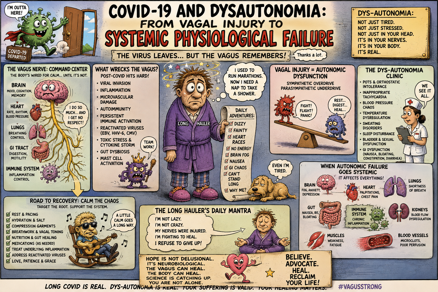
A narrative review of autonomic dysfunction, vagal disruption, pathophysiology and clinical course in Long COVID
Abstract
Background: Dysautonomia has emerged as one of the most consequential neurological and cardiovascular manifestations of post-COVID-19 condition. Patients may develop postural orthostatic tachycardia syndrome (POTS), orthostatic hypotension, inappropriate sinus tachycardia, impaired heart-rate variability, gastrointestinal dysmotility, sudomotor abnormalities, thermoregulatory disturbance, cognitive dysfunction, fatigue and exercise intolerance. Increasing evidence suggests that these manifestations may reflect more than nonspecific physiological deconditioning. SARS-CoV-2 can affect autonomic pathways through immune activation, endothelial dysfunction, small-fibre neuropathy, altered baroreflex function, autoantibodies, persistent viral material and, potentially, direct inflammation of the vagus nerve.
Objective: To synthesise current evidence concerning the aetiology, pathology, physiology, clinical manifestations and longitudinal course of COVID-19-associated dysautonomia, with particular emphasis on the vagus nerve and its effects across cardiovascular, respiratory, gastrointestinal, immune, endocrine and neurological systems.
Evidence: Observational studies, autonomic laboratory investigations, neurophysiological studies, histopathological examinations, imaging studies, systematic reviews and recent mechanistic investigations were reviewed, with particular attention to publications through August 2026.
Results: Cardiovascular autonomic dysfunction appears to affect a substantial proportion of highly symptomatic patients with post-COVID condition. Studies demonstrate impaired heart-rate variability, abnormal orthostatic responses, impaired cardiovagal function and, in selected cohorts, persistent abnormalities in baroreflexes, catecholamine physiology and sympathetic innervation. Histopathological studies have demonstrated SARS-CoV-2 RNA and inflammatory infiltrates within vagus nerves obtained at autopsy, with inflammatory transcriptional signatures in neurons, endothelial cells and Schwann cells. A clinical pilot study subsequently demonstrated structural and functional abnormalities of the vagus nerve in patients with Long COVID, accompanied by gastrointestinal dysmotility and respiratory-muscle weakness. More recent investigations provide evidence for autonomic small-fibre pathology and associations between functional autoantibodies directed against G-protein-coupled receptors and cardiovascular/autonomic phenotypes. These findings support a model in which dysautonomia is heterogeneous and may result from several interacting mechanisms rather than a single lesion.
Conclusions: COVID-19-associated dysautonomia should be regarded as a systemic physiological disorder involving interconnected neural, vascular, immune, endocrine and metabolic systems. Vagal dysfunction is particularly important because the vagus nerve constitutes a major bidirectional interface between brain, heart, lungs, gastrointestinal tract and immune system. The evidence does not establish vagal injury as the universal cause of Long COVID dysautonomia, but it provides a compelling mechanistic framework for a substantial subgroup. Future clinical trials should phenotype patients according to autonomic physiology, small-fibre function, immune abnormalities and vagal function rather than treating dysautonomia as a single disorder.
Introduction
The autonomic nervous system is the physiological infrastructure through which the body maintains internal equilibrium.
Heart rate must accelerate when a person stands.
Blood vessels must constrict when gravity suddenly draws blood toward the lower extremities.
The gastrointestinal tract must coordinate propulsion, secretion and absorption.
Respiration must adjust continuously to metabolic demand.
Sweat glands must regulate temperature.
The endocrine system must respond to stress.
The immune system must recognize danger while simultaneously limiting its own inflammatory response.
Most of these operations occur outside conscious awareness.
The autonomic nervous system therefore represents one of the body’s principal mechanisms for converting changing environmental and internal conditions into coordinated physiological responses.
COVID-19 can disrupt this system.
The resulting disorder has acquired several names—dysautonomia, autonomic dysfunction, orthostatic intolerance, postural orthostatic tachycardia syndrome and cardiovascular autonomic dysfunction—but these terms describe overlapping rather than identical phenomena.
The clinical importance is considerable.
Patients may report that standing causes their heart to race, their blood pressure to fall, their vision to blur and their cognition to deteriorate. Walking across a room may provoke disproportionate tachycardia and exhaustion. Meals may produce bloating, nausea or presyncope. Heat may become intolerable. Sleep may be disturbed. Sweating may become abnormal. The patient may feel simultaneously exhausted and physiologically overstimulated.
These symptoms can be profoundly disabling while conventional cardiac and neurological examinations remain apparently normal.
This apparent contradiction has contributed to delayed recognition.
The accumulating literature, however, increasingly demonstrates measurable abnormalities of autonomic physiology in a subset of patients with post-COVID condition. A major 2024 review concluded that cardiovascular autonomic dysfunction may affect approximately one-third of highly symptomatic patients with Long COVID, although prevalence estimates vary substantially according to population and methodology.[1] A 2026 systematic review and meta-analysis found a tendency towards altered neurocardiac autonomic function, while also emphasising substantial heterogeneity and a high risk of bias in many of the available studies.[2]
The most important emerging question is therefore no longer whether dysautonomia occurs after COVID-19.
It is:
What has happened to the autonomic system, and why does the disturbance persist?
The Autonomic Nervous System as an Integrated Organ
Classical physiology divides autonomic control into sympathetic and parasympathetic components.
The distinction remains useful but is incomplete.
Modern autonomic physiology encompasses a distributed network involving:
- the hypothalamus;
- brainstem autonomic nuclei;
- vagal pathways;
- spinal autonomic circuits;
- sympathetic ganglia;
- adrenal medulla;
- peripheral nerves;
- enteric nervous system;
- vascular endothelium;
- immune cells;
- endocrine organs.
The autonomic system therefore operates less like a two-wire electrical circuit than like an integrated regulatory network.
The vagus nerve is particularly important because it is bidirectional.
Approximately 80% of vagal fibres are afferent, carrying information from visceral organs toward the brain. Efferent fibres transmit parasympathetic commands from the brainstem to organs including the heart and gastrointestinal tract.
The vagus consequently functions as both sensor and regulator.
It reports physiological conditions to the brain and modifies those conditions in return.
This architecture makes the vagus particularly vulnerable to systemic inflammatory diseases and particularly important in understanding Long COVID.
The Vagus Nerve: The Missing Link in Long COVID?
The possibility that SARS-CoV-2 affects the vagus nerve is no longer merely theoretical.
A postmortem histopathological investigation published in 2023 examined vagus nerves from individuals who died with COVID-19. SARS-CoV-2 RNA was detected within vagal tissue together with inflammatory-cell infiltration, predominantly involving monocytes. Transcriptomic analysis demonstrated inflammatory responses in neurons, endothelial cells and Schwann cells, with the magnitude of inflammatory signalling related to viral RNA burden.[3]
The finding is important for two reasons.
First, it provides anatomical evidence that the vagus nerve can be involved directly during SARS-CoV-2 infection.
Second, it provides a potential route by which systemic infection could disturb autonomic regulation.
However, caution is necessary.
The study examined patients with acute, often severe, fatal COVID-19. It does not demonstrate that the same pathology persists in every patient with Long COVID.
The appropriate inference is therefore not:
SARS-CoV-2 destroys the vagus nerve and causes Long COVID.
It is:
SARS-CoV-2 can infect and inflame vagal tissue, providing a biologically credible mechanism through which acute infection could initiate persistent autonomic dysfunction in susceptible individuals.
That distinction is essential for scientific accuracy.
The Vagus and the Inflammatory Reflex
The vagus nerve is also a major component of the so-called inflammatory reflex.
Peripheral inflammation is sensed by afferent neural pathways. Central autonomic circuits integrate this information. Efferent vagal signalling can subsequently modulate immune activity through cholinergic mechanisms involving acetylcholine and nicotinic receptors on immune cells.
This creates a physiological feedback loop:
inflammation → vagal sensing → brainstem integration → vagal efferent activity → modulation of inflammation.
If this reflex becomes impaired, the system may lose part of its capacity to restrain excessive inflammation.
A 2023 analysis of the relationship between SARS-CoV-2, vagal infection and inflammatory-reflex dysfunction proposed that inflammation of the vagus and associated medullary structures could impair this regulatory mechanism, potentially contributing to the hyperinflammatory physiology of acute COVID-19 and perhaps persistent autonomic dysfunction afterwards.[4]
This model is particularly attractive because it links two major observations of COVID-19:
- dysregulated immunity; and
- autonomic dysfunction.
The vagus could represent a physiological bridge between them.
Evidence of Vagal Dysfunction in Long COVID
The most direct clinical evidence came from a pilot study of patients with post-COVID condition who had symptoms suggestive of vagal dysfunction.
Lladós and colleagues studied 30 patients with Long COVID and compared them with 14 recovered COVID controls and 16 uninfected controls.[5]
The Long COVID group was characterised by:
- cognitive dysfunction in 83%;
- dyspnoea in 80%;
- tachycardia in 80%.
Ultrasound demonstrated a tendency toward increased vagal nerve cross-sectional area and hyperechogenicity.
More strikingly, investigators identified abnormalities in gastrointestinal motility and respiratory mechanics.
Reduced oesophageal-gastric-intestinal peristalsis occurred in 34% of Long COVID patients compared with none of the recovered COVID controls.
Respiratory-muscle abnormalities were also prominent. Flattening of the hemidiaphragms occurred in 47% of patients, while maximum inspiratory pressure was reduced in 62%.
The authors concluded that vagal and phrenic nerve dysfunction could contribute to the multifactorial pathophysiology of post-COVID condition.[5]
The significance of these findings lies in their anatomical coherence.
The symptoms were not confined to the heart.
They extended from:
brain → vagus → oesophagus → stomach → intestine
and
brainstem → phrenic pathways → diaphragm.
A disorder affecting interconnected autonomic and cranial/spinal pathways could therefore produce apparently unrelated symptoms that are actually physiologically linked.
A New Anatomical Finding: Gastric Vagal Denervation
The evidence has recently moved from nerve imaging toward tissue pathology.
A 2026 case-control study examined gastric biopsies from 12 patients with Long COVID and eight controls.[6]
Investigators identified a substantial reduction in gastric mucosal cholinergic innervation.
In the gastric fundus, nerve-fibre density was approximately:
2.1 nm/µm³ in Long COVID
versus
3.9 nm/µm³ in controls.
A similar reduction was observed in the antrum.
Importantly, gastric nerve density correlated with heart-rate variability parameters.
This finding may be among the most intriguing recent observations in the field.
It suggests that autonomic dysfunction may have an anatomical substrate within peripheral organs themselves.
Rather than imagining dysautonomia as a purely functional failure of central autonomic control, the evidence raises the possibility of a distributed neuropathy involving autonomic fibres.
Again, however, the study was very small.
It should therefore be regarded as evidence of a potential mechanism, not as proof that gastric denervation is characteristic of Long COVID as a whole.
Small-Fibre Neuropathy
Small-fibre pathology provides another potential explanation.
Small fibres carry:
- pain;
- temperature;
- autonomic signals.
The autonomic fibres regulating sweating, vascular tone and visceral function are particularly vulnerable because many belong to the small-fibre population.
A 2025 cohort study evaluated patients with post-COVID neuropathic pain and dysautonomic symptoms using skin biopsy, quantitative sensory testing and other neurophysiological methods.[7]
Fourteen of 17 evaluable patients—82%—had abnormal skin biopsies.
Seventy percent reported autonomic symptoms, and every patient in that autonomic subgroup had abnormal biopsy findings.
At six months, 10 of 17 patients reported subjective improvement in pain and/or dysautonomia, with or without treatment.
A separate 2026 histological study provided additional evidence that autonomic small-fibre damage can occur even when conventional somatic intraepidermal nerve-fibre density remains preserved.[8]
This observation is important.
It means that a normal standard neurological examination—or even a normal test of somatic small fibres—does not necessarily exclude autonomic neuropathy.
The disease may selectively involve the autonomic component of the peripheral nervous system.
The Cardiovascular Phenotype
The cardiovascular manifestations of Long COVID dysautonomia are among the most recognisable.
They include:
- postural tachycardia;
- orthostatic hypotension;
- inappropriate sinus tachycardia;
- palpitations;
- presyncope;
- syncope;
- impaired heart-rate variability;
- abnormal blood-pressure regulation.
A 2023 prospective autonomic study compared patients with post-COVID condition, patients with POTS and healthy controls.[9]
Both PASC and POTS groups demonstrated:
- reduced respiratory sinus arrhythmia;
- exaggerated heart-rate increases during standing;
- substantially greater autonomic symptom burden;
- impaired quality of life.
Among the PASC cohort, 79% met established POTS criteria in that selected study population.
The figure should not be extrapolated to all people with Long COVID because the cohort was enriched for patients referred for autonomic evaluation.
Nevertheless, it demonstrates that autonomic testing can reveal abnormalities not apparent from symptoms alone.
Orthostatic Physiology
Standing is a major physiological stress.
Approximately 500–800 mL of blood can shift toward the lower body when a person moves from supine to upright.
Normally:
- venous return falls;
- arterial pressure briefly declines;
- baroreceptors detect the change;
- sympathetic activity rises;
- vagal cardiac inhibition occurs;
- heart rate increases;
- peripheral vessels constrict;
- venous return is restored.
In POTS, the heart rate increases excessively without the degree of sustained hypotension required for orthostatic hypotension.
The physiological equation is simple:
upright posture + inadequate circulatory compensation = excessive autonomic demand.
The patient experiences this demand as:
- tachycardia;
- dizziness;
- weakness;
- breathlessness;
- cognitive dysfunction;
- fatigue.
The brain may also receive less effective perfusion despite apparently normal systemic blood pressure.
Hypovolaemia
One proposed contributor is reduced effective circulating volume.
A patient who has inadequate plasma volume requires greater sympathetic activation to maintain blood pressure when upright.
This may produce a vicious cycle:
reduced blood volume → increased tachycardia → reduced exercise tolerance → inactivity → further cardiovascular deconditioning → worsening orthostatic intolerance.
However, deconditioning alone is unlikely to explain the entire Long COVID phenotype.
Some patients demonstrate objective autonomic abnormalities, immune abnormalities and small-fibre pathology.
The most plausible model is therefore multifactorial.
Venous Pooling and Vascular Dysfunction
The peripheral circulation is an essential part of autonomic physiology.
When upright, sympathetic activation should constrict resistance and capacitance vessels.
If this response is impaired, blood pools in the lower body.
COVID-19 may affect this system through:
- endothelial dysfunction;
- microvascular abnormalities;
- autonomic denervation;
- altered adrenergic signalling;
- abnormal vascular receptor antibodies.
Longitudinal vascular studies have demonstrated persistent alterations in vascular parameters after COVID-19, including increased intima-media thickness compared with controls.[10]
These observations do not prove that vascular disease causes POTS, but they reinforce the concept that autonomic and vascular dysfunction may be biologically interconnected.
Autoantibodies Against G-Protein-Coupled Receptors
One of the most actively investigated hypotheses concerns functional autoantibodies against G-protein-coupled receptors.
These receptors regulate:
- vascular tone;
- heart rate;
- sympathetic signalling;
- parasympathetic signalling;
- renin-angiotensin physiology.
Investigators have detected antibodies targeting receptors including:
- β1 adrenergic receptors;
- β2 adrenergic receptors;
- muscarinic receptors;
- angiotensin II receptors;
- endothelin receptors.
A 2023 study found higher concentrations of several autonomic and vasoregulatory receptor autoantibodies in Long COVID than in control groups and reported associations with neurological symptoms and fatigue.[11]
More recent work has strengthened the hypothesis.
A 2025 study found associations between autoantibodies against AT1/AT2 receptors, β1/β2 adrenergic receptors, muscarinic M1/M3 receptors and CXCR3 and alterations in heart-rate variability and blood-pressure responses.[12]
Another 2025 investigation of 194 patients reported very high rates of functional antibodies against β2 adrenergic, M2 muscarinic, AT1 and MAS receptors and found associations between β2 antibodies and dizziness, concentration problems and POTS.[13]
These findings are provocative.
They suggest that the autonomic system may be attacked not by conventional nerve destruction alone, but by functional interference with the receptors through which autonomic signals are transmitted.
Yet the evidence remains incomplete.
Autoantibodies are not necessarily pathogenic simply because they are detectable.
They may be:
- causal;
- contributory;
- epiphenomenal;
- markers of another immune process.
This distinction must be resolved experimentally.
The Baroreflex
The baroreflex is one of the most important rapid regulators of blood pressure.
Stretch receptors in the carotid sinus and aortic arch sense arterial pressure.
Signals travel centrally to the brainstem.
The brain then adjusts:
- sympathetic activity;
- parasympathetic activity;
- heart rate;
- vascular tone.
The result is continuous stabilisation of blood pressure.
The baroreflex can therefore be viewed as the body’s rapid-response blood-pressure computer.
Evidence from the NIH Neuro-PASC programme is particularly important.
Goldstein and colleagues followed a small group of patients longitudinally and found that 71% of initially abnormal physiological and laboratory findings remained abnormal after at least another year.[14]
Persistent abnormalities included:
- baroreflex-cardiovagal dysfunction;
- tilt-induced sudden hypotension;
- central catecholamine abnormalities;
- altered CSF findings;
- MRI abnormalities;
- immunological abnormalities.
Although only seven patients completed the longitudinal evaluation, the persistence of abnormalities is striking.
It argues against the idea that Long COVID dysautonomia is simply transient post-viral deconditioning in every case.
Sympathetic and Parasympathetic Imbalance
The autonomic system is not simply “overactive” or “underactive.”
A patient may simultaneously have excessive sympathetic activation and impaired parasympathetic modulation.
This creates a state of physiological instability.
The heart may accelerate excessively while heart-rate variability declines.
The patient may feel:
tired but wired.
The body behaves as though it is responding continuously to stress while simultaneously losing the flexibility required to adapt appropriately.
A 2023 prospective study found that parasympathetic abnormalities were particularly prominent in a cohort of severely affected Long COVID patients, with 31% demonstrating abnormal parasympathetic cardiac testing and significantly reduced Valsalva scores compared with controls.[15]
The findings support a model in which vagal dysfunction may be an important component of the disorder.
The Respiratory System
Respiration is partly autonomic.
The brainstem continuously integrates:
- carbon dioxide;
- oxygen;
- pH;
- metabolic demand.
The vagus provides sensory information from the lungs.
The phrenic nerves activate the diaphragm.
Thus respiratory dysfunction can arise from several levels of the system.
The vagus-nerve study described above found respiratory-muscle weakness and diaphragmatic abnormalities in Long COVID patients.[5]
This may help explain an otherwise puzzling clinical phenomenon:
Some patients report severe breathlessness despite relatively modest abnormalities on conventional pulmonary testing.
The problem may not be exclusively pulmonary.
It may involve:
respiratory control + autonomic regulation + respiratory muscle function + vascular physiology.
The Gastrointestinal System
The gastrointestinal tract is densely innervated by autonomic and enteric nerves.
The vagus regulates:
- gastric accommodation;
- motility;
- secretion;
- satiety;
- visceral signalling.
Dysautonomia can therefore produce:
- nausea;
- early satiety;
- reflux;
- bloating;
- constipation;
- diarrhoea;
- impaired gastric emptying;
- abdominal discomfort.
The finding of reduced gastric cholinergic innervation in Long COVID is particularly relevant because it provides an anatomical explanation for some of these symptoms.[6]
The gut may therefore be both:
victim of autonomic dysfunction
and
source of afferent signals that perpetuate autonomic disturbance.
This creates another feedback loop.
The Immune System
The relationship between autonomic function and immunity is bidirectional.
Inflammatory mediators affect autonomic circuits.
Autonomic signalling influences immune activity.
The vagal inflammatory reflex provides one pathway for this interaction.
If vagal function is impaired, inflammatory control may be altered.
Conversely, chronic inflammatory signalling may continue to impair autonomic function.
This produces a self-reinforcing loop:
persistent immune activation → autonomic dysfunction → impaired inflammatory regulation → continued immune activation.
This is a hypothesis rather than a universal mechanism, but it offers an elegant explanation for why autonomic symptoms can persist after the acute infection has resolved.
The Endocrine System
Autonomic physiology is inseparable from endocrine physiology.
The sympathetic nervous system interacts with:
- adrenal medulla;
- renin-angiotensin-aldosterone system;
- hypothalamic-pituitary-adrenal axis.
Orthostatic stress therefore produces coordinated changes in:
- norepinephrine;
- epinephrine;
- cortisol;
- renin;
- aldosterone.
Abnormalities in this coordination may contribute to orthostatic intolerance.
A 2024 study of patients with Long COVID undergoing active-standing testing found that 38% of selected patients with suspected orthostatic intolerance had a positive test and that endocrine differences, including cortisol abnormalities in some adults, accompanied the orthostatic phenotype.[16]
These findings reinforce the concept that Long COVID dysautonomia is not simply a disorder of heart rate.
It is a disorder of integrated physiological regulation.
Cognitive Dysfunction
Brain fog is among the most disabling symptoms of Long COVID.
It may involve:
- impaired attention;
- slowed processing;
- working-memory impairment;
- executive dysfunction;
- difficulty retrieving words.
Autonomic dysfunction can contribute through several mechanisms.
Orthostatic stress can alter cerebral perfusion.
Abnormal catecholamine signalling can affect cognition.
Sleep disruption can worsen executive function.
Inflammation can alter neuronal signalling.
Reduced autonomic flexibility may impair the brain’s ability to adapt to physiological stress.
Thus:
brain fog may be partly neurological, partly vascular, partly metabolic and partly autonomic.
This is another reason why attempts to divide Long COVID into strictly “neurological” and “cardiovascular” diseases are artificial.
Exercise Intolerance and Post-exertional Malaise
Perhaps the most consequential symptom is inability to tolerate exertion.
The patient may be able to walk modestly one day and then experience a dramatic deterioration after apparently trivial activity.
Potential mechanisms include:
- autonomic instability;
- impaired oxygen extraction;
- vascular dysregulation;
- mitochondrial dysfunction;
- abnormal skeletal-muscle metabolism;
- immune activation;
- post-exertional autonomic stress.
This phenotype requires particular care.
Exercise prescription should not be indiscriminate.
A patient with post-exertional malaise can worsen after excessive exertion.
The correct clinical approach is to identify the individual’s physiological tolerance and avoid repeated severe crashes.
Clinical Course
The course of Long COVID dysautonomia is heterogeneous.
Some patients improve within months.
Others experience persistent symptoms for years.
Some fluctuate dramatically.
A patient may have weeks of improvement followed by a relapse triggered by:
- reinfection;
- exertion;
- heat;
- poor sleep;
- stress;
- menstruation;
- dehydration;
- illness.
The 2026 two-year follow-up study of neurology outpatients with post-acute sequelae of COVID-19 provides further evidence that autonomic dysfunction can remain clinically relevant over prolonged periods.[17]
Longitudinal persistence is also supported by the NIH physiological studies, in which most initially abnormal findings remained abnormal after another year.[14]
The clinical course therefore resembles other chronic autonomic disorders more closely than an uncomplicated post-viral convalescence.
Why Some Patients Recover
Recovery probably occurs through several mechanisms.
Possible contributors include:
- resolution of immune activation;
- recovery of endothelial function;
- restoration of autonomic balance;
- regeneration or reorganisation of small fibres;
- improved blood-volume regulation;
- restoration of sleep;
- neural plasticity;
- reduction in inflammatory signalling.
The 2025 small-fibre cohort is encouraging in this regard: 10 of 17 patients reported improvement in pain and/or dysautonomia at six months.[7]
Recovery therefore remains possible even when symptoms have persisted for many months.
Why Others Remain Ill
Persistent disease may result when several pathological processes reinforce each other.
For example:
immune activation
↓
autonomic dysfunction
↓
orthostatic intolerance
↓
reduced physical activity
↓
vascular deconditioning
↓
greater orthostatic stress
↓
more sympathetic activation
↓
sleep disruption
↓
worsening autonomic function
The resulting system becomes self-perpetuating.
This may explain why simply waiting for the original infection to disappear does not necessarily restore normal physiology.
Diagnosis
Dysautonomia should be suspected when Long COVID patients report:
- tachycardia on standing;
- palpitations;
- dizziness;
- presyncope;
- syncope;
- heat intolerance;
- abnormal sweating;
- gastrointestinal dysmotility;
- unexplained fatigue;
- exercise intolerance;
- cognitive dysfunction.
Assessment may include:
Orthostatic vital signs
Heart rate and blood pressure should be recorded after adequate supine rest and during standing.
Active standing test
Useful for detecting orthostatic tachycardia and blood-pressure abnormalities.
Tilt-table testing
Particularly useful when diagnosis remains uncertain.
Autonomic reflex testing
May include:
- Valsalva manoeuvre;
- deep-breathing heart-rate variability;
- quantitative sudomotor testing.
Ambulatory monitoring
May document inappropriate sinus tachycardia and abnormal heart-rate variability.
Small-fibre evaluation
When appropriate:
- skin biopsy;
- quantitative sensory testing;
- autonomic sudomotor testing.
Laboratory evaluation
Should be directed toward excluding common mimics and identifying potentially treatable contributors.
Treatment
Treatment remains substantially empirical.
A 2025 scoping review identified only 11 effectiveness studies and concluded that the evidence base was dominated by small and uncontrolled studies.[18]
This is an important limitation.
Nevertheless, several interventions are widely used.
Non-pharmacological management
Depending on the phenotype, clinicians may recommend:
- adequate fluid intake;
- increased sodium intake when medically appropriate;
- compression garments;
- physical counter-manoeuvres;
- gradual recumbent conditioning;
- avoidance of prolonged standing;
- heat avoidance;
- careful management of post-exertional symptoms.
Treatment must be individualised, particularly in patients with heart failure, kidney disease or hypertension, where aggressive fluid or sodium loading may be inappropriate.
Pharmacological Management
Medications may be selected according to phenotype.
Potential approaches include:
- beta-blockers;
- ivabradine;
- midodrine;
- fludrocortisone;
- pyridostigmine;
- other agents used in conventional autonomic disorders.
No single medication has been established as a universally effective treatment for Long COVID dysautonomia.
The 2025 evidence review identified ivabradine and intravenous immunoglobulin among prominent ongoing investigational approaches.[18]
Vagus Nerve Stimulation
If vagal dysfunction contributes to Long COVID, neuromodulation is an intriguing therapeutic possibility.
Transcutaneous vagus nerve stimulation could theoretically:
- increase parasympathetic activity;
- improve heart-rate variability;
- influence inflammatory signalling;
- alter autonomic balance.
A 2026 review specifically examined transcutaneous vagus nerve stimulation for Long COVID-associated autonomic dysfunction and described a mechanistic rationale together with emerging clinical evidence.[19]
However, the evidence remains preliminary.
Vagus stimulation should therefore be considered investigational rather than established treatment.
A Unified Pathophysiological Model
The evidence supports a model with several potentially interacting pathways:
SARS-CoV-2 infection
↓
viral persistence / immune dysregulation / endothelial injury
↓
neuroinflammation
↓
vagal and autonomic pathway dysfunction
↓
small-fibre injury + baroreflex impairment + receptor dysfunction
↓
abnormal cardiovascular regulation
↓
orthostatic intolerance / POTS / hypotension
↓
cerebral perfusion abnormalities
↓
brain fog / fatigue / exercise intolerance
At the same time:
vagal dysfunction
↓
gastrointestinal dysmotility
↓
altered gut-brain signalling
↓
immune and metabolic disturbance
And:
autonomic dysfunction
↓
sleep disturbance + endocrine stress + impaired exercise tolerance
↓
fatigue and cognitive dysfunction
This model explains why the disorder is systemic.
The Vagus as a Physiological Hub
The most compelling conceptual insight emerging from this literature is that the vagus nerve may function as a physiological hub rather than merely another affected nerve.
It connects:
brain
with
heart
lungs
gastrointestinal tract
immune system
and
metabolic regulation.
Damage or dysfunction at this interface could therefore generate symptoms in multiple organs simultaneously.
This may explain why patients can present with a seemingly incoherent collection of symptoms:
- tachycardia;
- breathlessness;
- reflux;
- constipation;
- sweating abnormalities;
- dizziness;
- brain fog;
- fatigue;
- anxiety.
The symptoms are not necessarily unrelated.
They may represent different expressions of a common autonomic disturbance.
What the Evidence Establishes—and What It Does Not
Several conclusions can now be made with reasonable confidence.
Established
- Autonomic symptoms are common in Long COVID.
- Objective autonomic abnormalities occur in a subset of patients.
- POTS and orthostatic intolerance are important clinical phenotypes.
- Abnormal heart-rate variability and baroreflex function have been demonstrated.
- Autonomic abnormalities can persist for at least one to two years.
- Small-fibre abnormalities have been demonstrated in selected cohorts.
- Vagal structural and functional abnormalities have been reported.
- SARS-CoV-2 RNA and inflammation have been demonstrated in vagal tissue in fatal COVID-19.
- Gastrointestinal and respiratory abnormalities can accompany vagal dysfunction.
Strongly suggestive
- Vagal inflammation contributes to Long COVID dysautonomia.
- Autoantibodies against GPCRs contribute to autonomic dysfunction.
- Autonomic small-fibre injury is an important phenotype.
- Immune and autonomic abnormalities reinforce one another.
Not yet established
- That vagal injury causes all Long COVID dysautonomia.
- That GPCR autoantibodies are pathogenic in every patient.
- That persistent viral infection is necessary for dysautonomia.
- That immunotherapy will reverse the disorder.
- That vagus-nerve stimulation will provide clinically meaningful long-term benefit.
This distinction is crucial for a peer-reviewed clinical literature.
Implications for Clinical Medicine
The most important clinical implication is that autonomic testing should not be regarded as an esoteric investigation reserved for rare neurological disease.
For a patient with Long COVID who develops:
tachycardia + dizziness + fatigue + cognitive dysfunction + exercise intolerance
the autonomic nervous system should be considered early.
A simple orthostatic assessment can sometimes reveal the physiological abnormality.
More comprehensive autonomic testing can then determine whether the phenotype involves:
- tachycardia;
- hypotension;
- cardiovagal dysfunction;
- sudomotor dysfunction;
- sympathetic impairment;
- baroreflex abnormalities.
This approach converts an apparently subjective symptom complex into measurable physiology.
Implications for Research
Future Long COVID trials should stop treating dysautonomia as a single entity.
Patients should be phenotyped according to:
- POTS;
- inappropriate sinus tachycardia;
- orthostatic hypotension;
- neurogenic versus non-neurogenic mechanisms;
- small-fibre neuropathy;
- GPCR autoantibody status;
- vagal function;
- heart-rate variability;
- baroreflex sensitivity;
- blood volume;
- endothelial function;
- inflammatory phenotype.
Only then will it become possible to determine whether different biological subgroups require different therapies.
A patient with hypovolaemic POTS is unlikely to require precisely the same intervention as a patient with autonomic small-fibre neuropathy.
A patient with functional receptor autoantibodies may require a different strategy from one whose principal abnormality is persistent endothelial dysfunction.
The era of treating Long COVID as a single disease should give way to a precision-autonomic model.
Conclusion
Dysautonomia has become one of the clearest examples of the biological complexity of Long COVID.
It cannot adequately be explained as anxiety, deconditioning or a vague post-viral syndrome.
Nor should every autonomic symptom be attributed automatically to SARS-CoV-2.
The evidence instead points toward a heterogeneous disorder in which several mechanisms may coexist:
immune dysregulation, endothelial dysfunction, small-fibre neuropathy, altered baroreflex control, functional autoantibodies, metabolic abnormalities and persistent neural inflammation.
Within this network, the vagus nerve occupies a uniquely important position.
It is simultaneously:
- a sensory pathway;
- a motor pathway;
- a cardiovascular regulator;
- a gastrointestinal regulator;
- a respiratory sensor;
- a component of the inflammatory reflex;
- a conduit between the brain and the visceral organs.
Evidence of SARS-CoV-2-associated vagal inflammation provides a plausible initiating mechanism.[3]
Evidence of structural and functional vagal abnormalities in Long COVID provides a possible persistent phenotype.[5]
Evidence of reduced cholinergic innervation within the gastric mucosa provides a potential peripheral anatomical substrate.[6]
Evidence of autonomic small-fibre loss provides another.[7,8]
Evidence of persistent baroreflex and cardiovagal dysfunction demonstrates that physiological abnormalities can survive for years beyond acute infection.[14]
Evidence concerning GPCR autoantibodies suggests that, in at least some patients, the autonomic system may be functionally disrupted at the receptor level rather than simply anatomically damaged.[11-13]
The result is not a disorder confined to the cardiovascular system.
It is a disorder of physiological coordination.
The heart beats too rapidly.
The blood vessels fail to respond appropriately to gravity.
The brain may receive inadequate or unstable perfusion.
The gastrointestinal tract loses normal motility.
Sweating and thermoregulation become unreliable.
Respiratory muscles may weaken.
Sleep deteriorates.
Cognition becomes impaired.
Exercise becomes intolerable.
The immune system may remain activated.
The patient experiences all of these abnormalities as one illness.
That illness is increasingly understandable.
The central question for the next phase of Long COVID research is therefore not whether dysautonomia exists.
It does.
The question is which autonomic circuit has failed in which patient, why it failed, and whether the failure can be reversed.
Answering that question will require moving beyond symptom inventories toward physiological phenotyping, tissue-level investigation, longitudinal autonomic testing and biomarker-guided therapeutic trials.
The emerging evidence suggests that the autonomic nervous system—and particularly the vagus nerve—may prove to be one of the principal physiological gateways through which an acute viral infection becomes a chronic multisystem disease.
References and Footnotes
- Fedorowski A, Fanciulli A, Raj SR, Sheldon R, Shibao C, Sutton R, et al. Cardiovascular autonomic dysfunction in post-COVID-19 syndrome: a major health-care burden. Nat Rev Cardiol. 2024;21:379-395.
- Neurocardiac Autonomic Dysfunction in Patients With Post-COVID-19 Condition: A Systematic Review and Meta-Analysis. Eur J Neurol. 2026. The analysis found trends toward reduced heart-rate variability but substantial heterogeneity and high risk of bias in many studies.
- Vagus nerve inflammation contributes to dysautonomia in COVID-19. Histopathological examination demonstrated SARS-CoV-2 RNA and inflammatory-cell infiltration in vagal tissue, with inflammatory transcriptional responses involving neurons, endothelial cells and Schwann cells.
- Vagus nerve SARS-CoV-2 infection and inflammatory reflex dysfunction: is there a causal relationship? J Intern Med. 2023. The article discusses the potential relationship between vagal inflammation, impaired inflammatory reflexes and COVID-19 hyperinflammation.
- Lladós G, Massanella M, Coll-Fernández R, et al. Vagus nerve dysfunction in the post-COVID-19 condition: a pilot cross-sectional study. Clin Microbiol Infect. 2024;30:515-521.
- Acanfora D, Nolano M, Acanfora C, et al. Vagal cholinergic denervation of the gastric mucosa in Long-COVID-19: in vivo evidence of structural autonomic dysfunction. 2026. Gastric mucosal cholinergic innervation was significantly reduced in Long COVID compared with controls and correlated with heart-rate-variability parameters.
- Drobinska N, Nehme M, Assal F, Laffitte E, Guessous I, Lascano AM. Small Fiber Neuropathy in Long COVID: a cohort study with multimodal assessment and follow-up. Eur Neurol. 2025. Fourteen of 17 evaluable patients had abnormal skin biopsies; 70% reported autonomic symptoms.
- Falco P, Galosi E, Litewczuk D, et al. Autonomic small fiber involvement in painful long COVID: a histological and clinical study. Front Hum Neurosci. 2026. Reduced autonomic nerve-fibre density was found in piloerector muscles and sweat glands, including patients with preserved somatic intraepidermal nerve-fibre density.
- High Incidence of Autonomic Dysfunction and Postural Orthostatic Tachycardia Syndrome in Patients with Long COVID: implications for management and health care planning. The study demonstrated reduced respiratory sinus arrhythmia, exaggerated orthostatic tachycardia and substantial autonomic symptom burden in selected PASC patients.
- Long-term effects of COVID-19 on vascular parameters—a prospective longitudinal ultrasound clinical study. Post-COVID participants demonstrated persistent vascular differences, including increased intima-media thickness compared with controls.
- Severity of neurological Long-COVID symptoms correlates with increased level of autoantibodies targeting vasoregulatory and autonomic nervous system receptors. 2023. Higher concentrations and prevalence of selected adrenergic and muscarinic receptor autoantibodies were associated with Long COVID and neurological/fatigue phenotypes.
- Autonomic dysfunction and vasoregulation in long COVID-19 are linked to anti-GPCR autoantibodies. 2025. Associations were reported between several GPCR autoantibodies and heart-rate variability and blood-pressure phenotypes.
- Functional Autoantibodies Targeting G-Protein-Coupled Receptors and Their Clinical Phenotype in Patients with Long-COVID. 2025. In 194 patients, several functional GPCR autoantibodies were associated with distinct Long COVID symptom phenotypes, including POTS, dizziness, concentration difficulties and fatigue/post-exertional malaise.
- Goldstein DS, Mina Y, Walitt B, et al. Persistent Autonomic and Immunologic Abnormalities in Neurologic Post-Acute Sequelae of SARS-CoV2 Infection. Neurology. 2024;103. In a small longitudinal NIH cohort, 71% of initially abnormal findings remained abnormal after at least one additional year.
- Parasympathetic autonomic dysfunction is more often evidenced than sympathetic autonomic dysfunction in fluctuating and polymorphic symptoms of “long-COVID” patients. Sci Rep. 2023;13:8251. Six of 16 severely affected patients had abnormal autonomic testing; parasympathetic cardiac abnormalities were especially prominent.
- Clinical and endocrine features of orthostatic intolerance detected in patients with long COVID. Sci Rep. 2024. Among 86 selected Long COVID patients suspected of orthostatic intolerance, 33 (38%) had a positive standing test, accompanied by characteristic autonomic and endocrine findings.
- Ahmed S, Greenberg J, Kenney R, et al. Autonomic dysfunction and quality of life in a cohort of neurology outpatients with post-acute sequelae of COVID-19, a two-year follow-up study. J Clin Neurosci. 2026;143:111719.
- Treadwell JR, Wagner J, Reston JT, et al. Treatments for Long COVID autonomic dysfunction: a scoping review. Clin Auton Res. 2025;35:5-29. The review identified only 11 effectiveness studies and concluded that the evidence base remains dominated by small or uncontrolled studies.
- Roche F, Pichot V, Bory C, Bory N, Hupin D. Transcutaneous vagus nerve stimulation for long COVID-associated autonomic dysfunction: mechanistic rationale and emerging clinical evidence. Clin Auton Res. 2026.
- Goldstein DS. Post-COVID dysautonomias: what we know and (mainly) what we don’t know. Nat Rev Neurol. 2024;20:99-113. This review emphasises the heterogeneous nature of PASC dysautonomia and the limitations of existing studies, particularly the under-investigation of hormonal, enteric and broader autonomic components.
- Orthostatic tachycardia after COVID-19. BMJ. 2023;380. This BMJ clinical review describes orthostatic intolerance and POTS after COVID and highlights the limitations of existing diagnostic and therapeutic evidence.