John Murphy, CEO The COVID-19 Long haul Foundation
Etiology, pathology, genomics, physiology, and clinical course
Abstract
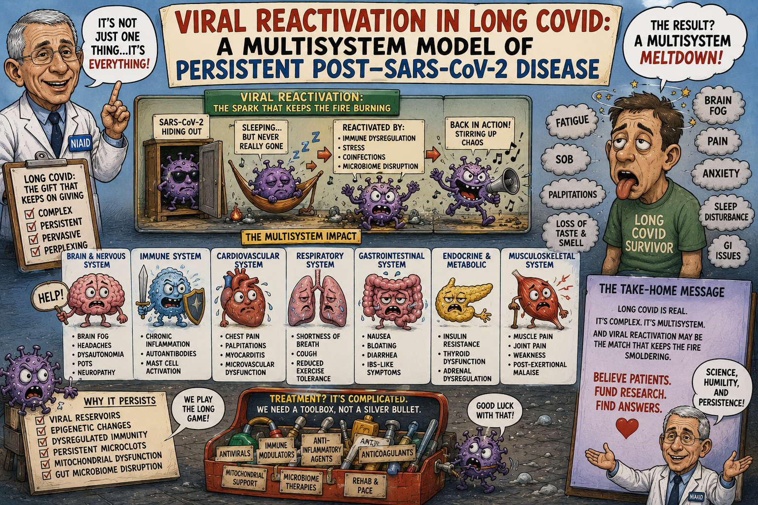
Post–COVID-19 condition, commonly termed Long COVID or post-acute sequelae of SARS-CoV-2 infection (PASC), is a heterogeneous disorder characterized by persistent, recurrent, or newly emergent symptoms after acute SARS-CoV-2 infection. Increasing evidence indicates that the syndrome is biologically heterogeneous and may arise through interacting mechanisms that include persistence of SARS-CoV-2 material, immune dysregulation, endothelial and microvascular injury, autonomic dysfunction, metabolic abnormalities, autoimmunity, and reactivation of latent viruses.
The viral-reactivation hypothesis has undergone an important transformation. Early studies relied predominantly on serologic evidence of Epstein–Barr virus (EBV) reactivation, whereas newer investigations have measured viral transcripts longitudinally and simultaneously with host transcriptomics, cytokines, cellular immunophenotyping, proteomics, and metabolomics. Most notably, a study published in Nature in August 2026 analyzed 1,154 hospitalized patients longitudinally for up to 12 months and demonstrated reactivation of multiple chronic viruses, particularly members of the Herpesviridae and Anelloviridae families. Reactivation correlated with disease severity, systemic inflammation, host immune states, and selected long-COVID outcomes. Importantly, however, the investigators did not establish causation.
These observations support a model in which SARS-CoV-2 infection perturbs an equilibrium between the host immune system and its persistent virome. In susceptible individuals, altered cellular immunity, inflammatory signaling, tissue injury, and metabolic stress may permit reactivation of latent viruses. Viral reactivation may then function as a biological amplifier, perpetuating immune activation after the acute SARS-CoV-2 infection has subsided. EBV is a particularly plausible participant because it establishes lifelong latency in B cells and is tightly controlled by cellular immunity. Yet the newest evidence suggests that the phenomenon is broader than EBV alone: herpesviruses and anelloviruses may participate in a complex host–virome interaction.
The clinical implications are substantial. If viral reactivation constitutes a causal endotype rather than merely a marker of severe illness or immune dysfunction, antiviral or immunologically directed treatment could become an important component of precision therapy. Establishing causality will require prospective studies demonstrating temporal precedence, tissue-specific viral activity, mechanistic linkage to host pathology, and clinical improvement after targeted suppression of the relevant virus.
Introduction
Long COVID presents one of the most consequential challenges to the conventional model of viral infection.
The classical sequence is straightforward:
infection → viral replication → immune clearance → resolution → immunological memory.
Long COVID demonstrates that resolution is not inevitable.
Some patients recover completely. Others develop persistent fatigue, post-exertional malaise, cognitive impairment, dyspnea, neuropathic symptoms, sleep disturbance, dysautonomia, gastrointestinal dysfunction, cardiovascular abnormalities, sensory disturbances, or combinations of these manifestations. Symptoms may persist for months or years and may fluctuate substantially over time.
Current evidence increasingly supports a biological, multisystem disorder rather than a single disease mechanism. A 2026 synthesis identified persistent viral material, immune dysregulation, autonomic dysfunction, microvascular pathology, and other interacting mechanisms as leading explanations for the syndrome.
Among these mechanisms, reactivation of viruses that ordinarily remain latent or persistently controlled by the immune system is particularly intriguing.
The human host is not virologically sterile.
EBV, cytomegalovirus (CMV), herpes simplex viruses, human herpesvirus 6, varicella-zoster virus, and numerous anelloviruses can persist for years or decades. Their continued containment depends upon a dynamic equilibrium involving innate immunity, T cells, B cells, natural-killer cells, interferon signaling, and tissue-specific immune surveillance.
SARS-CoV-2 may disrupt this equilibrium.
I. The Human Virome as a Component of Normal Physiology
Persistent viral infection is a normal feature of human biology.
EBV infects approximately 90–95% of adults worldwide and persists principally within B-cell compartments.
The virus alternates between latent and lytic states.
During latency, viral gene expression is restricted and the viral genome persists with minimal production of infectious virions. During reactivation, a coordinated lytic transcriptional program produces viral proteins, viral DNA replication, and potentially infectious virions.
This equilibrium is normally maintained by immune surveillance.
The consequence is that reactivation is not equivalent to new infection.
It represents a disturbance of an existing host–virus relationship.
That distinction is fundamental to understanding Long COVID.
II. SARS-CoV-2 as a Perturbation of the Host–Virome Equilibrium
Acute SARS-CoV-2 infection produces a profound systemic physiological disturbance.
Among the consequences are:
- activation of innate immune pathways;
- interferon responses;
- cytokine production;
- lymphocyte redistribution;
- T-cell activation and exhaustion;
- B-cell activation;
- endothelial dysfunction;
- complement activation;
- metabolic stress;
- tissue injury.
Longitudinal studies have subsequently demonstrated persistent immune abnormalities in some patients with Long COVID, including inflammatory signaling, complement activation, metabolic dysregulation, and immune-exhaustion programs lasting more than 180 days.
Such an environment provides a biologically plausible setting for reactivation of latent viruses.
The conceptual sequence becomes:
SARS-CoV-2 infection
→
systemic immune perturbation
→
altered antiviral surveillance
→
reactivation of persistent viruses
→
secondary antigenic stimulation
→
further immune and metabolic disturbance
→
persistent disease.
This is a mechanistic hypothesis rather than an established universal pathway.
III. The 2026 Multi-Omic Evidence
The most important recent development is the longitudinal Nature study by Maguire and colleagues, published August 5, 2026.
The investigators analyzed 1,154 hospitalized patients with COVID-19 enrolled in the IMPACC study.
Participants were followed longitudinally for up to 12 months after hospitalization.
The investigators examined:
- nasal swabs;
- peripheral-blood mononuclear cells;
- endotracheal aspirates in ventilated patients;
- viral transcriptomics;
- host transcriptomics;
- cytokines;
- cellular immunophenotyping;
- metabolomics;
- proteomics;
- clinical outcomes.
This design represents an important methodological advance over earlier studies that inferred viral reactivation primarily from antibody titers.
The study identified significant reactivation of chronic viruses, particularly among Herpesviridae and Anelloviridae, and found distinct temporal patterns for different viruses. Reactivation was associated with COVID-19 severity, systemic inflammation, immune alterations, and selected chronic outcomes.
The authors specifically concluded that immunosuppression alone did not explain the observed frequency of reactivation and that viral reactivation occurred in immunocompetent patients with severe COVID-19.
IV. Why Viral Transcripts Matter
Earlier EBV studies frequently relied on antibody measurements.
That approach has limitations.
An elevated EBV antibody titer may reflect:
- prior infection;
- altered antibody kinetics;
- immune dysregulation;
- or recent viral activity.
It does not necessarily demonstrate active viral replication.
The 2026 study instead examined viral transcripts, providing stronger evidence of active viral gene expression. The investigators also cross-validated EBV and CMV transcriptional findings against serologic responses and validated HSV-1 transcript detection against plasma proteomics.
This is an important conceptual advance.
The hierarchy of evidence can be considered:
serologic evidence
<
viral DNA/RNA detection
<
viral transcription
<
viral protein production
<
productive replication
<
demonstration that viral activity causes host pathology.
The last step remains incompletely established.
V. Epstein–Barr Virus
EBV remains the most extensively studied candidate.
Early work by Gold and colleagues reported an association between markers of EBV reactivation and Long-COVID symptoms, particularly fatigue and cognitive dysfunction.
Subsequent studies and reviews have continued to report associations between EBV reactivation and Long COVID, although findings have not been uniform.
A 2025 review concluded that the mechanistic relationship among COVID-19, EBV reactivation, and autoimmunity remains unresolved and emphasized the need to distinguish causality from coincidence.
This caution is particularly important because EBV is nearly ubiquitous.
A positive EBV test therefore cannot, by itself, identify an EBV-mediated Long-COVID endotype.
VI. EBV Reactivation and T-Cell Surveillance
EBV persistence is controlled substantially through cellular immunity.
CD8+ cytotoxic T cells recognize EBV-infected cells and limit their expansion.
Natural-killer cells contribute to early antiviral surveillance.
CD4+ T cells provide regulatory and helper functions.
Thus, perturbation of T-cell physiology can alter EBV control without producing conventional immunodeficiency.
This may explain an important observation from the 2026 study: chronic viral reactivation occurred frequently even among patients who were not pharmacologically immunosuppressed.
The implication is that functional immune dysregulation may be sufficient to alter viral control.
VII. Immune Exhaustion
One possible mechanism is T-cell exhaustion.
Persistent antigenic stimulation can cause T cells to remain activated while progressively losing aspects of their cytotoxic capacity.
This creates an apparently paradoxical state:
inflammation is excessive, but effective viral surveillance may be inadequate.
Such a state could facilitate viral reactivation while simultaneously maintaining chronic inflammatory signaling.
Recent Long-COVID studies have identified persistent immune-exhaustion programs together with inflammatory and complement activation.
The biological loop may therefore be:
viral persistence/reactivation
→
chronic antigen exposure
→
T-cell dysfunction
→
impaired viral control
→
further viral activity.
VIII. Viral Reactivation Is Broader Than EBV
Perhaps the most important conceptual advance from the 2026 study is that the phenomenon should not be reduced to EBV.
Multiple Herpesviridae members demonstrated evidence of reactivation.
Anelloviridae also emerged as important.
The latter are particularly interesting because anelloviruses are extraordinarily prevalent in humans but have historically had an uncertain pathogenic role.
The study found an association between post-hospitalization Anelloviridae transcripts and a physical long-COVID symptom group even after adjustment for age, chronic immunosuppressive medication, and acute COVID-19 severity.
The investigators noted that anelloviruses have previously been associated with immunosenescence and other chronic conditions.
This raises an alternative possibility:
anellovirus transcription may be a biomarker of impaired immune control rather than a direct pathogen.
That distinction remains unresolved.
IX. The Virome as an Integrated Biological System
These observations suggest that Long COVID should be considered in terms of a host virome, not merely one virus.
The virome can be conceptualized as a dynamic ecological system:
SARS-CoV-2
EBV
CMV
HSV
HHV-6
Anelloviridae
host immune system
→
a dynamically interacting biological network.
Perturbation of one component can affect others.
Acute SARS-CoV-2 infection may therefore behave as an ecological disturbance.
The resulting disease is potentially analogous to an ecosystem that fails to return to its previous equilibrium.
X. Pathology: From Viral Reactivation to Tissue Injury
How could a reactivated virus produce chronic disease?
Several pathways are plausible.
Direct cytopathic effects
Active viral replication can injure infected cells.
Innate immune activation
Viral nucleic acids activate pattern-recognition receptors and inflammatory pathways.
Cytokine amplification
Interferons, IL-6, TNF, chemokines, and other mediators can produce systemic effects.
Endothelial injury
Inflammation can alter vascular permeability, coagulation, and microvascular function.
Autoimmunity
Persistent antigen exposure may promote autoreactive immune responses.
Metabolic disruption
Inflammation alters mitochondrial function and cellular energy metabolism.
Neural effects
Systemic inflammation and vascular dysfunction can alter cerebral and autonomic physiology.
The biological consequence therefore need not depend upon extensive direct infection of every symptomatic organ.
XI. Complement and Thromboinflammation
Complement represents a particularly important interface between infection and vascular pathology.
Persistent complement activation has been documented in Long COVID, including evidence of thromboinflammatory abnormalities.
Complement can:
- activate endothelial cells;
- recruit leukocytes;
- promote platelet activation;
- increase vascular permeability;
- damage tissue.
Thus a reactivated virus could theoretically perpetuate a vascular inflammatory state even when SARS-CoV-2 itself is no longer replicating systemically.
This could contribute to abnormalities in:
- skeletal muscle;
- brain;
- myocardium;
- lungs.
XII. Endothelial and Microvascular Physiology
The microcirculation is particularly vulnerable to systemic inflammatory disturbances.
Adequate tissue function requires:
oxygen delivery + vascular regulation + mitochondrial utilization.
A defect at any level can impair function.
Consequently, patients may experience profound exercise intolerance even when conventional cardiopulmonary testing appears relatively preserved.
A microvascular mechanism could explain part of the disconnect between structural imaging and physiological disability.
Viral reactivation may therefore function upstream of a vascular pathway rather than directly producing the final symptom.
XIII. Autonomic Dysfunction
Dysautonomia is another possible downstream pathway.
Long-COVID patients may develop:
- orthostatic tachycardia;
- orthostatic hypotension;
- impaired vasoconstriction;
- abnormal sweating;
- gastrointestinal dysmotility;
- temperature dysregulation;
- exercise intolerance.
Potential mechanisms include:
- autoimmune autonomic injury;
- endothelial dysfunction;
- peripheral nerve injury;
- altered baroreflexes;
- inflammatory signaling;
- abnormal cerebral perfusion.
Whether EBV or other latent viruses directly cause this dysfunction remains unproven.
The more defensible hypothesis is that viral reactivation may contribute to an inflammatory environment capable of disturbing autonomic regulation.
XIV. Neurological Physiology
The neurological manifestations of Long COVID are particularly difficult to explain through a single mechanism.
Patients report:
- cognitive slowing;
- impaired attention;
- memory dysfunction;
- headache;
- dizziness;
- neuropathic pain;
- paresthesia;
- anosmia;
- dysgeusia;
- sleep disturbance.
Possible mechanisms include:
- neuroinflammation;
- microvascular dysfunction;
- blood–brain-barrier alteration;
- glial activation;
- autonomic dysregulation;
- peripheral nerve injury;
- immune-mediated dysfunction.
EBV reactivation could contribute through systemic immune activation without requiring widespread EBV infection of neurons.
XV. Metabolic and Mitochondrial Physiology
Long-COVID fatigue is not adequately explained by subjective exhaustion alone.
The physiology of exertional intolerance suggests that cellular energy production, oxygen utilization, vascular delivery, autonomic regulation, or combinations thereof may be abnormal in some patients.
Persistent inflammatory signaling can alter:
- mitochondrial oxidative phosphorylation;
- substrate utilization;
- redox balance;
- calcium homeostasis;
- reactive oxygen species;
- ATP production.
Recent multi-omic investigations have identified metabolic abnormalities accompanying persistent inflammatory and immune-exhaustion signatures in Long COVID.
Viral reactivation could amplify this state by imposing an additional metabolic burden.
XVI. Post-Exertional Malaise
Post-exertional malaise (PEM) represents a particularly distinctive clinical phenomenon.
A patient may perform an apparently modest amount of physical or cognitive activity and experience delayed deterioration hours or even a day later.
Potential mechanisms include:
exertion → metabolic stress → autonomic dysfunction → inflammatory amplification → delayed systemic deterioration.
A viral-reactivation model offers another possible component:
exertion → transient immune/metabolic perturbation → viral reactivation or enhanced transcription → inflammatory amplification.
This hypothesis is intriguing but has not been adequately tested longitudinally.
XVII. Gastrointestinal Biology
The gastrointestinal tract may represent an important interface between persistent viruses, immunity, and systemic physiology.
SARS-CoV-2 persistence in gastrointestinal tissues has been reported, and contemporary reviews have proposed interactions among intestinal-barrier dysfunction, viral persistence, immune dysregulation, microbiome changes, and neuroinflammation.
The intestine contains a large proportion of the body’s immune tissue.
Consequently:
intestinal inflammation → altered permeability → microbial translocation → systemic immune activation
could provide another mechanism by which a localized biological abnormality becomes systemic.
Viral reactivation could potentially reinforce this pathway.
XVIII. Genomics and Host Susceptibility
The fact that only a subset of infected individuals develop Long COVID strongly suggests host susceptibility.
Relevant genetic systems include:
- HLA antigen presentation;
- interferon signaling;
- innate viral sensing;
- T-cell regulation;
- B-cell differentiation;
- complement;
- cytokine pathways;
- mitochondrial biology.
The genetics of EBV persistence are especially instructive.
A 2026 Nature study identified both genetic and nongenetic determinants of host control of persistent EBV infection, emphasizing the importance of immune regulation and the host–virus relationship.
This provides a plausible framework for Long COVID:
host genotype → quality of antiviral surveillance → probability of viral reactivation → probability of persistent inflammation.
But specific genetic variants that predict an EBV-driven Long-COVID endotype have not yet been established.
XIX. Epigenetic Reprogramming
Genetic susceptibility is only part of the equation.
Viral infection can alter chromatin accessibility, transcription-factor activity, DNA methylation, and cellular metabolic state.
If these changes persist after acute infection, immune cells may remain in an altered functional state.
Such epigenetic reprogramming could help explain why Long COVID may continue after the initiating infection is no longer detectable in blood.
It also provides a possible explanation for relapses:
a biologically altered host remains hypersensitive to subsequent physiological stressors.
XX. Clinical Course
The clinical trajectory is heterogeneous.
Persistent disease
Symptoms continue directly from acute COVID-19.
Delayed disease
The patient initially improves before developing persistent symptoms.
Relapsing–remitting disease
Symptoms fluctuate, sometimes dramatically.
Progressive multisystem disease
Additional organ systems become involved over time.
Partial recovery
Symptoms gradually diminish but fail to disappear completely.
These patterns suggest that Long COVID is not simply a fixed structural injury.
The relapsing phenotype, in particular, is compatible with a dynamic process involving immune activation, autonomic instability, metabolic stress, persistent antigen, or intermittent viral activity.
XXI. What the 2026 Nature Study Changes
The new study substantially changes the evidentiary landscape.
Previously, the proposition was approximately:
SARS-CoV-2 may reactivate EBV.
The new evidence is broader:
SARS-CoV-2 infection is associated with reactivation of multiple persistent viruses, with measurable temporal dynamics and systemic immunological consequences.
The study found:
- multiple chronic viruses reactivating;
- distinct timing among viral species;
- associations with acute disease severity;
- associations with systemic inflammation;
- persistence of some viral reactivation into convalescence;
- an association between Anelloviridae transcripts and a physical Long-COVID phenotype.
That is considerably stronger evidence than the earlier literature based primarily on EBV antibody titers.
XXII. What the Study Does Not Prove
The investigators were explicit about an important limitation.
The study does not establish that viral reactivation causes Long COVID.
Several possibilities remain.
Causal model
Viral reactivation contributes directly to persistent disease.
Amplification model
SARS-CoV-2 initiates Long COVID and viral reactivation perpetuates or worsens it.
Consequence model
Severe systemic inflammation produces viral reactivation, which is largely an epiphenomenon.
Biomarker model
Viral reactivation reflects an underlying state of immune dysfunction.
Mixed model
Different mechanisms operate in different patients.
The mixed model currently best accommodates the evidence.
XXIII. An Important Negative Finding Regarding EBV
The 2026 study contains an especially important nuance.
The investigators did not find a higher rate of EBV transcripts during acute COVID-19 among patients who subsequently developed Long COVID. They noted that this result does not necessarily exclude an EBV contribution because their convalescent patient-reported outcomes were collected before modern Long-COVID definitions were established and may not have captured the full phenotype.
This is precisely the kind of finding that should temper premature conclusions.
It suggests that:
acute EBV transcription ≠ necessarily the determinant of subsequent Long COVID.
Later immune or viral events may matter more than the initial acute-phase measurement.
XXIV. The Timing Problem
Temporal biology may ultimately determine causality.
Suppose:
EBV reactivation precedes symptoms.
That supports causality.
Suppose instead:
Long-COVID symptoms and systemic inflammation precede EBV reactivation.
That favors EBV as a consequence.
Suppose:
both occur simultaneously.
The causal relationship becomes more difficult to resolve.
Future studies therefore require much more frequent sampling than the six acute time points available in the 2026 IMPACC analysis. The authors themselves identified sampling frequency, tissue coverage, participant attrition, and the study population as important limitations.
XXV. Tissue Compartment Matters
Blood may not reveal what is occurring in tissues.
A patient could have:
- no detectable EBV DNAemia;
- no striking serum antibody change;
yet have biologically important viral activity in a tissue compartment.
Similarly, SARS-CoV-2 RNA or protein could persist in tissues without producing detectable systemic viraemia.
Therefore, future studies must distinguish:
blood virology
from
tissue virology.
This distinction may ultimately explain apparently contradictory studies.
XXVI. Toward Molecular Endotyping
The future of Long-COVID medicine is likely to involve endotyping rather than treating the syndrome as one disease.
Potential endotypes include:
Persistent SARS-CoV-2 endotype
Evidence of persistent viral antigen or replication.
Herpesvirus-reactivation endotype
Evidence of active EBV, CMV, HHV-6, HSV, or related viral transcription.
Anellovirus-associated immune-dysfunction endotype
High or persistent anellovirus transcription accompanying immune dysregulation.
Autoimmune endotype
Pathogenic autoreactive antibodies or cellular responses.
Vascular endotype
Objective endothelial or microvascular dysfunction.
Autonomic endotype
Objective dysautonomia.
Metabolic endotype
Abnormal cellular energy production or exercise physiology.
Patients may occupy several categories simultaneously.
XXVII. Implications for Clinical Trials
The therapeutic implications are substantial.
A trial of antiviral therapy in an unselected Long-COVID population may fail because only a fraction of participants have virus-driven disease.
The appropriate design would instead identify patients with molecular evidence of the relevant viral process.
For example:
EBV transcription positive
→ targeted anti-EBV intervention.
SARS-CoV-2 antigen positive
→ SARS-CoV-2-directed antiviral strategy.
No viral activity but autoimmune phenotype
→ immunomodulatory strategy.
Predominantly autonomic phenotype
→ autonomic intervention.
The therapeutic trial itself then becomes a test of biological causality.
XXVIII. What Would Establish Causation?
Five criteria would provide unusually strong evidence.
1. Temporal precedence
Viral reactivation occurs before clinical deterioration.
2. Dose-response relationship
Greater viral activity predicts greater disease severity.
3. Tissue localization
The relevant virus is demonstrable in biologically relevant tissue.
4. Mechanistic linkage
Viral activity produces measurable downstream immune, vascular, metabolic, or neural abnormalities.
5. Therapeutic reversal
Targeted suppression of the virus reduces viral activity and produces meaningful clinical improvement.
The fifth criterion is particularly powerful.
A successful intervention would transform a plausible hypothesis into a demonstrated mechanism.
XXIX. A Unified Pathophysiological Model
The most coherent model presently available can be expressed as:
SARS-CoV-2 infection
↓
viral replication and tissue injury
↓
innate immune activation
↓
adaptive immune perturbation
↓
altered control of persistent viruses
↓
EBV/CMV/HHV-6/HSV/anellovirus reactivation
↓
additional antigenic and inflammatory stimulation
↓
complement and endothelial activation
↓
microvascular dysfunction
↓
autonomic and metabolic disturbance
↓
neurological, cardiopulmonary, gastrointestinal and systemic symptoms
↓
persistent disease.
The model is intentionally pluralistic.
It does not require every patient to follow every pathway.
XXX. Clinical Implications
The recognition of viral reactivation should not presently lead to indiscriminate antiviral treatment.
Routine EBV seropositivity is not evidence of active EBV disease.
Nor does detection of viral DNA automatically establish pathogenic replication.
The clinical priority remains careful phenotyping and exclusion of alternative causes of persistent symptoms.
Nevertheless, the emerging virological evidence argues strongly for expanding clinical research beyond SARS-CoV-2 itself.
The relevant question is increasingly:
What is the state of the patient’s entire host–virus ecosystem after SARS-CoV-2 infection?
That question may ultimately prove more informative than SARS-CoV-2 testing alone.
XXXI. Research Priorities
A definitive longitudinal Long-COVID cohort should simultaneously measure:
- whole-genome host variation;
- viral metagenomics;
- viral transcriptomics;
- viral proteins;
- tissue viral reservoirs;
- T-cell receptor repertoires;
- B-cell receptor repertoires;
- cytokines;
- complement;
- autoantibodies;
- metabolomics;
- mitochondrial function;
- endothelial function;
- autonomic physiology;
- exercise physiology;
- neurocognitive function.
Samples should be obtained during acute infection, early recovery, symptom emergence, relapse, remission, and sustained recovery.
Such a design would permit investigators to reconstruct the causal sequence rather than merely compare patients who are already chronically ill with controls.
XXXII. Conclusions
The hypothesis that viral reactivation contributes to Long COVID has entered a substantially more sophisticated phase.
The evidence is no longer limited to observations that patients with Long COVID have elevated EBV antibodies.
The August 2026 Nature study by Maguire and colleagues, involving 1,154 hospitalized patients followed longitudinally with multi-omic measurements, demonstrated widespread reactivation of chronic viruses, particularly Herpesviridae and Anelloviridae, and linked these phenomena to disease severity, systemic inflammation, immune states, and selected long-COVID outcomes.
The study also provides an important conceptual correction: viral reactivation is not necessarily simply a manifestation of pharmacological immunosuppression. Reactivation occurred in immunocompetent patients during severe COVID-19 and was associated with systemic inflammatory states.
Yet the central scientific question remains unanswered.
Does viral reactivation cause Long COVID, amplify it, or merely mark the immune dysfunction produced by SARS-CoV-2?
The answer may be all three, depending on the patient.
EBV remains an especially compelling candidate because it establishes lifelong latency within B cells and depends heavily on cellular immune surveillance for containment. But the new evidence indicates that the phenomenon is broader, encompassing multiple herpesviruses and anelloviruses.
This suggests that Long COVID may represent a disorder of the host–virome equilibrium.
SARS-CoV-2 may initiate the disturbance. Persistent SARS-CoV-2 antigen, tissue injury, immune dysregulation, autoimmunity, endothelial dysfunction, metabolic stress, and latent-virus reactivation may then interact to prevent restoration of physiological homeostasis.
The result is not necessarily persistent infection with a single pathogen.
It may instead be a self-reinforcing pathological network in which the initiating virus becomes only one component of a persistent biological state.
The therapeutic implication is profound.
If this model is correct, the future of Long-COVID medicine will not be based upon treating “Long COVID” as one disease. It will depend upon identifying molecular endotypes, determining which patients harbor active viral reservoirs or viral reactivation, and matching treatment to mechanism.
The decisive experiments are therefore now clear:
identify the virus → demonstrate temporal and tissue-specific activity → establish its physiological consequences → suppress it → determine whether the patient improves.
Until that sequence has been demonstrated, viral reactivation should be regarded as a strong and increasingly compelling mechanistic hypothesis, but not yet a universal explanation for Long COVID.
Numbered References
- Maguire C, Chen J, Rouphael N, et al.; IMPACC Network. Virus reactivation in acute and long COVID-19. Nature. 2026;656:700-711. doi:10.1038/s41586-026-10740-z. Published August 5, 2026.
- Davis HE, McCorkell L, Vogel JM, Topol EJ. Long COVID: major findings, mechanisms and recommendations. Nat Rev Microbiol. 2023;21:133-146.
- Altmann DM, Whettlock EM, Liu S, Arachchillage DJ, Boyton RJ. The immunology of long COVID. Nat Rev Immunol. 2023;23:618-634.
- Su Y, Yuan D, Chen DG, et al. Multiple early factors anticipate post-acute COVID-19 sequelae. Cell. 2022;185:881-895.
- Klein J, Wood J, Jaycox JR, et al. Distinguishing features of long COVID identified through immune profiling. Nature. 2023;623:139-148.
- Phetsouphanh C, Darley DR, Wilson DB, et al. Immunological dysfunction persists for 8 months following initial mild-to-moderate SARS-CoV-2 infection. Nat Immunol. 2022;23:210-216.
- Gold JE, Okyay RA, Licht WE, Hurley DJ. Investigation of long COVID prevalence and its relationship to Epstein-Barr virus reactivation. Pathogens. 2021;10:763.
- Peluso MJ, Deveau TM, Munter SE, et al. Chronic viral coinfections differentially affect the likelihood of developing long COVID. J Clin Invest. 2023;133:e163669.
- Stein SR, Ramelli SC, Grazioli A, et al. SARS-CoV-2 infection and persistence in the human body and brain at autopsy. Nature. 2022;612:758-766.
- Swank Z, Senussi Y, Manickas-Hill Z, et al. Persistent circulating severe acute respiratory syndrome coronavirus 2 spike is associated with post-acute COVID-19 sequelae. Clin Infect Dis. 2023;76:e487-e490.
- Cervia-Hasler C, et al. Persistent complement dysregulation with signs of thromboinflammation in active long Covid. Science. 2024;383:eadg7942.
- Damania B, Kenney SC, Raab-Traub N. Epstein-Barr virus: biology and clinical disease. Cell. 2022;185:3652-3670.
- Gáspár Z, et al. Human herpesvirus reactivation and its potential role in the pathogenesis of post-acute sequelae of SARS-CoV-2 infection. GeroScience. 2025.
- Tarasco MC, et al. COVID-19, Epstein-Barr virus reactivation and autoimmunity. 2025.
- Hashimoto K. The role of Epstein-Barr virus and the gut-brain axis in long COVID. Mol Psychiatry. 2023.
- Ling J, et al. What could cause the reactivation of Epstein-Barr virus in long COVID? 2025.
- Schmidt A, et al. Host control of persistent Epstein-Barr virus infection. Nature. 2026.
- Aid M, et al. Long COVID involves activation of proinflammatory and immune exhaustion pathways. Nat Immunol. 2025/2026.
- Thompson RC, et al. Molecular states during acute COVID-19 reveal distinct etiologies of long-term sequelae. Nat Med. 2023;29:236-246.
- Ozonoff A, et al. Features of acute COVID-19 associated with post-acute sequelae of SARS-CoV-2 phenotypes: results from the IMPACC study. Nat Commun. 2024;15:216.
- Goetzke CC, et al. TGFβ links EBV to multisystem inflammatory syndrome in children. Nature. 2025;640:762-771.
- Leclerc L, et al. Intestinal barrier compromise, viral persistence, and immune dysregulation in Long COVID. 2026.
- Faghy MA, et al. Current status and future perspectives on the mechanistic understanding of Long COVID. 2026.