Authors: Nicholas L. Li,1P. Toby Coates,2 and Brad H. Rovin1,∗
Kidney Int. 2021 Nov; 100(5): 959–965.Published online 2021 Sep 14. doi: 10.1016/j.kint.2021.09.002 PMCID: PMC8437826PMID: 34534551
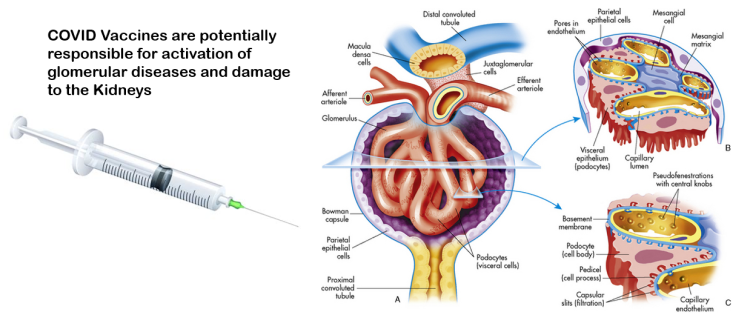
To date, >4 billion doses of the various severe acute respiratory syndrome coronavirus 2 (SARS-CoV-2) vaccines have been administered worldwide in response to the coronavirus disease 2019 (COVID-19) pandemic. Even as widespread vaccination campaigns have contributed to declining case rates, adverse events are appearing beyond those originally reported in the clinical trials of vaccine efficacy and safety. Of particular relevance to the kidney is the increasing number of reports of de novo or reactivation of glomerular diseases (Table 1 1, 2, 3, 4, 5, 6, 7, 8, 9, 10, 11, 12, 13, 14, 15, 16, 17, 18, 19, 20, 21, 22, 23, 24, 25). The occurrence of glomerular disease after immunization against influenza, pneumococcus, and hepatitis B has been reported in the past.26, 27, 28 The reported patients developed acute onset nephrotic syndrome following vaccination, and kidney biopsies were consistent with a minimal change disease (MCD) pattern of injury. Although temporal association (median onset of 12 days) with vaccination and disease onset suggested a vaccine-related induction of immune injury, the pathophysiological mechanisms responsible have not been determined.
Table 1
Summary of reported cases of glomerular disease activation with COVID-19 vaccination
| Disease | Age, yr, median (range) | % Female (n) | Vaccine type | No. of cases | De novo or flarea | Maintenance immune therapy | Temporal association to vaccination, d | Treatment | Outcome | COVID-IgG response | References |
|---|---|---|---|---|---|---|---|---|---|---|---|
| IgAN | 38 (13–52) | 58 (7 of 12) | Pfizer–BioNTech, Moderna | 12 | 5 De novo, 7 flare | No, or steroids, mycophenolic acid, calcineurin inhibitor in transplant patient | 1–2 | RASi, steroids, cyclophosphamide | Spontaneous resolution, renal response to immunotherapy | Positive | 1, 2, 3, 4, 5, 6, 7 |
| MCD | 61 (22–”early 80s”) | 36 (4 of 11) | Pfizer–BioNTech, Moderna, Astra Zeneca | 11 | 7 De novo, 4 flare | No, or steroids, calcineurin inhibitor, rituximab | 1–13 (median, 7) | Steroids, calcineurin inhibitor | Renal response to immunotherapy in most cases | Positive | 8, 9, 10, 11, 12, 13, 14, 15, 16, 17 |
| MN | 68 (66–70) | 50 (1 of 2) | Pfizer–BioNTech, Sinovac | 2 | 1 De novo (anti-THSD7A+), 1 flare (anti-PLA2R+) | No | 7–14 | RASi | NR | Positive | 18,19 |
| AAN | 78 (52–81) | 33 (1 of 3) | Moderna, Pfizer–BioNTech | 3 | De novo | No | 14 | Steroids, cyclophosphamide, plasma exchange | Renal response | Positive | 3,20,21 |
| Anti-GBM | 60 (60–”older female”) | 100 (2 of 2) | Moderna | 2 | De novo | No | 1–14 | Steroids, cyclophosphamide, plasma exchange | No recovery | NR | 6,22 |
| IgG4-RD | 66 | 0 (0 of 1) | Pfizer–BioNTech | 1 | Flare | Rituximab | 14 | Steroids, rituximab | Renal response | Positive | 23 |
| LN | 42 | 100 (1 of 1) | Pfizer–BioNTech | 1 | Flare | Hydroxychloroquine | 7 | Steroids, mycophenolate mofetil | Partial response | Positive | 24 |
| Scleroderma renal crisis | 34 | 100 (1 of 1) | Pfizer–BioNTech | 1 | De novo | No | 1 | RASi | Response | Positive | 25 |
AAN, anti–neutrophil cytoplasmic antibody–associated nephritis; anti-GBM, anti–glomerular basement membrane antibody disease; COVID, coronavirus; COVID-19, coronavirus disease 2019; IgAN, IgA nephropathy; IgG4-RD, IgG4-related disease; LN, lupus nephritis; MCD, minimal change disease; MN, membranous nephropathy; NR, not reported; PLA2R, phospholipase A2 receptor; RASi, renin-angiotensin system inhibitor; THSD7a, thrombospondin type-1 domain-containing 7A.aDe novo indicates disease development in a patient not known to have a prior glomerular disease; flare indicates activation of a known, but controlled, glomerular disease.
After vaccination against COVID-19, reports of exacerbation, and in some cases, new onset of glomerular diseases began arriving at Kidney International and other nephrology journals. Although the development of de novo glomerular disease is intriguing, increased patient awareness of symptoms after vaccination may have prompted medical attention, revealing a previously undiagnosed kidney disease as opposed to a de novo disease. Indeed, chronicity on the kidney biopsy may suggest the glomerular disease preceded COVID-19 vaccination. Although nearly all approved vaccine platforms have been implicated, cases have been far more common after the mRNA-based vaccines, Pfizer–BioNTech BNT162b2 and Moderna mRNA1273 (Table 1). Of course, this may simply reflect more widespread use of these mRNA vaccines. Another interesting feature of COVID-19 vaccine-associated glomerular disease (CVAGD) is that most cases appear to be either IgA nephropathy (IgAN) or MCD (Table 1). The timing of IgAN activation is generally within a day or two after receiving the second dose of BNT162b2 or mRNA1273, whereas MCD appears to occur at a median of 7 days after the first dose (Table 1). Although these associations do not prove causation, we suggest that the volume of cases of MCD and IgAN and the consistent time course of events indicate a direct role of the mRNA vaccines in these 2 glomerular diseases. Several other glomerular diseases have occurred in smaller numbers following vaccination, sometimes quickly (scleroderma renal crisis), but more often after about 2 weeks (e.g., membranous nephropathy, anti–neutrophil cytoplasmic antibody–associated vasculitis, anti–glomerular basement membrane disease, and IgG4 renal disease). Given the small number of cases of these immune-mediated glomerular diseases, and the longer time to their appearance, it is difficult to be certain that they were activated by the vaccines. Nonetheless, considering these cases in aggregate, it appears that the COVID-19 vaccines can (re)activate autoantibody-mediated kidney disease.
It is not clear how COVID-19 vaccines, and in particular the mRNA vaccines, induce MCD, IgAN, and other autoimmune kidney diseases. mRNA-based vaccine technology has been available for some time, although the SARS-CoV-2 vaccines were the first to be investigated in large-scale phase 3 randomized trials. It has been previously demonstrated that this vaccine technology promotes more potent immune responses than inactivated viral vaccines and even natural infection. A comparison of the immune responses to the COVID-19 vaccine platforms is given in Table 2 29, 30, 31, 32, 33, 34, 35. This ability of the mRNA vaccines to enhance virus-specific responses over and above more traditional vaccines has likely contributed to the high efficacy in preventing disease from SARS-CoV-2, as well as the viral variants that have evolved during this pandemic. BNT162b2 or mRNA1273 deliver lipid nanoparticle encapsulated mRNA encoding the full-length SARS-CoV-2 spike protein. These vaccines were found to be safe and efficacious in preventing severe COVID-19 in both clinical trial and real-world conditions, although patients with known autoimmune diseases were not included in the initial trials.36 These lipid nanoparticle–mRNA vaccines stimulate robust antigen-specific T-cell responses, including T follicular helper (Tfh) cells, and potent germinal center B-cell responses, leading to durable neutralizing antibody production.37
Table 2
Immune responses to SARS-CoV-2 vaccine platforms
| Vaccine | Example manufacturer | T-cell responses | B-cell responses | Cytokine responses | References |
|---|---|---|---|---|---|
| LNP-mRNA | Pfizer–BioNTech, Moderna | Antigen-specific Th1-biased CD4+ response, CD8+ IFNɣ, IL-2 | Prolonged S-specific germinal center B-cell responses | IFNɣ, IL-2, type I interferon via toll-like receptor-7 | 29, 30, 31 |
| Adenovirus-DNA | AstraZeneca, Janssen/Johnson & Johnson | Antigen-specific Th1-biased CD4+ response, monofunctional and cytotoxic CD8+ response | IgG1/IgG3 predominant, low IgG2/IgG4 | IFNɣ, TNFα, IL-2, type 1 interferon via toll-like receptor-9 | 31,32 |
| Inactivated whole virus | Sinovac Biotech | Th1-biased response with minimal Th2 | RBD-specific binding antibody and neutralizing antibody production | IFNɣ, TNFα, IL-2 | 33,34 |
| Recombinant protein subunit | Novavax | Th1-biased response with minimal Th2 | S-binding antibody and neutralizing antibody production | IFNɣ, TNFα, IL-2 | 35 |
IFNγ, interferon gamma; IL-2, interleukin 2; LNP, lipid nanoparticle; RBD, receptor-binding domain; SARS-CoV-2, severe acute respiratory syndrome coronavirus 2; Th1, T-helper cell 1; Th2, T-helper cell 2; TNFα, tumor necrosis factor alpha.
In the cases of IgAN, disease symptoms occurred right after vaccination, suggesting a rapid immune mechanism, such as a memory recall response or mobilization of cells positioned to secrete galactose-deficient IgA1 antibodies. Although purely speculative, we wonder if the COVID-19 vaccines can robustly stimulate the gut-associated lymphoid tissue (Peyer patches) responsible for IgA1 production, as they do in other lymphoid tissues. IgA1 hyperresponsiveness has been observed in patients with IgAN following influenza vaccination.38 In the case of COVID-19 vaccination, circulating IgA responses following administration have been observed to be similar in kinetics to IgG responses, with levels reaching a plateau 18 to 21 days after first mRNA dose, and further increases after a second dose peaking at 7 days after dose.39 The temporal associations with hematuria onset following vaccination on the order of days argues against the contribution of spike protein-specific IgA molecules from participating in disease. However, it is known that patients with IgAN have increased circulating galactose-deficient IgA1, and perhaps bystander activation of the immune system with mRNA COVID-19 vaccination may act as a trigger for the formation of immune complexes and subsequent glomerular injury.
In contrast, the development of MCD following vaccination takes some time, suggesting a role for cellular immunity. Central to the pathogenesis of MCD is the development of podocyte injury due to dysregulated T-cell activation.29 The COVID-19 mRNA vaccines trigger enhanced Tfh responses that peak 7 days after immunization. A potential contribution to the pathogenesis of MCD by Tfh cells has been suggested by observations that circulating subsets of Tfh cells are increased in patients with MCD, and the frequency of these populations is reduced in patients who are successfully treated with steroids.40 Given these findings, and the reported onset of disease at a time point that correlates with Tfh response, perhaps mRNA vaccine-induced alterations in the Tfh population and/or their associated cytokine profile in a susceptible individual could promote podocyte injury and the development of nephrotic syndrome and MCD.
The later appearing cases of autoantibody-mediated glomerular disease may be due to the induction of vaccine-associated autoimmunity. Vaccine-associated autoimmunity has been postulated to occur by antigen-specific and nonspecific mechanisms. Antigen-specific triggers for vaccine-mediated autoimmunity are thought to be secondary to molecular mimicry. That is, exposure to a non–self-antigen, such as SARS-CoV-2 spike protein, could elicit responses directed against host tissues if there was sufficient sequence homology to allow for cross-recognition. The SARS-CoV-2 spike protein shares homology with several human proteins, which may then be subject to off-target immune attack after vaccination.41 Consistent with the mimicry hypothesis, it has been suggested that homologous sequences between human alveolar surfactant-related proteins and SARS-CoV-2 spike glycoproteins contribute to host immune attack and the subsequent pulmonary pathology seen with COVID-19 infection.42 Similarly, mimicry of viral antigens with host proteins has been proposed to contribute to immune attack in the central nervous system, exacerbating neurologic complications in COVID-19.43
Antigen nonspecific mechanisms of autoimmunity with vaccination are thought to occur through bystander activation. In this model, the vaccine-stimulated immune response may trigger cellular damage and exposure of normally hidden self-antigens, which are then recognized by host immunity. Alternatively, by this model, innate immune responses may upregulate cytokine signaling and self-antigen presentation by antigen-presenting cells to potentially autoreactive T cells. Either of these mechanisms could conceivably contribute to the development of glomerular disease in response to vaccination, with perhaps different disease phenotypes resulting from each.
Interestingly, to date, there has been only one report of an exacerbation of lupus nephritis (LN) after COVID-19 vaccination, and this was with the BNT162b2 vaccine. This paucity of cases is somewhat unexpected. Tfh cells, robustly activated by mRNA-based COVID-19 vaccines, are important for autoantibody development in lupus.44 Germinal center and peripheral leukocyte cytokine profiles after vaccination are reminiscent of cytokine profiles from lupus patients, with especially high levels of interferon-α, interleukin-6, and tumor necrosis factor-α.45 In the reported case, a patient with known class V LN in remission developed nephrotic syndrome following the first vaccine injection, and kidney biopsy revealed International Society of Nephrology/Renal Pathology Society class II and V LN with an activity index of 0. Given the robust immune activation achieved with the mRNA vaccines, it is surprising that in this case immune complex deposits were limited to the subepithelial compartment and there was no development of proliferative LN. The absence of proliferative LN cases may arguably be because many patients who have lupus nephritis are maintained on long-term immunosuppression. Most patients who developed CVAGD were not on immunosuppression (Table 1). Perhaps a baseline level of immunosuppression is sufficient to blunt the immune response to mRNA vaccination and prevent autoimmune reactions. This is supported by the observation that solid organ transplant patients on various forms of immune suppression, including those typically used in lupus nephritis, such as glucocorticoids and mycophenolate mofetil, demonstrate a weaker response to 2 doses of BNT162b2 vaccination.46 However, considering the few reports of patients on immunosuppression who still developed glomerular injury after vaccination, including one kidney transplant patient, being on immunosuppression is clearly not the only factor determining who will develop kidney disease with these vaccines. Ultimately, there are likely individual patient-specific factors involved that determine whether vaccination results in immune protection or autoimmune injury.
In the published cases of CVAGD, glomerulonephritis was often managed with the usual therapeutic options for these diseases, frequently leading to a clinical response (Table 1). Although evidence is limited, we support a management strategy of CVAGD that is consistent with the conventional therapy of glomerular diseases not associated with vaccination, including the use of immunosuppression if typical indications develop. It is not unreasonable to extrapolate from the management of glomerulonephritis in general, given the presumption in CVAGD that the same disease mechanisms and pathways of vaccine-independent glomerular disease are activated by COVID-19 vaccination. However, management decisions should be tailored to individual cases given the rarity of these events.
As the worldwide COVID-19 vaccination campaign continues to accelerate, it is probable that we will continue to see CVAGD. Not all cases have been, or will be, reported, there is likely reporting bias, and the number of patients with known glomerular disease who have been vaccinated is not known, so the true incidence of CVAGD will be difficult to determine. As multiple doses of vaccines are now being offered, close observation to watch for an increase in CVAGD will be needed. However, in the context of the billions of doses of COVID-19 vaccine that have been administered, the relatively small number of cases thus far suggests a low incidence. Care providers should consider the possibility of glomerulonephritis in patients who develop gross hematuria or edema after vaccination to aid in the prompt diagnosis and management of these diseases. The possibility of CVAGD should not, however, prompt vaccine hesitancy. Most reported cases were easily managed and resolved on their own or responded to typical therapy. Also, COVID-19 infection itself has been linked to the development of immune-mediated kidney diseases.47 The benefits of COVID-19 vaccination appear to greatly outweigh the risks of glomerular disease occurrence or recurrence, and vaccination remains the best method of preventing the morbidity and mortality associated with SARS-CoV-2 infection. Therefore, we are offering vaccination to all of our patients with glomerular diseases, with the following considerations.
Patients in remission and off all immunosuppression should be followed up closely after vaccination and be told to report hematuria or swelling immediately for early intervention. For patients undergoing active immunosuppressive treatment with anti-metabolites (e.g., mycophenolate mofetil or azathioprine), cytotoxic drugs (e.g., cyclophosphamide), anti–B-cell therapies (e.g., rituximab), and costimulation blockers (e.g., abatacept), antibody response to COVID-19 vaccines is likely to be poor.48 , 49 It is probably reasonable to postpone vaccination until these intensive therapies have been tapered or completed. Timing is also important for anti-CD20 B-cell therapies as these have prolonged effects after dosing. For such patients, it is important to continue all preventative measures in place before vaccines were available, and all individuals within the patient’s “bubble” should be vaccinated to provide an additional layer of protection. Finally, it is difficult to speculate on the management of patients who develop CVAGD after the first injection of an mRNA-based vaccine. Checking SARS-CoV-2 antibody response after the first dose may provide some confidence that the patient developed an immune response and may not need the second dose, but of course this does not equate with protection against COVID-19. A change in vaccine platform could also be considered for a second dose. Alternatively, if the CVAGD was mild and readily resolved, administration of the follow-up dose could be considered.
The Immunonephrology Working Group of the European Renal Association–European Dialysis and Transplant Association recently published recommendations on the use of COVID-19 vaccines in patients with autoimmune kidney diseases and supports the vaccination of all individuals without known contraindications.50 However, these recommendations did not advise on whether vaccination with one vaccine platform was preferable to another. Despite the higher number of reports of glomerular disease activation or reactivation with mRNA COVID-19 vaccines compared with the traditional vaccines, it remains difficult to make a recommendation against the mRNA platform. As CVAGD has been seen with non-mRNA vaccines, avoiding Pfizer–BioNTech or Moderna vaccines does not completely eliminate autoimmune risk. Furthermore, the differences in efficacy between the various vaccines cannot be overlooked. Ultimately, as with all decisions in medicine, theoretical risks must be balanced against known benefits of interventions, and discussions between care providers and patients in this regard are important.Go to:
Disclosure
All the authors declared no competing interests.
See the article “A case of gross hematuria and IgA nephropathy flare-up following SARS-CoV-2 vaccination” in Kidney Int, volume 100 on page 238.See the article “Minimal change disease and acute kidney injury following the Pfizer-BioNTech COVID-19 vaccine” in Kidney Int, volume 100 on page 461.See the article “Relapse of primary membranous nephropathy after inactivated SARS-CoV-2 virus vaccination” in Kidney Int, volume 100 on page 464.See the article “Post-vaccinal minimal change disease” in Kidney Int, volume 100 on page 459.See the article “Minimal change disease following the Moderna mRNA-1273 SARS-CoV-2 vaccine” in Kidney Int, volume 100 on page 463.See the article “Histologic correlates of gross hematuria following Moderna COVID-19 vaccine in patients with IgA nephropathy” in Kidney Int, volume 100 on page 468.See the article “Anti-GBM nephritis with mesangial IgA deposits after SARS-CoV-2 mRNA vaccination” in Kidney Int, volume 100 on page 471.See the article “Relapse of minimal change disease following the AstraZeneca COVID-19 vaccine” in Kidney Int, volume 100 on page 459.See the article “Relapse of class V lupus nephritis after vaccination with COVID-19 mRNA vaccine” in Kidney Int, volume 100 on page 941.See the article “Letter regarding “Minimal change disease relapse following SARS-CoV-2 mRNA vaccine”” in Kidney Int, volume 100 on page 458.See the article “A case of membranous nephropathy following Pfizer–BioNTech mRNA vaccination against COVID-19” in Kidney Int, volume 100 on page 938.See the article “Minimal change disease relapse following SARS-CoV-2 mRNA vaccine” in Kidney Int, volume 100 on page 457.See the article “Gross hematuria following vaccination for severe acute respiratory syndrome coronavirus 2 in 2 patients with IgA nephropathy” in Kidney Int, volume 99 on page 1487.See the article “ANCA glomerulonephritis after the Moderna COVID-19 vaccination” in Kidney Int, volume 100 on page 473.See the article “Relapse of IgG4-related nephritis following mRNA COVID-19 vaccine” in Kidney Int, volume 100 on page 465.See the article “IgA nephropathy presenting as macroscopic hematuria in 2 pediatric patients after receiving the Pfizer COVID-19 vaccine” in Kidney Int, volume 100 on page 705.See the article “Gross hematuria following SARS-CoV-2 vaccination in patients with IgA nephropathy” in Kidney Int, volume 100 on page 466.See the article “Is COVID-19 vaccination unmasking glomerulonephritis?” in Kidney Int, volume 100 on page 469.See the article “De novo vasculitis after mRNA-1273 (Moderna) vaccination” in Kidney Int, volume 100 on page 474.See the article “Scleroderma renal crisis following mRNA vaccination against SARS-CoV-2” in Kidney Int, volume 100 on page 940.This article has been cited by other articles in PMC.
References
1. Hanna C., Herrera Hernandez L.P., Bu L. IgA nephropathy presenting as macroscopic hematuria in 2 pediatric patients after receiving the Pfizer COVID-19 vaccine. Kidney Int. 2021;100:705–706. [PMC free article] [PubMed] [Google Scholar]
2. Kudose S., Friedmann P., Albajrami O., D’Agati V.D. Histologic correlates of gross hematuria following Moderna COVID-19 vaccine in patients with IgA nephropathy. Kidney Int. 2021;100:468–469. [PMC free article] [PubMed] [Google Scholar]
3. Anderegg M.A., Liu M., Saganas C. De novo vasculitis after mRNA-1273 (Moderna) vaccination. Kidney Int. 2021;100:474–476. [PMC free article] [PubMed] [Google Scholar]
4. Negrea L., Rovin B.H. Gross hematuria following vaccination for severe acute respiratory syndrome coronavirus 2 in 2 patients with IgA nephropathy. Kidney Int. 2021;99:1487. [PMC free article] [PubMed] [Google Scholar]
5. Perrin P., Bassand X., Benotmane I., Bouvier N. Gross hematuria following SARS-CoV-2 vaccination in patients with IgA nephropathy. Kidney Int. 2021;100:466–468. [PMC free article] [PubMed] [Google Scholar]
6. Tan H.Z., Tan R.Y., Choo J.C.J. Is COVID-19 vaccination unmasking glomerulonephritis? Kidney Int. 2021;100:469–471. [PMC free article] [PubMed] [Google Scholar]
7. Rahim S.E.G., Lin J.T., Wang J.C. A case of gross hematuria and IgA nephropathy flare-up following SARS-CoV-2 vaccination. Kidney Int. 2021;100:238. [PMC free article] [PubMed] [Google Scholar]
8. Weijers J., Alvarez C., Hermans M.M.H. Post-vaccinal minimal change disease. Kidney Int. 2021;100:459–461. [PMC free article] [PubMed] [Google Scholar]
9. Morlidge C., El-Kateb S., Jeevaratnam P., Thompson B. Relapse of minimal change disease following the AstraZeneca COVID-19 vaccine. Kidney Int. 2021;100:459. [PMC free article] [PubMed] [Google Scholar]
10. D’Agati V.D., Kudose S., Bomback A.S. Minimal change disease and acute kidney injury following the Pfizer-BioNTech COVID-19 vaccine. Kidney Int. 2021;100:461–463. [PMC free article] [PubMed] [Google Scholar]
11. Schwotzer N., Kissling S., Fakhouri F. Letter regarding “Minimal change disease relapse following SARS-CoV-2 mRNA vaccine.” Kidney Int. 2021;100:458–459. [PMC free article] [PubMed] [Google Scholar]
12. Holzworth A., Couchot P., Cruz-Knight W., Brucculeri M. Minimal change disease following the Moderna mRNA-1273 SARS-CoV-2 vaccine. Kidney Int. 2021;100:463–464. [PMC free article] [PubMed] [Google Scholar]
13. Kervella D., Jacquemont L., Chapelet-Debout A. Minimal change disease relapse following SARS-CoV-2 mRNA vaccine. Kidney Int. 2021;100:457–458. [PMC free article] [PubMed] [Google Scholar]
14. Leclerc S., Royal V., Lamarche C., Laurin L.-P. Minimal change disease with severe acute kidney injury following the Oxford-AstraZeneca COVID-19 vaccine: a case report. Am J Kidney Dis. 2021;78:607–610. [PMC free article] [PubMed] [Google Scholar]
15. Komaba H., Wada T., Fukagawa M. Relapse of minimal change disease following the Pfizer-BioNTech COVID-19 vaccine. Am J Kidney Dis. 2021;78:469–470. [PMC free article] [PubMed] [Google Scholar]
16. Maas R.J., Gianotten S., van der Meijden W.A.G. An additional case of minimal change disease following the Pfizer-BioNTech COVID-19 vaccine. Am J Kidney Dis. 2021;78:312. [PMC free article] [PubMed] [Google Scholar]
17. Lebedev L., Sapojnikov M., Wechsler A. Minimal change disease following the Pfizer-BioNTech COVID-19 vaccine. Am J Kidney Dis. 2021;78:142–145. [PMC free article] [PubMed] [Google Scholar]
18. Da Y., Goh G.H., Khatri P. A case of membranous nephropathy following Pfizer–BioNTech mRNA vaccination against COVID-19. Kidney Int. 2021;100:938–939. [PMC free article] [PubMed] [Google Scholar]
19. Aydın M.F., Yıldız A., Oruç A. Relapse of primary membranous nephropathy after inactivated SARS-CoV-2 virus vaccination. Kidney Int. 2021;100:464–465. [PMC free article] [PubMed] [Google Scholar]
20. Sekar A., Campbell R., Tabbara J., Rastogi P. ANCA glomerulonephritis after the Moderna COVID-19 vaccination. Kidney Int. 2021;100:473–474. [PMC free article] [PubMed] [Google Scholar]
21. Shakoor M.T., Birkenbach M.P., Lynch M. ANCA-associated vasculitis following Pfizer-BioNTech COVID-19 vaccine. Am J Kidney Dis. 2021;78:611–613. [PMC free article] [PubMed] [Google Scholar]
22. Sacker A., Kung V., Andeen N. Anti-GBM nephritis with mesangial IgA deposits after SARS-CoV-2 mRNA vaccination. Kidney Int. 2021;100:471–472. [PMC free article] [PubMed] [Google Scholar]
23. Masset C., Kervella D., Kandel-Aznar C. Relapse of IgG4-related nephritis following mRNA COVID-19 vaccine. Kidney Int. 2021;100:465–466. [PMC free article] [PubMed] [Google Scholar]
24. Tuschen K., Bräsen J.H., Schmitz J. Relapse of class V lupus nephritis after vaccination with COVID-19 mRNA vaccine. Kidney Int. 2021;100:941–944. [PMC free article] [PubMed] [Google Scholar]
25. Oniszczuk J., Pagot E., Limal N. Scleroderma renal crisis following mRNA vaccination against SARS-CoV-2. Kidney Int. 2021;100:940–941. [PMC free article] [PubMed] [Google Scholar]
26. Gutierrez S., Dotto B., Petiti J.P. Minimal change disease following influenza vaccination and acute renal failure: just a coincidence? Nefrologia. 2012;32:414–415. [PubMed] [Google Scholar]
27. Kikuchi Y., Imakiire T., Hyodo T. Minimal change nephrotic syndrome, lymphadenopathy and hyperimmunoglobulinemia after immunization with a pneumococcal vaccine. Clin Nephrol. 2002;58:68–72. [PubMed] [Google Scholar]
28. Ozdemir S., Bakkaloglu A., Oran O. Nephrotic syndrome associated with recombinant hepatitis B vaccination: a causal relationship or just a mere association? Nephrol Dial Transplant. 1998;13:1888–1889. [PubMed] [Google Scholar]
29. Sahin U., Muik A., Derhovanessian E. COVID-19 vaccine BNT162b1 elicits human antibody and TH1 T cell responses. Nature. 2020;586:594–599. [PubMed] [Google Scholar]
30. Turner J.S., O’Halloran J.A., Kalaidina E. SARS-CoV-2 mRNA vaccines induce persistent human germinal centre responses. Nature. 2021;596:109–113. [PubMed] [Google Scholar]
31. Teijaro J.R., Farber D.L. COVID-19 vaccines: modes of immune activation and future challenges. Nat Rev Immunol. 2021;21:195–197. [PMC free article] [PubMed] [Google Scholar]
32. Ewer K.J., Barrett J.R., Belij-Rammerstorfer S. T cell and antibody responses induced by a single dose of ChAdOx1 nCoV-19 (AZD1222) vaccine in a phase 1/2 clinical trial. Nat Med. 2021;27:270–278. [PubMed] [Google Scholar]
33. Ella R., Reddy S., Jogdand H. Safety and immunogenicity of an inactivated SARS-CoV-2 vaccine, BBV152: interim results from a double-blind, randomised, multicentre, phase 2 trial, and 3-month follow-up of a double-blind, randomised phase 1 trial. Lancet Infect Dis. 2021;21:950–961. [PMC free article] [PubMed] [Google Scholar]
34. Wu Z., Hu Y., Xu M. Safety, tolerability, and immunogenicity of an inactivated SARS-CoV-2 vaccine (CoronaVac) in healthy adults aged 60 years and older: a randomised, double-blind, placebo-controlled, phase 1/2 clinical trial. Lancet Infect Dis. 2021;21:803–812. [PMC free article] [PubMed] [Google Scholar]
35. Keech C., Albert G., Cho I. Phase 1-2 trial of a SARS-CoV-2 recombinant spike protein nanoparticle vaccine. N Engl J Med. 2020;383:2320–2332. [PMC free article] [PubMed] [Google Scholar]
36. Thompson M.G., Burgess J.L., Naleway A.L. Prevention and attenuation of Covid-19 with the BNT162b2 and mRNA-1273 vaccines. N Engl J Med. 2021;385:320–329. [PMC free article] [PubMed] [Google Scholar]
37. Lederer K., Castano D., Gomez Atria D. SARS-CoV-2 mRNA vaccines foster potent antigen-specific germinal center responses associated with neutralizing antibody generation. Immunity. 2020;53:1281–1295.e1285. [PMC free article] [PubMed] [Google Scholar]
38. van den Wall Bake A.W., Beyer W.E., Evers-Schouten J.H. Humoral immune response to influenza vaccination in patients with primary immunoglobulin A nephropathy: an analysis of isotype distribution and size of the influenza-specific antibodies. J Clin Invest. 1989;84:1070–1075. [PMC free article] [PubMed] [Google Scholar]
39. Wisnewski A.V., Campillo Luna J., Redlich C.A. Human IgG and IgA responses to COVID-19 mRNA vaccines. PLoS One. 2021;16 [PMC free article] [PubMed] [Google Scholar]
40. Li T., Shi Y., Sun W. Increased PD-1(+)CD154(+) Tfh cells are possibly the most important functional subset of PD-1(+) T follicular helper cells in adult patients with minimal change disease. Mol Immunol. 2018;94:98–106. [PubMed] [Google Scholar]
41. Vojdani A., Vojdani E., Kharrazian D. Reaction of human monoclonal antibodies to SARS-CoV-2 proteins with tissue antigens: implications for autoimmune diseases. Front Immunol. 2020;11:617089. [PMC free article] [PubMed] [Google Scholar]
42. Kanduc D., Shoenfeld Y. On the molecular determinants of the SARS-CoV-2 attack. Clin Immunol. 2020;215:108426. [PMC free article] [PubMed] [Google Scholar]
43. Ellul M.A., Benjamin L., Singh B. Neurological associations of COVID-19. Lancet Neurol. 2020;19:767–783. [PMC free article] [PubMed] [Google Scholar]
44. Gensous N., Schmitt N., Richez C. T follicular helper cells, interleukin-21 and systemic lupus erythematosus. Rheumatology (Oxford) 2017;56:516–523. [PubMed] [Google Scholar]
45. Jacob N., Stohl W. Cytokine disturbances in systemic lupus erythematosus. Arthritis Res Ther. 2011;13:228. [PMC free article] [PubMed] [Google Scholar]
46. Kamar N., Abravanel F., Marion O. Three doses of an mRNA Covid-19 vaccine in solid-organ transplant recipients. N Engl J Med. 2021;385:661–662. [PMC free article] [PubMed] [Google Scholar]
47. Kudose S., Batal I., Santoriello D. Kidney biopsy findings in patients with COVID-19. J Am Soc Nephrol. 2020;31:1959–1968. [PMC free article] [PubMed] [Google Scholar]
48. Boyarsky B.J., Werbel W.A., Avery R.K. Antibody response to 2-dose SARS-CoV-2 mRNA vaccine series in solid organ transplant recipients. JAMA. 2021;325:2204–2206. [PMC free article] [PubMed] [Google Scholar]
49. Kant S., Kronbichler A., Salas A. Timing of COVID-19 vaccine in the setting of anti-CD20 therapy: a primer for nephrologists. Kidney Int Rep. 2021;6:1197–1199. [PMC free article] [PubMed] [Google Scholar]
50. Kronbichler A., Anders H.J., Fernandez-Juarez G.M. Recommendations for the use of COVID-19 vaccines in patients with immune-mediated kidney diseases [e-pub ahead of print]. Nephrol Dial Transplant. https://doi.org/10.1093/ndt/gfab064 Accessed August 15, 2021.