Authors: Amar D. Desai,*Michael Lavelle,*Brian C. Boursiquot, and Elaine Y. Wan
Am J Physiol Cell Physiol. 2022 Jan 1; 322(1): C1–C11.Published online 2021 Nov 24. doi: 10.1152/ajpcell.00375.2021PMCID: PMC8721906PMID: 34817268
Abstract
SARS-CoV-2 has rapidly spread across the globe and infected hundreds of millions of people worldwide. As our experience with this virus continues to grow, our understanding of both short-term and long-term complications of infection with SARS-CoV-2 continues to grow as well. Just as there is heterogeneity in the acute infectious phase, there is heterogeneity in the long-term complications seen following COVID-19 illness. The purpose of this review article is to present the current literature with regards to the epidemiology, pathophysiology, and proposed management algorithms for the various long-term sequelae that have been observed in each organ system following infection with SARS-CoV-2. We will also consider future directions, with regards to newer variants of the virus and their potential impact on the long-term complications observed.
OVERVIEW AND EPIDEMIOLOGY
The severe acute respiratory syndrome coronavirus 2 (SARS-CoV-2), responsible for coronavirus disease 2019 (COVID-19), has spread across the world. Despite vaccination efforts, SARS-CoV-2 has infected over 1.1 million individuals in the United States alone, with a new wave of increasing cases partially due to novel variants, such as the Delta variant of the virus, which are more easily transmissible (1–3). Although the highest mortality rates had been seen primarily in the elderly population, as more of the vulnerable population became vaccinated, the spread of the virus shifted toward an unvaccinated, younger demographic (4).
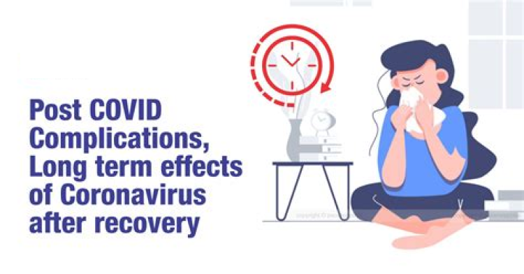
The clinical presentation of COVID-19 has been shown to vary widely, often with respiratory complications as a major feature. SARS-CoV-2 is notable in that a number of patients have gone on to develop long-term complications of the virus (Fig. 1). Beyond initial reports of patients feeling fatigued for months following initial infection, long-haul COVID-19 has come to represent wide complications and sequelae of symptoms that may arise (5). Previous studies have shown a number of potential late complications possible for COVID-19 infection; these include lung fibrosis, venous thromboembolism (VTE), arterial thromboses, cardiac thrombosis and inflammation, stroke, “brain fog,” dermatological complications, and overall mood dysfunctions (6). Although the scope of these long-term complications is wide, specific attributes of patients have been shown to be predictive of which symptoms they develop and for how long (7). Herein, we evaluate the pathophysiology of the virus and development of long-haul COVID-19. We also explore several key complications that may arise including cardiovascular, neurological and psychological, hematological, pulmonary, dermatological, and other injuries.

Schematic representation of long-term sequelae observed following COVID-19 infection. Created with BioRender.com.
PATHOPHYSIOLOGY
Although the exact mechanisms responsible for long-term complications of COVID-19 infection remain unknown, there are a number of pathophysiological mechanisms of the virus that may account for these longer-term complications and sequelae. Possible pathophysiological mechanisms may include direct viral tissue damage; the entry receptor for SARS-CoV-2, angiotensin-converting enzyme 2 (ACE2), is expressed in a variety of locations in the body allowing the virus to enter target cells through activation of its spike protein by transmembrane serine protease 2 (8, 9). These receptors are expressed in epithelial cells, nasal goblet cells, gastrointestinal epithelial cells, pancreatic β cells, and renal podocytes suggesting that direct tissue damage may be a primary mechanism of the presentation of SARS-CoV-2 infection, which may also contribute to its longer-term complications (10–12). Studies early on in the pandemic revealed that endothelial cells had high expression of ACE2 and that COVID-19 infection led to substantial alteration to the integrity of the vessel barrier and promotion of a procoagulative state (13). The long-term sequelae of these changes have been observed in follow-up studies of survivors of COVID-19, revealing pulmonary radiological abnormalities in 71% of patients and functional abnormalities in 25% of patients 3 mo following COVID-19 infection (14).
Beyond direct cellular infection, several other mechanisms exist which may explain the pathophysiology leading to COVID-19 multiorgan systemic disorder. Other suggested pathways leading to long-term COVID-19 infection complications include endothelial injury, immune system dysregulation, and hypercoagulability often leading to thrombosis (5). Immune system dysregulation has been suggested due to the finding of autoreactive T cells in autopsies of deceased individuals infected with COVID-19, likely due to mechanisms similar to those in autoimmune disease (15).
Similar studies on the long-term complications of SARS-CoV-1 virus, the predecessor of the SARS-CoV-2 virus, which emerged in 2003, reported similar complications and sequelae and long-term complications (16). Both these viruses share the same host cell receptor in ACE2, suggesting similar mechanisms of cell entry; however, SARS-CoV-2 has a stronger affinity to the receptor and has an additional cleavage site, which may allow for more effective infection, and possibly, more severe longer-term complications (17).
Patients with symptomatic long COVID-19 have been found to have either PCR-negative or trace PCR-positive persistent low-level detection of SARS-CoV-2 infection (16). In cases of persistent PCR-positive infection, a possible mechanism that may explain longer-term symptoms is a SARS-CoV-2-specific CD8 T cell response of increased breadth and magnitude, which was found in a Danish study of patients with postsymptomatic COVID-19 (17). Notably, some of these individuals were found to have PCR-positive shedding of the virus over 3 mo following initial infection (17, 18).
PULMONARY COMPLICATIONS AND SEQUELAE
Although SARS-CoV-2 can have wide-ranging impacts throughout the body, COVID-19 remains predominantly a respiratory illness. There have been many long-term pulmonary complications described following COVID-19 infection. These include, but are not limited to, dyspnea, ventilator dependence, oxygen dependence, pulmonary function test (PFT) abnormalities, and fibrotic lung disease. The most common pulmonary symptom reported following COVID-19 is dyspnea, which can persist in 22.9%–53% of patients ∼2 mo after symptom onset (18–20). In addition to subjective symptoms, infection with SARS-CoV-2 can result in long-term objective changes in pulmonary physiology. Dependence on oxygen has been reported in up to 6.6% of survivors to hospital discharge (19). In those with respiratory failure requiring a tracheostomy, long-term weaning of ventilator dependence is not often successful. In 1,890 patients requiring a tracheostomy in Spain, weaning from mechanical ventilation at 1 mo follow-up was successful in only 48% of patients (21). Abnormalities in lung function as assessed by pulmonary function testing (PFT) have also been described previously. In a study of 55 noncritically ill patients with COVID-19 in China, PFT assessment during a 3-mo follow-up period revealed abnormalities in 25% of patients, with a reduction in diffusing capacity for carbon monoxide (DLCO) being the most common (16%) (14). Similar observations were noted by Salem et al. (22) with increased rates of restrictive lung findings when compared with matched controls. Radiographic abnormalities also persist in a significant number of patients who have recovered from COVID-19. When assessed ∼3 mo after discharge following severe COVID-19 pneumonia (defined as respiratory rate >30, SpO2 < 90% on room air or severe respiratory distress, and clinical evidence of pneumonia), 81% had abnormal findings on CT chest (23). The ongoing degree of pulmonary fibrosis and long-term worsening will continue to be better defined and understood as our long-term experience with SARS-CoV-2 continues.
There are various mechanisms through which SARS-CoV-2 is able to cause pulmonary injury. Many of the changes that occur as a result of acute infection can promote fibrosis and result in long-term complications. Direct viral entry into cells, specifically type II alveolar epithelial cells that stabilize the epithelial barrier, leads to cell death, which, in turn, leads to an increase in proinflammatory cytokines. The resultant diffuse alveolar damage and cytokines recruit lymphocytes, macrophages, and neutrophils, which recruit fibroblasts that ultimately result in fibrosis (24). In addition to direct damage to the lung parenchyma, damage to the pulmonary vasculature has also been described. Early autopsy studies showed the presence of microthrombi within the small vessels of the pulmonary vasculature (25).
The majority of management considerations following discharge of the patient with COVID-19 involve anticipating, monitoring, and supporting the complications outlined previously. In a proposed blueprint for the post-COVID-19 disease recovery phase published by Lutchmansigh et al. (26), an early and comprehensive assessment following hospital discharge is recommended. This includes serial assessments in the clinic for symptoms, as well as obtaining laboratory markers, serial PFTs, 6-min walk test, and high-resolution computed tomography imaging of the lungs within 3 mo. A patient may return to the routine care of their primary care practice with resolution of symptoms or normalization of any abnormalities observed during diagnostic evaluation over the course of a year of recovery. Alternatively, in patients with ongoing symptoms or abnormalities, referral to a specialist in the management of advanced lung disease should be considered (26). Patients who required hospitalization in an intensive care unit may require more careful assessment and diagnostic investigation following infection as they are at increased risk for many of the long-term complications described in Fig. 1.
CARDIOVASCULAR COMPLICATIONS AND SEQUELAE
Cardiac symptoms are a common complaint following discharge from the hospital after COVID-19. Carfi et al. (18) described chest pain in as many as 21% of patients 60 days after discharge from the hospital. Palpitations have also been described as a frequent symptom at 60-day follow-up in as many as 9% of patients. Outside of subjective cardiac symptoms described as part of the long-term consequences of infection with SARS-CoV-2, there have also been several measurable observed outcomes. An increased incidence of postural tachycardia syndrome (POTS) has been reported following infection with SARS-CoV-2, with ongoing investigation into the exact prevalence (27). Some of the long-term cardiovascular effects of COVID-19 are observed as a consequence of acute infection. In a large study of young, healthy, competitive college athletes, myocarditis was identified in as many as 2.3% of participants, with the majority of those being subclinical myocarditis as detected by cardiac magnetic resonance imaging. Furthermore, follow-up routine cardiac magnetic resonance imaging in 100 patients discharged with COVID-19 discovered ongoing inflammation in 60% of patients with ongoing elevation in high-sensitivity troponin T in as many as 71% of patients (28). The clinical relevance of this finding has been debated, as similar studies in other respiratory illnesses have not been reported, but it does seem to suggest that the cardiac effects of infection with SARS-CoV-2 can be prolonged (28). Finally, the acute phase of COVID-19 has been shown to be associated with high rates of abnormal findings on echocardiography (29). In a prospective study of 1,216 patients admitted with COVID-19, 55% of patients had an abnormal echocardiogram, including abnormalities in 46% of the 901 patients without preexisting cardiac disease (29). Although data were not published on echocardiographic findings at long-term follow-up, the complications observed in the acute setting must be considered in the long-term care of these patients.
There have been several different proposed mechanisms for the pathogenesis of cardiac injury from SARS-CoV-2. Compiled data of early descriptive autopsy studies have observed various findings on histological analysis of the heart, including interstitial inflammatory infiltration, myocardial hypertrophy and necrosis, and RT-PCR positivity for SARS-CoV-2 (30). As has been well described, SARS-CoV-2 has affinity for the ACE2 receptor located on the surface cells, which is the mechanism through which direct viral entry occurs (17). In a study of single-cell RNA sequencing, ACE2 expression has been shown to be present in 7.5% of myocytes, making it an organ at increased risk for direct viral injury (31). This has been supported by autopsy studies in which SARS-CoV-2 viral RNA was present in the myocardium of 61.5% of patients (32). In those patients with the highest viral load (>1,000 copies), there was also increased expression of cytokines (32). This increased expression of cytokines has also been proposed as a mechanism of cardiac injury leading to endothelial dysfunction, plaque instability, myocardial infarction, and myocardial damage (33). As it specifically relates to the cause of myocarditis, after the virus gains entry into the cell, viral antigen presented results in cell-mediated toxicity by CD8+ T lymphocytes with associated myocardial inflammation (34). Both the long-term and short-term cardiovascular complications and sequelae of COVID-19 are manifested through these various mechanisms.
Any patient with known cardiovascular complication of the acute infection and those who go on to develop cardiovascular complaints in the late phase, several weeks to months following acute infection, of the disease should be referred for evaluation by a subspecialist in cardiology (35). Evaluation of the patient should include comprehensive assessment with a history and physical exam and a 12-lead ECG, with consideration of cardiac biomarkers and cardiovascular imaging as each clinical scenario dictates (35). As the experience with COVID-19 complications and sequelae continues to grow, it has been proposed to draw on our experiences in non-COVID-19 illnesses to guide our practices (36). For any patient with a diagnosis of myocarditis, a resting echocardiogram, 24-h Holter monitor, and exercise ECG should be performed no less than 3–6 mo following diagnosis with a return to activity only after there has been normalization of cardiac biomarkers and inflammatory markers, resolution of arrhythmia, and normalization of ventricular function (37). Similarly, patients who develop left ventricular systolic dysfunction should be managed according to current heart failure guidelines. Although there were initial concerns about the use of renin-angiotensin-aldosterone system (RAAS) inhibition due to the virus’ affinity for ACE2 receptors, randomized data of continuation or discontinuation of RAAS inhibition in hospitalized patients with COVID-19 showed no difference in outcomes (38). Consequently, society guidelines currently recommend to continue RAAS-related treatments as indicated for patients with COVID-19 (39). There was also recent investigation into the benefit of the addition of statins for critically ill patients with COVID-19 with the hypothesized mechanism of benefit being their antithrombotic and anti-inflammatory properties. The INSPIRATION-S trial, enrolled ICU patients with confirmed COVID-19 and randomized patients to receive atorvastatin 20 mg daily or placebo. When assessed for the primary composite endpoint of all-cause mortality at 30 days, venous or arterial thrombosis, or treatment with extracorporeal membrane oxygenation (ECMO), atorvastatin was not shown to have any benefit over placebo (40).
HEMATOLOGICAL COMPLICATIONS AND SEQUELAE
Acute COVID-19 has been associated with an increased risk of thrombotic events, especially in critically ill patients (41, 42). The etiology of this coagulopathy is multifactorial, including microvascular dysfunction and increased expression of tissue factors in response to inflammatory cytokines, as well as the effects of hypoxia on upregulation of hypoxia-inducible transcription factors (43, 44). Given the increased risk of thrombosis seen in this patient population, the routine use of intermediate-dose anticoagulation (enoxaparin 1 mg/kg daily) compared with standard prophylactic dose anticoagulation (enoxaparin 40 mg daily) was recently tested in a randomized controlled fashion in critically ill patients with COVID-19 in the INSPIRATION trial. Intermediate-dose anticoagulation was not shown to reduce the composite endpoint of arterial or venous thrombosis, treatment with ECMO, or mortality at 30 days when compared with standard prophylactic-dose anticoagulation (45). Bleeding events are also seen, but given the low risk of major bleeding, the benefits of inpatient VTE prophylaxis outweigh the risks for most patients (46, 47).
The exact duration of hypercoagulability is unknown, but most VTE appears to occur within 2–4 wk of infection (48–50). Extending VTE prophylaxis for hospitalized patients with COVID-19 beyond discharge and into the outpatient setting has been proposed, but current guidelines do not support this as a routine measure (51). In general, extended duration of pharmacological thromboprophylaxis has unclear net benefit in acutely ill medical patients, as studies have shown a reduced risk of VTE, but increased risk of hemorrhage (52). An expert panel on behalf of the American College of Chest Physicians stated that there would be a net benefit of prophylaxis in patients with COVID-19 at low bleeding risk if the rate of symptomatic VTE was above 1.8% at 35 to 42 days after discharge (53), but based on current studies, VTE rates do not appear to be this high.
In retrospective studies, the rates of VTE in patients with COVID-19 who were discharged from the hospital ranged from 0.48% to 1.9% (48–50, 54, 55). Notably, these rates may be similar to rates of symptomatic VTE in other patient populations discharged from the hospital (56). In a large prospective study designed to better assess various postdischarge hematological outcomes in patients with COVID-19, the risks were 1.55% for VTE, 1.71% for arterial thromboembolism, and 1.73% for major bleeds. These authors found that postdischarge pharmacological thromboprophylaxis was protective against a composite of all-cause mortality, VTE, and arterial thromboembolism in patients with COVID-19; but the authors did not assess whether these interventions significantly worsened bleeding (55). Ongoing randomized clinical trials will better answer to address this question and may provide more information that can be used for risk stratification (NCT04416048, NCT04508439).
NEUROPSYCHIATRIC COMPLICATIONS AND SEQUELAE
There have been various neurological and psychiatric long-term complications associated with infection with SARS-CoV-2. Long-term symptom data from multiple different sources reported ongoing neurological findings in patients 2 mo after acute infection, including, fatigue, muscle weakness, sleep difficulties, myalgia, and headache (18, 57). Such symptoms have become the hallmark of the long-COVID syndrome. Loss of smell and taste has also been a feature of infection with SARS-CoV-2 that is unique compared with other viral infections. Long-term follow-up at 2 mo found ongoing loss of taste and smell in anywhere from 11% to 13.1% of patients (19, 57). With the significant burden of severe, critical illness, and acute respiratory distress syndrome (ARDS) associated with COVID-19, similar perturbations in cognition can be expected to those observed in prior studies in patients with ARDS. Impairments in memory (13%), verbal fluency (16%), and executive function (49%) have been described in ARDS survivors from other causes at 1 yr of follow-up (58).
Long-term psychiatric sequelae are also experienced by those infected with SARS-CoV-2. Huang et al. (57) reported 23% of patients with anxiety/depression at 60-day follow-up following hospitalization with COVID-19. In a study of 402 patients discharged from the hospital following COVID-19 with follow-up at 1 mo, Mazza et al. (59) reported rates of post-traumatic stress disorder (PTSD) at 28%, depression at 31%, anxiety at 42%, and insomnia at 40%. A large-scale cohort study performed in the United States showed an increased incidence of a new psychiatric diagnosis in 14 to 90 days following infection with SARS-CoV-2 when compared with several other health events (e.g., other respiratory illnesses, skin infection, bone fracture, etc.) (60).
Just as in the pathology observed in other organs, the neurological complications of COVID-19 are thought to occur through a few proposed mechanisms. A few of the proposed mechanisms include direct viral injury, systemic inflammation, and cerebrovascular changes, with the most likely scenario involving a combination of all the aforementioned (61). Autopsy studies involving the first SARS-CoV-1 epidemic identified genome sequences in the brain, though autopsy studies confirming the same finding in SARS-CoV-2 have not yet been published to date (62). In fact, cerebrospinal fluid analysis with RT-PCR for SARS-CoV-2 in critically ill patients failed to identify the virus (63). These findings support the hypothesis that systemic inflammatory damage plays a significant role in the development of neurocognitive complications following SARS-CoV-2 infection (61). With regards to loss of smell and taste, the pathophysiology has not been well defined. Possible mechanisms include direct viral injury to olfactory cells in the olfactory epithelium, as well as direct injury to additional cells in the olfactory epithelium that are required for smell and express ACE2 (64). Likewise, the loss of taste experienced has been postulated to be due to ACE2 expression diffusely on mucous membranes of the mouth, including the tongue (65). As the two senses are so intimately connected, it has also been suggested that the loss of smell contributes significantly to the loss of taste (65).
Further evaluation with formal neuropsychological testing should be considered in any patient with neurological or psychiatric symptoms following infection with SARS-CoV-2. Standard screening tools for anxiety and depression should be utilized, as well as specifically screening for PTSD in those at highest risk (i.e., severe illness), as these may, in part, be contributing to cognitive findings (66). Any patient experiencing impaired smell and taste should be considered for olfactory training, which typically involves exposure to intense smells on a daily basis for 3 mo (64).
DERMATOLOGICAL COMPLICATIONS AND SEQUELAE
Of the reported cutaneous manifestations of COVID-19 infection, a literature review found that 36.1% of 72 documented patients across 18 studies reported maculopapular exanthem (morbilliform) as the most common cutaneous manifestation of COVID-19, followed by papulovesicular rash (34.7%), urticaria (9.7%), and painful red acral purple papules (15.3%), with 19.4% of these manifestations being in the hands and feet (67). Another international study of 2,560 patients found that pernio-like lesions were the most common cutaneous manifestation (51.5%), with the latency time between upper-respiratory infections and cutaneous findings being 1.5 days in children versus 7.9 days in adults (68). In the Chinese post-acute COVID-19 study of hospitalized patients, only 47 of 1,655 patients (3%) reported skin rashes 6 mo after infection onset (57); instead, hair loss was a far more commonly reported symptom for patients months after COVID-19 infection reported in 24 of 120 patients (20.0%) as a postdischarge symptom 110 days after hospital discharge (69). Still, other more rare presentations have been reported in case reports, suggesting that manifestations in different patients may be different despite being infected with the same virus (70). Although it has been suggested that vesicular rashes may be indicative of an initial diagnosis of COVID-19 and vascular rashes may be indicative of disease prognosis, the precise use of these symptoms in this fashion has not yet been validated and should be a subject of future prospective studies (71).
Although the exact mechanisms for many of these symptoms are unknown and likely due to the same mechanisms responsible for other symptoms of long-haul COVID-19, there are some unique viral mechanisms that may be applicable. For instance, telogen effluvium describes the phenomenon of temporary hair loss in the form of nonscarring alopecia, which occurs following shock, a traumatic event, postpartum hormonal changes, or acute febrile illness/viral infection, typically for a period under 6 mo (72). Patients experiencing hair-loss symptoms following COVID-19 infection can likely find symptoms to be reversible with administration of medications like minoxidil, finasteride, and topical corticosteroids (73).
CHILDREN
Children are diagnosed with COVID-19 at lower rates than adults and generally have a milder illness in the acute phase (74). However, complications and sequelae of infection include multisystem inflammatory syndrome in children (MIS-C), which is characterized by fever and multiorgan dysfunction in the weeks following SARS-CoV-2 infection (75, 76). The incidence of MIS-C is 316 cases per 1,000,000 infections, predominantly affecting children of racial/ethnic minority backgrounds (77, 78). Features of MIS-C overlap with both severe acute COVID-19 and Kawasaki disease, and nearly 75% of patients require ICU admission (77, 79, 80). The most common manifestations are gastrointestinal symptoms, cardiovascular complications, respiratory symptoms, mucocutaneous manifestations, and neurological complications (77, 81). Among cardiovascular complications, nearly half have myocarditis with some reduction in left ventricular ejection fraction, and over 20% have coronary artery dilatation or aneurysm (77). To treat MIS-C, over 75% of patients receive intravenous immunoglobulin, over half require inotropic support, one-quarter are intubated, and one-quarter are ventilated noninvasively. Other common treatment modalities include aspirin, steroids, and other immune-modulating therapies. The prognosis is overall favorable, with a low mortality rate of 1.9%, and typically cardiovascular complications resolve within weeks (77). However, persistently reduced ejection fraction and unresolved coronary artery aneurysms are still seen over a month later in a minority of patients, and the long-term consequences of MIS-C are uncertain (77, 80).
DIABETES COMPLICATIONS AND SEQUELAE
Preexisting diabetes mellitus has been associated with worse COVID-19 outcomes. At the same time, COVID-19 has been associated with new-onset hyperglycemia and acute decompensation of diabetes, including diabetic ketoacidosis in both patients with type 1 and type 2 diabetes (82). Besides iatrogenic hyperglycemia from steroid use, proposed mechanisms for hyperglycemia following infection include insulin resistance as a result of the inflammatory state, and insulin secretory deficits from impaired β cells—either due to direct viral damage or indirect effects (82, 83). It is unclear how many patients who are newly diagnosed with diabetes after COVID-19 already had unrecognized diabetes before infection, and simply had their diabetes unmasked or exacerbated. It is also unclear whether new-onset diabetes following hospitalization for COVID-19 is permanent. Therefore, the global CoviDiab Registry was created to further study the relationship between COVID-19 and diabetes, and to better characterize the duration of post-COVID-19 diabetes (84).
RENAL COMPLICATIONS AND SEQUELAE
Acute kidney injury (AKI) is common in acute COVID-19, and 5% of all hospitalized patients require inpatient renal replacement therapy (85). The etiology of AKI is multifactorial, with contributing factors including direct viral damage, systemic hypoxia, effects of inflammatory cytokines, and abnormal coagulation (86–89). Acute tubular necrosis is the most common histopathological finding, but glomerulopathy and microvascular thrombi are seen as well (87–93). AKI is associated with increased hospital mortality, and those who survive hospital discharge may have residual renal dysfunction (85). In one retrospective study, 35% of those with AKI still had abnormal renal function at discharge, and 30% of those who required inpatient dialysis continued to require dialysis after discharge. At follow-up in the same study (median 21 days), 36% of those with residual kidney disease at discharge had recovered, but 14% of those who recovered before discharge had recurrent kidney disease (94). In another study, 35% of survivors of COVID-19 had renal dysfunction at 6 mo, and 13% had new-onset renal dysfunction after having had normal kidney function during their initial illness (57). Given the improved outcomes associated with close follow-up, those with kidney disease after acute COVID-19 should establish care with a nephrologist (95, 96).
GASTROINTESTINAL COMPLICATIONS AND SEQUELAE
Gastrointestinal symptoms are common in acute COVID-19 and are also seen later in recovery. In one systematic review of postacute COVID-19 manifestations, diarrhea was among the top 10 most common complaints, with a prevalence of 6%. Other long-term symptoms include nausea, vomiting, abdominal pain, and loss of appetite (97). These persistent symptoms may be related to ongoing viral replication in the gastrointestinal tract, given the prolonged fecal shedding of SARS-CoV-2 seen even after respiratory samples become negative (98–101). COVID-19 is also associated with alterations in the gut microbiome, although the effects of this on long-term symptoms are not clear (102, 103).
Abnormal liver function tests are frequently seen in the acute phase, corresponding to hepatocellular injury and/or biliary stasis (104, 105). The mechanisms of injury include direct viral cytotoxicity, particularly in the biliary tree, as well as the effects of systemic inflammation, hypoxia, coagulopathy, and adverse effects of drugs (105–108). In autopsy studies, common findings include hepatic steatosis, hepatic congestion, vascular thrombosis, fibrosis, portal inflammation, and lobular inflammation (109). In survivors of COVID-19 with acute liver injury, abnormalities in liver function may persist but gradually improve over weeks to months (106).Go to:
MUSCULOSKELETAL COMPLICATIONS AND SEQUELAE
Musculoskeletal symptoms are among the most common complaints in COVID-19, both at disease onset and in the postacute phase (97, 110, 111). Unsurprisingly, ACE 2 receptors are found in skeletal muscle and synovial tissue, suggesting that viral invasion of these tissues contributes to symptoms (112). Although arthritis is associated with some viral illnesses, COVID-19 more typically causes myalgias and arthralgias without true inflammatory arthritis (113, 114). Reports of new-onset rheumatological diseases after COVID-19 are mostly limited to case reports (114). Not unlike other critical illnesses, a major complication of severe COVID-19 is catabolic muscle wasting as a result of systemic inflammation, prolonged bed rest, and malnutrition (115). Physical impairments after critical illness can last for months or even years (116); and among survivors of COVID-19, those who had more severe acute illness had more muscle weakness, more problems with mobility, and had shorter distances walked in 6 min at 6 mo follow-up (57). This highlights the importance of optimizing nutrition and rehabilitation, both in the early and post-acute phases of COVID-19.
GENITOURINARY COMPLICATIONS AND SEQUELAE
Genitourinary symptoms have not typically been described in patients with postsymptomatic COVID-19; however, male infertility, in particular, has arisen a possible long-term health concern of COVID-19 infection (117). Notably, viral mRNA has been detected in the semen of infected men, with evidence that male gonads may also be particularly susceptible due to an increased expression of ACE2 receptors (8, 118). Studies have also shown significant reductions in testosterone and dihydrotestosterone, as well as testicular signs of pathological inflammation in several patients, with the suggested incidence of viral orchitis being reported as being as high as 19% (118, 119). In female patients, suggestions have been made as to raised luteinizing hormone as well as preterm delivery without the risk of vertical transmission, however, larger studies are needed to better understand the validity of these hypotheses (118, 120).
FUTURE RESEARCH
There are a number of considerations that are yet to be addressed in the COVID-19 pandemic. As feared early on in the pandemic, inability to control the virus has led to the development of novel variants of the virus, including but not limited to the Delta (B.1.617.2 lineage) variant first identified in India, Gamma (P.1 lineage) first identified in Brazil, and the Lambda (C.37 lineage) variant first identified in Peru (2). Several of these new variants have also been deemed variants of interest (VOIs) by the World Health Organization, suggesting a reduction in the effectiveness of therapeutics or vaccination in neutralizing the virus. The Delta variant has notably begun to spread in the United States with a number of breakthrough cases found even among vaccinated individuals, likely due to the Delta variant’s ability to partially, but not completely, avoid the neutralizing monoclonal or polyclonal antibodies elicited by immunity to SARS-CoV-2, either through vaccination or previous infection (3).
Given the relatively recent onset of these novel variants, it remains unclear how the presentation of long-haul COVID-19 will be impacted in individuals infected with these strains of the virus. The recent onset of the Delta variant has not allowed for large, published studies regarding differences in symptom presentation and patient follow-up over time. However, a preprint of an analysis of the Delta variant in China by Kang et al. (121) states that individuals with the strain are more likely to spread the virus given a higher presymptomatic viral load, with 73.9% of transmissions from Delta cases occurring before symptom onset. In these individuals, however, vaccination still seemed effective, as unvaccinated individuals (OR: 2.84) or those with only one dose of vaccination (OR: 6.02) were more likely to spread infection than recipients of two doses of the vaccine. Specifics in terms of symptoms of patients with these novel variants stratified by age and race also remain to be seen and will depend on larger studies.
Finally, previous studies on the Alpha variant of the virus suggested racial discrepancies in terms of treatment received duration of symptoms and likelihood of contracting COVID-19 as well as long COVID-19 symptoms (5). It remains unclear whether these differences are due to discrepancies in care received or molecular mechanisms behind why certain individuals may be more predisposed to development of longer-term symptoms. The impact of these novel strains on the development of long COVID-19 and which patients will be most affected will require further study.
M.L. prepared figures; A.D.D., M.L., B.C.B., and E.Y.W. drafted manuscript; A.D.D., M.L., B.C.B., and E.Y.W. edited and revised manuscript; A.D.D., M.L., B.C.B., and E.Y.W. approved final version of manuscript.Go to:
REFERENCES
1. Dong E, Du H, Gardner L. An interactive web-based dashboard to track COVID-19 in real time. Lancet Infect Dis 20: 533–534, 2020. doi:10.1016/S1473-3099(20)30120-1. [PMC free article] [PubMed] [CrossRef] [Google Scholar]
2. Faria NR, Mellan TA, Whittaker C, Claro IM, Candido DDS, Mishra S, et al. . Genomics and epidemiology of a novel SARS-CoV-2 lineage in Manaus, Brazil. medRxiv, 2021. doi:10.1101/2021.02.26.21252554. [PMC free article] [PubMed] [CrossRef]
3. Planas D, Veyer D, Baidaliuk A, Staropoli I, Guivel-Benhassine F, Rajah MM, Planchais C, Porrot F, Robillard N, Puech J, Prot M, Gallais F, Gantner P, Velay A, Le Guen J, Kassis-Chikhani N, Edriss D, Belec L, Seve A, Courtellemont L, Péré H, Hocqueloux L, Fafi-Kremer S, Prazuck T, Mouquet H, Bruel T, Simon-Lorière E, Rey FA, Schwartz O. Reduced sensitivity of SARS-CoV-2 variant delta to antibody neutralization. Nature 596: 276–280, 2021. doi:10.1038/s41586-021-03777-9. [PubMed] [CrossRef] [Google Scholar]
4. Centers for Disease Control and Prevention. COVID-19 Weekly Cases and Deaths per 100,000 Population by Age, Race/Ethnicity, and Sex. 2021. https://www.cdc.gov/nchs/nvss/vsrr/covid_weekly/index.htm.
5. Nalbandian A, Sehgal K, Gupta A, Madhavan MV, McGroder C, Stevens JS, Cook JR, Nordvig AS, Shalev D, Sehrawat TS, Ahluwalia N, Bikdeli B, Dietz D, Der-Nigoghossian C, Liyanage-Don N, Rosner GF, Bernstein EJ, Mohan S, Beckley AA, Seres DS, Choueiri TK, Uriel N, Ausiello JC, Accili D, Freedberg DE, Baldwin M, Schwartz A, Brodie D, Garcia CK, Elkind MSV, Connors JM, Bilezikian JP, Landry DW, Wan EY. Post-acute COVID-19 syndrome. Nat Med 27: 601–615, 2021. doi:10.1038/s41591-021-01283-z. [PMC free article] [PubMed] [CrossRef] [Google Scholar]
6. SeyedAlinaghi S, Afsahi AM, MohsseniPour M, Behnezhad F, Salehi MA, Barzegary A, Mirzapour P, Mehraeen E, Dadras O. Late complications of COVID-19; a systematic review of current evidence. Arch Acad Emerg Med 9: e14, 2021. doi:10.22037/aaem.v9i1.1058. [PMC free article] [PubMed] [CrossRef] [Google Scholar]
7. Sudre CH, Murray B, Varsavsky T, Graham MS, Penfold RS, Bowyer RC, Pujol JC, Klaser K, Antonelli M, Canas LS, Molteni E, Modat M, Jorge Cardoso M, May A, Ganesh S, Davies R, Nguyen LH, Drew DA, Astley CM, Joshi AD, Merino J, Tsereteli N, Fall T, Gomez MF, Duncan EL, Menni C, Williams FMK, Franks PW, Chan AT, Wolf J, Ourselin S, Spector T, Steves CJ. Attributes and predictors of long COVID. Nat Med 27: 626–631, 2021. [Erratum in Nat Med 27: 1116, 2021]. doi:10.1038/s41591-021-01292-y. [PMC free article] [PubMed] [CrossRef] [Google Scholar]
8. Gupta A, Madhavan MV, Sehgal K, Nair N, Mahajan S, Sehrawat TS, Bikdeli B, Ahluwalia N, Ausiello JC, Wan EY, Freedberg DE, Kirtane AJ, Parikh SA, Maurer MS, Nordvig AS, Accili D, Bathon JM, Mohan S, Bauer KA, Leon MB, Krumholz HM, Uriel N, Mehra MR, Elkind MSV, Stone GW, Schwartz A, Ho DD, Bilezikian JP, Landry DW. Extrapulmonary manifestations of COVID-19. Nat Med 26: 1017–1032, 2020. doi:10.1038/s41591-020-0968-3. [PubMed] [CrossRef] [Google Scholar]
9. Hoffmann M, Kleine-Weber H, Schroeder S, Krüger N, Herrler T, Erichsen S, Schiergens TS, Herrler G, Wu NH, Nitsche A, Müller MA, Drosten C, Pöhlmann S. SARS-CoV-2 cell entry depends on ACE2 and TMPRSS2 and is blocked by a clinically proven protease inhibitor. Cell 181: 271–280.e8, 2020. doi:10.1016/j.cell.2020.02.052. [PMC free article] [PubMed] [CrossRef] [Google Scholar]
10. Qi F, Qian S, Zhang S, Zhang Z. Single cell RNA sequencing of 13 human tissues identify cell types and receptors of human coronaviruses. Biochem Biophys Res Commun 526: 135–140, 2020. doi:10.1016/j.bbrc.2020.03.044. [PMC free article] [PubMed] [CrossRef] [Google Scholar]
11. Pan X-W, Xu D, Zhang H, Zhou W, Wang L-H, Cui X-G. Identification of a potential mechanism of acute kidney injury during the COVID-19 outbreak: a study based on single-cell transcriptome analysis. Intensive Care Med 46: 1114–1116, 2020. doi:10.1007/s00134-020-06026-1. [PMC free article] [PubMed] [CrossRef] [Google Scholar]
12. Ziegler CGK, Allon SJ, Nyquist SK, Mbano IM, Miao VN, Tzouanas CN, et al. . SARS-CoV-2 receptor ACE2 is an interferon-stimulated gene in human airway epithelial cells and is detected in specific cell subsets across tissues. Cell 181: 1016–1035.e19, 2020. doi:10.1016/j.cell.2020.04.035. [PMC free article] [PubMed] [CrossRef] [Google Scholar]
13. Jin Y, Ji W, Yang H, Chen S, Zhang W, Duan G. Endothelial activation and dysfunction in COVID-19: from basic mechanisms to potential therapeutic approaches. Signal Transduct Target Ther 5: 293, 2020. doi:10.1038/s41392-020-00454-7. [PMC free article] [PubMed] [CrossRef] [Google Scholar]
14. Zhao Y-M, Shang Y-M, Song W-B, Li Q-Q, Xie H, Xu Q-F, Jia JL, Li LM, Mao HL, Zhou XM, Luo H, Gao YF, Xu AG. Follow-up study of the pulmonary function and related physiological characteristics of COVID-19 survivors three months after recovery. EClinicalMedicine 25: 100463, 2020. doi:10.1016/j.eclinm.2020.100463. [PMC free article] [PubMed] [CrossRef] [Google Scholar]
15. Ehrenfeld M, Tincani A, Andreoli L, Cattalini M, Greenbaum A, Kanduc D, Alijotas-Reig J, Zinserling V, Semenova N, Amital H, Shoenfeld Y. Covid-19 and autoimmunity. Autoimmun Rev 19: 102597, 2020. doi:10.1016/j.autrev.2020.102597. [PMC free article] [PubMed] [CrossRef] [Google Scholar]
16. Ngai JC, Ko FW, Ng SS, To K-W, Tong M, Hui DS. The long-term impact of severe acute respiratory syndrome on pulmonary function, exercise capacity and health status. Respirology 15: 543–550, 2010. doi:10.1111/j.1440-1843.2010.01720.x. [PMC free article] [PubMed] [CrossRef] [Google Scholar]
17. Shang J, Ye G, Shi K, Wan Y, Luo C, Aihara H, Geng Q, Auerbach A, Li F. Structural basis of receptor recognition by SARS-CoV-2. Nature 581: 221–224, 2020. doi:10.1038/s41586-020-2179-y. [PMC free article] [PubMed] [CrossRef] [Google Scholar]
18. Carfì A, Bernabei R, Landi F; Gemelli Against COVID-19 Post-Acute Care Study Group. Persistent symptoms in patients after acute COVID-19. JAMA 324: 603–605, 2020. doi:10.1001/jama.2020.12603. [PMC free article] [PubMed] [CrossRef] [Google Scholar]
19. Chopra V, Flanders SA, O’Malley M, Malani AN, Prescott HC. Sixty-day outcomes among patients hospitalized with COVID-19. Ann Intern Med 174: 576–578, 2021. doi:10.7326/M20-5661. [PMC free article] [PubMed] [CrossRef] [Google Scholar]
20. Mandal S, Barnett J, Brill SE, Brown JS, Denneny EK, Hare SS, Heightman M, Hillman TE, Jacob J, Jarvis HC, Lipman MCI, Naidu SB, Nair A, Porter JC, Tomlinson GS, Hurst JR; ARC Study Group. ‘Long-COVID’: a cross-sectional study of persisting symptoms, biomarker and imaging abnormalities following hospitalisation for COVID-19. Thorax 76: 396–398, 2021. doi:10.1136/thoraxjnl-2020-215818. [PMC free article] [PubMed] [CrossRef] [Google Scholar]
21. Martin-Villares C, Perez Molina-Ramirez C, Bartolome-Benito M, Bernal-Sprekelsen M; COVID ORL ESP Collaborative Group (*). Outcome of 1890 tracheostomies for critical COVID-19 patients: a national cohort study in Spain. Eur Arch Otorhinolaryngol 278: 1605–1612, 2021. doi:10.1007/s00405-020-06220-3. [PMC free article] [PubMed] [CrossRef] [Google Scholar]
22. Salem AM, Al Khathlan N, Alharbi AF, Alghamdi T, AlDuilej S, Alghamdi M, Alfudhaili M, Alsunni A, Yar T, Latif R, Rafique N, Al Asoom L, Sabit H. The long-term impact of COVID-19 pneumonia on the pulmonary function of survivors. Int J Gen Med 14: 3271–3280, 2021. doi:10.2147/IJGM.S319436. [PMC free article] [PubMed] [CrossRef] [Google Scholar]
23. Balbi M, Conti C, Imeri G, Caroli A, Surace A, Corsi A, Mercanzin E, Arrigoni A, Villa G, Di Marco F, Bonaffini PA, Sironi S. Post-discharge chest CT findings and pulmonary function tests in severe COVID-19 patients. Eur J Radiol 138: 109676, 2021. doi:10.1016/j.ejrad.2021.109676. [PMC free article] [PubMed] [CrossRef] [Google Scholar]
24. McDonald LT. Healing after COVID-19: are survivors at risk for pulmonary fibrosis? Am J Physiol Lung Cell Mol Physiol 320: L257–L265, 2021. doi:10.1152/ajplung.00238.2020. [PMC free article] [PubMed] [CrossRef] [Google Scholar]
25. Wichmann D. Autopsy findings and venous thromboembolism in patients with COVID-19. Ann Intern Med 173: 1030, 2020. doi:10.7326/L20-1206. [PubMed] [CrossRef] [Google Scholar]
26. Lutchmansingh DD, Knauert MP, Antin-Ozerkis DE, Chupp G, Cohn L, Dela Cruz CS, Ferrante LE, Herzog EL, Koff J, Rochester CL, Ryu C, Singh I, Tickoo M, Winks V, Gulati M, Possick JD. A clinic blueprint for post-coronavirus disease 2019 RECOVERY: learning from the past, looking to the future. Chest 159: 949–958, 2021. doi:10.1016/j.chest.2020.10.067. [PMC free article] [PubMed] [CrossRef] [Google Scholar]
27. Raj SR, Arnold AC, Barboi A, Claydon VE, Limberg JK, Lucci V-EM, Numan M, Peltier A, Snapper H, Vernino S; American Autonomic Society. Long-COVID postural tachycardia syndrome: an American Autonomic Society statement. Clin Auton Res 31: 365–368, 2021. doi:10.1007/s10286-021-00798-2. [PMC free article] [PubMed] [CrossRef] [Google Scholar]
28. Puntmann VO, Carerj ML, Wieters I, Fahim M, Arendt C, Hoffmann J, Shchendrygina A, Escher F, Vasa-Nicotera M, Zeiher AM, Vehreschild M, Nagel E. Outcomes of cardiovascular magnetic resonance imaging in patients recently recovered from coronavirus disease 2019 (COVID-19). JAMA Cardiol 5: 1265–1273, 2020. doi:10.1001/jamacardio.2020.3557. [PMC free article] [PubMed] [CrossRef] [Google Scholar
]29. Dweck MR, Bularga A, Hahn RT, Bing R, Lee KK, Chapman AR, White A, Salvo GD, Sade LE, Pearce K, Newby DE, Popescu BA, Donal E, Cosyns B, Edvardsen T, Mills NL, Haugaa K. Global evaluation of echocardiography in patients with COVID-19. Eur Heart J Cardiovasc Imaging 21: 949–958, 2020. doi:10.1093/ehjci/jeaa178. [PMC free article] [PubMed] [CrossRef] [Google Scholar]
30. Kang Y, Chen T, Mui D, Ferrari V, Jagasia D, Scherrer-Crosbie M, Chen Y, Han Y. Cardiovascular manifestations and treatment considerations in COVID-19. Heart 106: 1132–1141, 2020. doi:10.1136/heartjnl-2020-317056. [PMC free article] [PubMed] [CrossRef] [Google Scholar]
31. Zou X, Chen K, Zou J, Han P, Hao J, Han Z. Single-cell RNA-seq data analysis on the receptor ACE2 expression reveals the potential risk of different human organs vulnerable to 2019-nCoV infection. Front Med 14: 185–192, 2020. doi:10.1007/s11684-020-0754-0. [PMC free article] [PubMed] [CrossRef] [Google Scholar]
32. Lindner D, Fitzek A, Bräuninger H, Aleshcheva G, Edler C, Meissner K, Scherschel K, Kirchhof P, Escher F, Schultheiss HP, Blankenberg S, Püschel K, Westermann D. Association of cardiac infection With SARS-CoV-2 in confirmed COVID-19 autopsy cases. JAMA Cardiol 5: 1281–1285, 2020. doi:10.1001/jamacardio.2020.3551. [PMC free article] [PubMed] [CrossRef] [Google Scholar]
33. Guzik TJ, Mohiddin SA, Dimarco A, Patel V, Savvatis K, Marelli-Berg FM, Madhur MS, Tomaszewski M, Maffia P, D’Acquisto F, Nicklin SA, Marian AJ, Nosalski R, Murray EC, Guzik B, Berry C, Touyz RM, Kreutz R, Wang DW, Bhella D, Sagliocco O, Crea F, Thomson EC, McInnes IB. COVID-19 and the cardiovascular system: implications for risk assessment, diagnosis, and treatment options. Cardiovasc Res 116: 1666–1687, 2020. doi:10.1093/cvr/cvaa106. [PMC free article] [PubMed] [CrossRef] [Google Scholar]
34. Siripanthong B, Nazarian S, Muser D, Deo R, Santangeli P, Khanji MY, Cooper LT Jr, Chahal CAA. Recognizing COVID-19-related myocarditis: the possible pathophysiology and proposed guideline for diagnosis and management. Heart Rhythm 17: 1463–1471, 2020. doi:10.1016/j.hrthm.2020.05.001. [PMC free article] [PubMed] [CrossRef] [Google Scholar]
35. Richter D, Guasti L, Koehler F, Squizzato A, Nistri S, Christodorescu R, Dievart F, Gaudio G, Asteggiano R, Ferrini M. Late phase of COVID-19 pandemic in general cardiology. A position paper of the ESC Council for Cardiology Practice. ESC Heart Fail 8: 3483–3494, 2021. doi:10.1002/ehf2.13466. [PMC free article] [PubMed] [CrossRef] [Google Scholar]
36. Hendren NS, Drazner MH, Bozkurt B, Cooper LT Jr.. Description and proposed management of the acute COVID-19 cardiovascular syndrome. Circulation 141: 1903–1914, 2020. doi:10.1161/CIRCULATIONAHA.120.047349. [PMC free article] [PubMed] [CrossRef] [Google Scholar]
37. Maron BJ, Udelson JE, Bonow RO, Nishimura RA, Ackerman MJ, Estes NAM 3rd, Cooper LT Jr, Link MS, Maron MS; American Heart Association Electrocardiography and Arrhythmias Committee of Council on Clinical Cardiology, Council on Cardiovascular Disease in Young, Council on Cardiovascular and Stroke Nursing, Council on Functional Genomics and Translational Biology, and American College of Cardiology. Eligibility and disqualification recommendations for competitive athletes with cardiovascular abnormalities: task force 3: hypertrophic cardiomyopathy, arrhythmogenic right ventricular cardiomyopathy and other cardiomyopathies, and myocarditis: a scientific statement from the American Heart Association and American College of Cardiology. Circulation 132: e273–e280, 2015. doi:10.1161/CIR.0000000000000239. [PubMed] [CrossRef] [Google Scholar]
38. Lopes RD, Macedo AVS, de Barros E Silva PGM, Moll-Bernardes RJ, Dos Santos TM, Mazza L, Feldman A, D’Andréa Saba Arruda G, de Albuquerque DC, Camiletti AS, de Sousa AS, de Paula TC, Giusti KGD, Domiciano RAM, Noya-Rabelo MM, Hamilton AM, Loures VA, Dionísio RM, Furquim TAB, De Luca FA, Dos Santos Sousa ÍB, Bandeira BS, Zukowski CN, de Oliveira RGG, Ribeiro NB, de Moraes JL, Petriz JLF, Pimentel AM, Miranda JS, de Jesus Abufaiad BE, Gibson CM, Granger CB, Alexander JH, de Souza OF; BRACE CORONA Investigators. Effect of discontinuing vs continuing angiotensin-converting enzyme inhibitors and angiotensin II receptor blockers on days alive and out of the hospital in patients admitted with COVID-19: a randomized clinical trial. JAMA 325: 254–264, 2021. doi:10.1001/jama.2020.25864. [PMC free article] [PubMed] [CrossRef] [Google Scholar]
39. Bozkurt B, Kovacs R, Harrington B. Joint HFSA/ACC/AHA statement addresses concerns Re: using RAAS antagonists in COVID-19. J Card Fail 26: 370, 2020. doi:10.1016/j.cardfail.2020.04.013. [PMC free article] [PubMed] [CrossRef] [Google Scholar]
40. Bikdeli B. Intermediate versus standard-dose prophylactic anticoagulation in critically ill patients with COVID-19 – INSPIRATION-S. In: American College of Cardiology Virtual Annual Scientific Session (ACC 2021), 2021. https://www.acc.org/latest-in-cardiology/clinical-trials/2021/05/14/03/14/inspiration-s. [Google Scholar]
41. Helms J, Tacquard C, Severac F, Leonard-Lorant I, Ohana M, Delabranche X, Merdji H, Clere-Jehl R, Schenck M, Fagot Gandet F, Fafi-Kremer S, Castelain V, Schneider F, Grunebaum L, Anglés-Cano E, Sattler L, Mertes PM, Meziani F; CRICS TRIGGERSEP Group (Clinical Research in Intensive Care and Sepsis Trial Group for Global Evaluation and Research in Sepsis). High risk of thrombosis in patients with severe SARS-CoV-2 infection: a multicenter prospective cohort study. Intensive Care Med 46: 1089–1098, 2020. doi:10.1007/s00134-020-06062-x. [PMC free article] [PubMed] [CrossRef] [Google Scholar]
42. Klok FA, Kruip MJHA, van der Meer NJM, Arbous MS, Gommers D, Kant KM, Kaptein FHJ, van Paassen J, Stals MAM, Huisman MV, Endeman H. Confirmation of the high cumulative incidence of thrombotic complications in critically ill ICU patients with COVID-19: An updated analysis. Thromb Res 191: 148–150, 2020. doi:10.1016/j.thromres.2020.04.041. [PMC free article] [PubMed] [CrossRef] [Google Scholar]
43. Hadid T, Kafri Z, Al-Katib A. Coagulation and anticoagulation in COVID-19. Blood Rev 47: 100761, 2021. doi:10.1016/j.blre.2020.100761. [PMC free article] [PubMed] [CrossRef] [Google Scholar]
44. Lazzaroni MG, Piantoni S, Masneri S, Garrafa E, Martini G, Tincani A, Andreoli L, Franceschini F. Coagulation dysfunction in COVID-19: the interplay between inflammation, viral infection and the coagulation system. Blood Rev 46: 100745, 2021. doi:10.1016/j.blre.2020.100745. [PMC free article] [PubMed] [CrossRef] [Google Scholar]
45. INSPIRATION Investigators; Sadeghipour P, Talasaz AH, Rashidi F, Sharif-Kashani B, Beigmohammadi MT. Effect of intermediate-dose vs standard-dose prophylactic anticoagulation on thrombotic events, extracorporeal membrane oxygenation treatment, or mortality among patients with COVID-19 admitted to the intensive care unit: the INSPIRATION randomized clinical trial. JAMA 325: 1620–1630, 2021. doi:10.1001/jama.2021.4152. [PMC free article] [PubMed] [CrossRef] [Google Scholar]
46. Paranjpe I, Fuster V, Lala A, Russak AJ, Glicksberg BS, Levin MA, Charney AW, Narula J, Fayad ZA, Bagiella E, Zhao S, Nadkarni GN. Association of treatment dose anticoagulation with in-hospital survival among hospitalized patients with COVID-19. J Am Coll Cardiol 76: 122–124, 2020. doi:10.1016/j.jacc.2020.05.001. [PMC free article] [PubMed] [CrossRef] [Google Scholar]
47. Al-Samkari H, Karp Leaf RS, Dzik WH, Carlson JCT, Fogerty AE, Waheed A, Goodarzi K, Bendapudi PK, Bornikova L, Gupta S, Leaf DE, Kuter DJ, Rosovsky RP. COVID-19 and coagulation: bleeding and thrombotic manifestations of SARS-CoV-2 infection. Blood 136: 489–500, 2020. doi:10.1182/blood.2020006520. [PMC free article] [PubMed] [CrossRef] [Google Scholar]
48. Roberts LN, Whyte MB, Georgiou L, Giron G, Czuprynska J, Rea C, Vadher B, Patel RK, Gee E, Arya R. Postdischarge venous thromboembolism following hospital admission with COVID-19. Blood 136: 1347–1350, 2020. doi:10.1182/blood.2020008086. [PMC free article] [PubMed] [CrossRef] [Google Scholar]
49. Patell R, Bogue T, Koshy A, Bindal P, Merrill M, Aird WC, Bauer KA, Zwicker JI. Postdischarge thrombosis and hemorrhage in patients with COVID-19. Blood 136: 1342–1346, 2020. doi:10.1182/blood.2020007938. [PMC free article] [PubMed] [CrossRef] [Google Scholar]
50. Salisbury R, Iotchkova V, Jaafar S, Morton J, Sangha G, Shah A, Untiveros P, Curry N, Shapiro S. Incidence of symptomatic, image-confirmed venous thromboembolism following hospitalization for COVID-19 with 90-day follow-up. Blood Adv 4: 6230–6239, 2020. doi:10.1182/bloodadvances.2020003349. [PMC free article] [PubMed] [CrossRef] [Google Scholar]
51. Leentjens J, van Haaps TF, Wessels PF, Schutgens REG, Middeldorp S. COVID-19-associated coagulopathy and antithrombotic agents-lessons after 1 year. Lancet Haematol 8: e524–e533, 2021. doi:10.1016/S2352-3026(21)00105-8. [PMC free article] [PubMed] [CrossRef] [Google Scholar]
52. Liew AY, Piran S, Eikelboom JW, Douketis JD. Extended-duration versus short-duration pharmacological thromboprophylaxis in acutely Ill hospitalized medical patients: a systematic review and meta-analysis of randomized controlled trials. J Thromb Thrombolysis 43: 291–301, 2017. doi:10.1007/s11239-016-1461-1. [PubMed] [CrossRef] [Google Scholar]
53. Moores LK, Tritschler T, Brosnahan S, Carrier M, Collen JF, Doerschug K, Holley AB, Jimenez D, Le Gal G, Rali P, Wells P. Prevention, diagnosis, and treatment of VTE in patients with coronavirus disease 2019: CHEST guideline and expert panel report. Chest 158: 1143–1163, 2020. doi:10.1016/j.chest.2020.05.559. [PMC free article] [PubMed] [CrossRef] [Google Scholar]
54. Bourguignon A, Beaulieu C, Belkaid W, Desilets A, Blais N. Incidence of thrombotic outcomes for patients hospitalized and discharged after COVID-19 infection. Thromb Res 196: 491–493, 2020. doi:10.1016/j.thromres.2020.10.017. [PMC free article] [PubMed] [CrossRef] [Google Scholar]
55. Giannis D, Allen SL, Tsang J, Flint S, Pinhasov T, Williams S, Tan G, Thakur R, Leung C, Snyder M, Bhatia C, Garrett D, Cotte C, Isaacs S, Gugerty E, Davidson A, Marder GS, Schnitzer A, Goldberg B, McGinn T, Davidson KW, Barish MA, Qiu M, Zhang M, Goldin M, Matsagkas M, Arnaoutoglou E, Spyropoulos AC. Postdischarge thromboembolic outcomes and mortality of hospitalized patients with COVID-19: the CORE-19 registry. Blood 137: 2838–2847, 2021. doi:10.1182/blood.2020010529. [PMC free article] [PubMed] [CrossRef] [Google Scholar]
56. Hull RD, Schellong SM, Tapson VF, Monreal M, Samama M-M, Nicol P, Vicaut E, Turpie AG, Yusen RD; EXCLAIM (Extended Prophylaxis for Venous ThromboEmbolism in Acutely Ill Medical Patients With Prolonged Immobilization) study. Extended-duration venous thromboembolism prophylaxis in acutely ill medical patients with recently reduced mobility: a randomized trial. Ann Intern Med 153: 8–18, 2010. doi:10.7326/0003-4819-153-1-201007060-00004. [PubMed] [CrossRef] [Google Scholar]
57. Huang C, Huang L, Wang Y, Li X, Ren L, Gu X, Kang L, Guo L, Liu M, Zhou X, Luo J, Huang Z, Tu S, Zhao Y, Chen L, Xu D, Li Y, Li C, Peng L, Li Y, Xie W, Cui D, Shang L, Fan G, Xu J, Wang G, Wang Y, Zhong J, Wang C, Wang J, Zhang D, Cao B. 6-month consequences of COVID-19 in patients discharged from hospital: a cohort study. Lancet 397: 220–232, 2021. doi:10.1016/S0140-6736(20)32656-8. [PMC free article] [PubMed] [CrossRef] [Google Scholar]
58. Mikkelsen ME, Christie JD, Lanken PN, Biester RC, Thompson BT, Bellamy SL, Localio AR, Demissie E, Hopkins RO, Angus DC. The adult respiratory distress syndrome cognitive outcomes study: long-term neuropsychological function in survivors of acute lung injury. Am J Respir Crit Care Med 185: 1307–1315, 2012. doi:10.1164/rccm.201111-2025OC. [PMC free article] [PubMed] [CrossRef] [Google Scholar]
59. Mazza MG, De Lorenzo R, Conte C, Poletti S, Vai B, Bollettini I, Melloni EMT, Furlan R, Ciceri F, Rovere-Querini P; COVID-19 BioB Outpatient Clinic Study group. Anxiety and depression in COVID-19 survivors: role of inflammatory and clinical predictors. Brain Behav Immun 89: 594–600, 2020. doi:10.1016/j.bbi.2020.07.037. [PMC free article] [PubMed] [CrossRef] [Google Scholar]
60. Taquet M, Luciano S, Geddes JR, Harrison PJ. Bidirectional associations between COVID-19 and psychiatric disorder: retrospective cohort studies of 62 354 COVID-19 cases in the USA. Lancet Psychiatry 8: 130–140, 2021. doi:10.1016/S2215-0366(20)30462-4. [PMC free article] [PubMed] [CrossRef] [Google Scholar]
61. Heneka MT, Golenbock D, Latz E, Morgan D, Brown R. Immediate and long-term consequences of COVID-19 infections for the development of neurological disease. Alzheimers Res Ther 12: 69, 2020. doi:10.1186/s13195-020-00640-3. [PMC free article] [PubMed] [CrossRef] [Google Scholar]
62. Gu J, Gong E, Zhang B, Zheng J, Gao Z, Zhong Y, Zou W, Zhan J, Wang S, Xie Z, Zhuang H, Wu B, Zhong H, Shao H, Fang W, Gao D, Pei F, Li X, He Z, Xu D, Shi X, Anderson VM, Leong AS. Multiple organ infection and the pathogenesis of SARS. J Exp Med 202: 415–424, 2005. doi:10.1084/jem.20050828. [PMC free article] [PubMed] [CrossRef] [Google Scholar]
63. Helms J, Kremer S, Merdji H, Clere-Jehl R, Schenck M, Kummerlen C, Collange O, Boulay C, Fafi-Kremer S, Ohana M, Anheim M, Meziani F. Neurologic features in severe SARS-CoV-2 infection. N Engl J Med 382: 2268–2270, 2020. doi:10.1056/NEJMc2008597. [PMC free article] [PubMed] [CrossRef] [Google Scholar]
64. Kanjanaumporn J, Aeumjaturapat S, Snidvongs K, Seresirikachorn K, Chusakul S. Smell and taste dysfunction in patients with SARS-CoV-2 infection: a review of epidemiology, pathogenesis, prognosis, and treatment options. Asian Pac J Allergy Immunol 38: 69–77, 2020. doi:10.12932/AP-030520-0826. [PubMed] [CrossRef] [Google Scholar]
65. Vaira LA, Salzano G, Fois AG, Piombino P, De Riu G. Potential pathogenesis of ageusia and anosmia in COVID-19 patients. Int Forum Allergy Rhinol 10: 1103–1104, 2020. doi:10.1002/alr.22593. [PMC free article] [PubMed] [CrossRef] [Google Scholar]
66. Kaseda ET, Levine AJ. Post-traumatic stress disorder: a differential diagnostic consideration for COVID-19 survivors. Clin Neuropsychol 34: 1498–1514, 2020. doi:10.1080/13854046.2020.1811894. [PubMed] [CrossRef] [Google Scholar]
67. Sachdeva M, Gianotti R, Shah M, Bradanini L, Tosi D, Veraldi S, Ziv M, Leshem E, Dodiuk-Gad RP. Cutaneous manifestations of COVID-19: report of three cases and a review of literature. J Dermatol Sci 98: 75–81, 2020. doi:10.1016/j.jdermsci.2020.04.011. [PMC free article] [PubMed] [CrossRef] [Google Scholar]
68. Mirza FN, Malik AA, Omer SB, Sethi A. Dermatologic manifestations of COVID-19: a comprehensive systematic review. Int J Dermatol 60: 418–450, 2021. doi:10.1111/ijd.15168. [PubMed] [CrossRef] [Google Scholar]
69. Garrigues E, Janvier P, Kherabi Y, Le Bot A, Hamon A, Gouze H, Doucet L, Berkani S, Oliosi E, Mallart E, Corre F, Zarrouk V, Moyer JD, Galy A, Honsel V, Fantin B, Nguyen Y. Post-discharge persistent symptoms and health-related quality of life after hospitalization for COVID-19. J Infect 81: e4–e6, 2020. doi:10.1016/j.jinf.2020.08.029. [PMC free article] [PubMed] [CrossRef] [Google Scholar]
70. Killion L, Beatty PE, Salim A. Rare cutaneous manifestation of COVID-19. BMJ Case Rep 14: e240863, 2021. doi:10.1136/bcr-2020-240863. [PMC free article] [PubMed] [CrossRef] [Google Scholar]
71. Daneshgaran G, Dubin DP, Gould DJ. Cutaneous manifestations of COVID-19: an evidence-based review. Am J Clin Dermatol 21: 627–639, 2020. doi:10.1007/s40257-020-00558-4. [PMC free article] [PubMed] [CrossRef] [Google Scholar]
72. Asghar F, Shamim N, Farooque U, Sheikh H, Aqeel R. Telogen effluvium: a review of the literature. Cureus 12: e8320, 2020. doi:10.7759/cureus.8320. [PMC free article] [PubMed] [CrossRef] [Google Scholar]
73. Harrison S, Sinclair R. Telogen effluvium. Clin Exp Dermatol 27: 389–385, 2002. doi:10.1046/j.1365-2230.2002.01080.x. [PubMed] [CrossRef] [Google Scholar]
74. Nikolopoulou GB, Maltezou HC. COVID-19 in children: where do we stand? Arch Med Res S0188-4409(21)00148-X, 2021. doi:10.1016/j.arcmed.2021.07.002. [PMC free article] [PubMed] [CrossRef] [Google Scholar]
75. Centers for Disease Control and Prevention (CDC). Health advisory on multisystem inflammatory syndrome in children (MIS-C) associated with coronavirus disease 2019, 2019. https://emergency.cdc.gov/han/2020/han00432.asp?deliveryName=USCDC_511-DM28431.
76. World Health Organization. Multisystem Inflammatory Syndrome in Children and Adolescents with COVID-19: scientific brief. https://apps.who.int/iris/handle/10665/332095 [2020 May 15]
77. Hoste L, Van Paemel R, Haerynck F. Multisystem inflammatory syndrome in children related to COVID-19: a systematic review. Eur J Pediatr 180: 2019–2034, 2021. doi:10.1007/s00431-021-03993-5. [PMC free article] [PubMed] [CrossRef] [Google Scholar]
78. Payne AB, Gilani Z, Godfred-Cato S, Belay ED, Feldstein LR, Patel MM, et al. . Incidence of multisystem inflammatory syndrome in children among US persons infected with SARS-CoV-2. JAMA Netw Open 4: e2116420, 2021. doi:10.1001/jamanetworkopen.2021.16420. [PMC free article] [PubMed] [CrossRef] [Google Scholar]
79. Godfred-Cato S, Bryant B, Leung J, Oster ME, Conklin L, Abrams J, Roguski K, Wallace B, Prezzato E, Koumans EH, Lee EH, Geevarughese A, Lash MK, Reilly KH, Pulver WP, Thomas D, Feder KA, Hsu KK, Plipat N, Richardson G, Reid H, Lim S, Schmitz A, Pierce T, Hrapcak S, Datta D, Morris SB, Clarke K, Belay E; California MIS-C Response Team. COVID-19-associated multisystem inflammatory syndrome in children – United States, March-July 2020. MMWR Morb Mortal Wkly Rep 69: 1074–1080, 2020. [Erratum in MMWR Morb Mortal Wkly Rep 69: 1229, 2020]. doi:10.15585/mmwr.mm6932e2. [PMC free article] [PubMed] [CrossRef] [Google Scholar]
80. Feldstein LR, Tenforde MW, Friedman KG, Newhams M, Rose EB, Dapul H, et al. . Characteristics and outcomes of US children and adolescents with Multisystem Inflammatory Syndrome in Children (MIS-C) compared with severe acute COVID-19. JAMA 325: 1074–1087, 2021. doi:10.1001/jama.2021.2091. [PMC free article] [PubMed] [CrossRef] [Google Scholar]
81. Feldstein LR, Rose EB, Horwitz SM, Collins JP, Newhams MM, Son MBF, et al. . Multisystem inflammatory syndrome in U.S. Children and Adolescents. N Engl J Med 383: 334–346, 2020. doi:10.1056/NEJMoa2021680. [PMC free article] [PubMed] [CrossRef] [Google Scholar]
82. Singh AK, Khunti K. COVID-19 and diabetes. Annu Rev 73: 2.1–2.19, 2022. doi: 10.1146/annurev-med-042220-011857. [CrossRef] [Google Scholar]
83. Unnikrishnan R, Misra A. Diabetes and COVID19: a bidirectional relationship. Nutr Diabetes 11: 21, 2021. doi:10.1038/s41387-021-00163-2. [PMC free article] [PubMed] [CrossRef] [Google Scholar]
84. Rubino F, Amiel SA, Zimmet P, Alberti G, Bornstein S, Eckel RH, Mingrone G, Boehm B, Cooper ME, Chai Z, Del Prato S, Ji L, Hopkins D, Herman WH, Khunti K, Mbanya JC, Renard E. New-onset diabetes in Covid-19. N Engl J Med 383: 789–790, 2020. doi:10.1056/NEJMc2018688. [PMC free article] [PubMed] [CrossRef] [Google Scholar]
85. Robbins-Juarez SY, Qian L, King KL, Stevens JS, Husain SA, Radhakrishnan J, Mohan S. Outcomes for patients with COVID-19 and acute kidney injury: a systematic review and meta-analysis. Kidney Int Rep 5: 1149–1160, 2020. doi:10.1016/j.ekir.2020.06.013. [PMC free article] [PubMed] [CrossRef] [Google Scholar]
86. Su H, Yang M, Wan C, Yi L-X, Tang F, Zhu H-Y, Yi F, Yang HC, Fogo AB, Nie X, Zhang C. Renal histopathological analysis of 26 postmortem findings of patients with COVID-19 in China. Kidney Int 98: 219–227, 2020. doi:10.1016/j.kint.2020.04.003. [PMC free article] [PubMed] [CrossRef] [Google Scholar]
87. Velez JCQ, Caza T, Larsen CP. COVAN is the new HIVAN: the re-emergence of collapsing glomerulopathy with COVID-19. Nat Rev Nephrol 16: 565–567, 2020. [Erratum in Nat Rev Nephrol 16: 614, 2020]. doi:10.1038/s41581-020-0332-3. [PMC free article] [PubMed] [CrossRef] [Google Scholar]
88. Peleg Y, Kudose S, D’Agati V, Siddall E, Ahmad S, Nickolas T, Kisselev S, Gharavi A, Canetta P. Acute kidney injury due to collapsing glomerulopathy following COVID-19 infection. Kidney Int Rep 5: 940–945, 2020. doi:10.1016/j.ekir.2020.04.017. [PMC free article] [PubMed] [CrossRef] [Google Scholar]
89. Jhaveri KD, Meir LR, Flores Chang BS, Parikh R, Wanchoo R, Barilla-LaBarca ML, Bijol V, Hajizadeh N. Thrombotic microangiopathy in a patient with COVID-19. Kidney Int 98: 509–512, 2020. doi:10.1016/j.kint.2020.05.025. [PMC free article] [PubMed] [CrossRef] [Google Scholar]
90. Kudose S, Batal I, Santoriello D, Xu K, Barasch J, Peleg Y, Canetta P, Ratner LE, Marasa M, Gharavi AG, Stokes MB, Markowitz GS, D’Agati VD. Kidney biopsy findings in patients with COVID-19. J Am Soc Nephrol 31: 1959–1968, 2020. doi:10.1681/ASN.2020060802. [PMC free article] [PubMed] [CrossRef] [Google Scholar]
91. Sharma P, Uppal NN, Wanchoo R, Shah HH, Yang Y, Parikh R, Khanin Y, Madireddy V, Larsen CP, Jhaveri KD, Bijol V; Northwell Nephrology COVID-19 Research Consortium. COVID-19-associated kidney injury: a case series of kidney biopsy findings. J Am Soc Nephrol 31: 1948–1958, 2020. doi:10.1681/ASN.2020050699. [PMC free article] [PubMed] [CrossRef] [Google Scholar]
92. Golmai P, Larsen CP, DeVita MV, Wahl SJ, Weins A, Rennke HG, Bijol V, Rosenstock JL. Histopathologic and ultrastructural findings in postmortem kidney biopsy material in 12 patients with AKI and COVID-19. J Am Soc Nephrol 31: 1944–1947, 2020. doi:10.1681/ASN.2020050683. [PMC free article] [PubMed] [CrossRef] [Google Scholar]
93. Santoriello D, Khairallah P, Bomback AS, Xu K, Kudose S, Batal I, Barasch J, Radhakrishnan J, D’Agati V, Markowitz G. Postmortem kidney pathology findings in patients with COVID-19. J Am Soc Nephrol 31: 2158–2167, 2020. doi:10.1681/ASN.2020050744. [PMC free article] [PubMed] [CrossRef] [Google Scholar]
94. Chan L, Chaudhary K, Saha A, Chauhan K, Vaid A, Zhao S, Paranjpe I, Somani S, Richter F, Miotto R, Lala A, Kia A, Timsina P, Li L, Freeman R, Chen R, Narula J, Just AC, Horowitz C, Fayad Z, Cordon-Cardo C, Schadt E, Levin MA, Reich DL, Fuster V, Murphy B, He JC, Charney AW, Böttinger EP, Glicksberg BS, Coca SG, Nadkarni GN; Mount Sinai COVID Informatics Center (MSCIC). AKI in hospitalized patients with COVID-19. J Am Soc Nephrol 32: 151–160, 2021. doi:10.1681/ASN.2020050615. [PMC free article] [PubMed] [CrossRef] [Google Scholar]
95. Harel Z, Wald R, Bargman JM, Mamdani M, Etchells E, Garg AX, Ray JG, Luo J, Li P, Quinn RR, Forster A, Perl J, Bell CM. Nephrologist follow-up improves all-cause mortality of severe acute kidney injury survivors. Kidney Int 83: 901–908, 2013. doi:10.1038/ki.2012.451. [PubMed] [CrossRef] [Google Scholar]
96. Meier P, Bonfils RM, Vogt B, Burnand B, Burnier M. Referral patterns and outcomes in noncritically ill patients with hospital-acquired acute kidney injury. Clin J Am Soc Nephrol 6: 2215–2225, 2011. doi:10.2215/CJN.01880211. [PMC free article] [PubMed] [CrossRef] [Google Scholar]
97. Aiyegbusi OL, Hughes SE, Turner G, Rivera SC, McMullan C, Chandan JS, Haroon S, Price G, Davies EH, Nirantharakumar K, Sapey E, Calvert MJ; TLC Study Group. Symptoms, complications and management of long COVID: a review. J R Soc Med 114: 428–442, 2021. doi:10.1177/01410768211032850. [PMC free article] [PubMed] [CrossRef] [Google Scholar]
98. Cheung KS, Hung IFN, Chan PPY, Lung KC, Tso E, Liu R, Ng YY, Chu MY, Chung TWH, Tam AR, Yip CCY, Leung KH, Fung AY, Zhang RR, Lin Y, Cheng HM, Zhang AJX, To KKW, Chan KH, Yuen KY, Leung WK. Gastrointestinal manifestations of SARS-CoV-2 infection and virus load in fecal samples from a Hong Kong Cohort: systematic review and meta-analysis. Gastroenterology 159: 81–95, 2020. doi:10.1053/j.gastro.2020.03.065. [PMC free article] [PubMed] [CrossRef] [Google Scholar]
99. Wu Y, Guo C, Tang L, Hong Z, Zhou J, Dong X, Yin H, Xiao Q, Tang Y, Qu X, Kuang L, Fang X, Mishra N, Lu J, Shan H, Jiang G, Huang X. Prolonged presence of SARS-CoV-2 viral RNA in faecal samples. Lancet Gastroenterol Hepatol 5: 434–435, 2020. doi:10.1016/S2468-1253(20)30083-2. [PMC free article] [PubMed] [CrossRef] [Google Scholar]
100. Xiao F, Tang M, Zheng X, Liu Y, Li X, Shan H. Evidence for gastrointestinal infection of SARS-CoV-2. Gastroenterology 158: 1831–1833.e3, 2020. doi:10.1053/j.gastro.2020.02.055. [PMC free article] [PubMed] [CrossRef] [Google Scholar]
101. Xu Y, Li X, Zhu B, Liang H, Fang C, Gong Y, Guo Q, Sun X, Zhao D, Shen J, Zhang H, Liu H, Xia H, Tang J, Zhang K, Gong S. Characteristics of pediatric SARS-CoV-2 infection and potential evidence for persistent fecal viral shedding. Nat Med 26: 502–505, 2020. doi:10.1038/s41591-020-0817-4. [PMC free article] [PubMed] [CrossRef] [Google Scholar]
102. Zuo T, Zhang F, Lui GCY, Yeoh YK, Li AYL, Zhan H, Wan Y, Chung ACK, Cheung CP, Chen N, Lai CKC, Chen Z, Tso EYK, Fung KSC, Chan V, Ling L, Joynt G, Hui DSC, Chan FKL, Chan PKS, Ng SC. Alterations in gut microbiota of patients with COVID-19 during time of hospitalization. Gastroenterology 159: 944–955.e8, 2020. doi:10.1053/j.gastro.2020.05.048. [PMC free article] [PubMed] [CrossRef] [Google Scholar]
103. Donati Zeppa S, Agostini D, Piccoli G, Stocchi V, Sestili P. Gut microbiota status in COVID-19: an unrecognized player? Front Cell Infect Microbiol 10: 576551, 2020. doi:10.3389/fcimb.2020.576551. [PMC free article] [PubMed] [CrossRef] [Google Scholar]
104. Zhang C, Shi L, Wang FS. Liver injury in COVID-19: management and challenges. Lancet Gastroenterol Hepatol 5: 428–430, 2020. doi:10.1016/S2468-1253(20)30057-1. [PMC free article] [PubMed] [CrossRef] [Google Scholar]
105. Kanmaniraja D, Kurian J, Holder J, Gunther MS, Chernyak V, Hsu K, Lee J, Mcclelland A, Slasky SE, Le J, Ricci ZJ. Review of COVID-19, part 1: abdominal manifestations in adults and multisystem inflammatory syndrome in children. Clin Imaging 80: 88–110, 2021. doi:10.1016/j.clinimag. 2021.06.025. [PMC free article] [PubMed] [CrossRef] [Google Scholar]
106. An Y-W, Song S, Li W-X, Chen Y-X, Hu X-P, Zhao J, Li ZW, Jiang GY, Wang C, Wang JC, Yuan B, Liu HQ. Liver function recovery of COVID-19 patients after discharge, a follow-up study. Int J Med Sci 18: 176–186, 2021. doi:10.7150/ijms.50691. [PMC free article] [PubMed] [CrossRef] [Google Scholar]
107. Wang Y, Liu S, Liu H, Li W, Lin F, Jiang L, Li X, Xu P, Zhang L, Zhao L, Cao Y, Kang J, Yang J, Li L, Liu X, Li Y, Nie R, Mu J, Lu F, Zhao S, Lu J, Zhao J. SARS-CoV-2 infection of the liver directly contributes to hepatic impairment in patients with COVID-19. J Hepatol 73: 807–816, 2020. doi:10.1016/j.jhep.2020.05.002. [PMC free article] [PubMed] [CrossRef] [Google Scholar]
108. Spearman CW, Aghemo A, Valenti L, Sonderup MW. COVID-19 and the liver: a 2021 update. Liver Int 41: 1988–1998, 2021. doi:10.1111/liv.14984. [PubMed] [CrossRef] [Google Scholar]
109. Díaz LA, Idalsoaga F, Cannistra M, Candia R, Cabrera D, Barrera F, Soza A, Graham R, Riquelme A, Arrese M, Leise MD, Arab JP. High prevalence of hepatic steatosis and vascular thrombosis in COVID-19: a systematic review and meta-analysis of autopsy data. World J Gastroenterol 26: 7693–7706, 2020. doi:10.3748/wjg.v26.i48.7693. [PMC free article] [PubMed] [CrossRef] [Google Scholar]
110. Stokes EK, Zambrano LD, Anderson KN, Marder EP, Raz KM, El Burai Felix S, Tie Y, Fullerton KE. Coronavirus Disease 2019 Case Surveillance – United States, January 22-May 30, 2020. MMWR Morb Mortal Wkly Rep 69: 759–765, 2020. doi:10.15585/mmwr.mm6924e2. [PMC free article] [PubMed] [CrossRef] [Google Scholar]
111. Abdullahi A, Candan SA, Abba MA, Bello AH, Alshehri MA, Afamefuna Victor E, Umar NA, Kundakci B. Neurological and musculoskeletal features of COVID-19: a systematic review and meta-analysis. Front Neurol 11: 687, 2020. doi:10.3389/fneur.2020.00687. [PMC free article] [PubMed] [CrossRef] [Google Scholar]
112. Li MY, Li L, Zhang Y, Wang XS. Expression of the SARS-CoV-2 cell receptor gene ACE2 in a wide variety of human tissues. Infect Dis Poverty 9: 45, 2020. doi:10.1186/s40249-020-00662-x. [PMC free article] [PubMed] [CrossRef] [Google Scholar]
113. Marks M, Marks JL. Viral arthritis. Clin Med (Lond) 16: 129–134, 2016. doi:10.7861/clinmedicine.16-2-129. [PMC free article] [PubMed] [CrossRef] [Google Scholar]
114. Zacharias H, Dubey S, Koduri G, D’Cruz D. Rheumatological complications of Covid 19. Autoimmun Rev 20: 102883, 2021. doi:10.1016/j.autrev.2021.102883. [PMC free article] [PubMed] [CrossRef] [Google Scholar]
115. Piotrowicz K, Gąsowski J, Michel J-P, Veronese N. Post-COVID-19 acute sarcopenia: physiopathology and management. Aging Clin Exp Res 33: 2887–2898, 2021. doi:10.1007/s40520-021-01942-8. [PMC free article] [PubMed] [CrossRef] [Google Scholar]
116. Fan E, Dowdy DW, Colantuoni E, Mendez-Tellez PA, Sevransky JE, Shanholtz C, Himmelfarb CRD, Desai SV, Ciesla N, Herridge MS, Pronovost PJ, Needham DM. Physical complications in acute lung injury survivors: a two-year longitudinal prospective study. Crit Care Med 42: 849–859, 2014. doi:10.1097/CCM.0000000000000040. [PMC free article] [PubMed] [CrossRef] [Google Scholar]
117. Mosleh H, Moradi F, Mehdizadeh M, Ajdary M, Moeinzadeh A, Shabani R. Health concerns regarding the effect of the COVID-19 pandemic on male fertility. Clin Exp Reprod Med 48: 189–193, 2021. doi:10.5653/cerm.2021.04378. [PMC free article] [PubMed] [CrossRef] [Google Scholar]
118. Khalili MA, Leisegang K, Majzoub A, Finelli R, Panner Selvam MK, Henkel R, Mojgan M, Agarwal A. Male fertility and the COVID-19 pandemic: systematic review of the literature. World J Mens Health 38: 506–520, 2020. doi:10.5534/wjmh.200134. [PMC free article] [PubMed] [CrossRef] [Google Scholar]
119. Delle Fave RF, Polisini G, Giglioni G, Parlavecchio A, Dell’Atti L, Galosi AB. COVID-19 and male fertility: taking stock of one year after the outbreak began. Arch Ital Urol Androl 93: 115–119, 2021. doi:10.4081/aiua.2021.1.115. [PubMed] [CrossRef] [Google Scholar]
120. Li F, Lu H, Zhang Q, Li X, Wang T, Liu Q, Yang Q, Qiang L. Impact of COVID-19 on female fertility: a systematic review and meta-analysis protocol. BMJ Open 11: e045524, 2021. doi:10.1136/bmjopen-2020-045524. [PMC free article] [PubMed] [CrossRef] [Google Scholar]
121. Kang M, Xin H, Yuan J, Taslim Ali Z, Liang Z, Zhang J, Hu T, Lau EHY, Zhang Y, Zhang M, Cowling BJ, Li Y, Wu P. Transmission dynamics and epidemiological characteristics of delta variant infections in China. medRxiv. 2021. doi:10.1101/2021.08.12.21261991. [CrossRef]