Ali Bodaghi,a Nadia Fattahi,b,c and Ali Ramazanib,d,∗ Heliyon. 2023 Feb; 9(2): e13323. Published online 2023 Jan 30. doi: 10.1016/j.heliyon.2023.e13323 PMCID: PMC9884646 PMID: 36744065
The use of biomarkers as early warning systems in the evaluation of disease risk has increased markedly in the last decade. Biomarkers are indicators of typical biological processes, pathogenic processes, or pharmacological reactions to therapy. The application and identification of biomarkers in the medical and clinical fields have an enormous impact on society. In this review, we discuss the history, various definitions, classifications, characteristics, and discovery of biomarkers. Furthermore, the potential application of biomarkers in the diagnosis, prognosis, and treatment of various diseases over the last decade are reviewed. The present review aims to inspire readers to explore new avenues in biomarker research and development.
1. Introduction
In recent years, biomarkers have become increasingly important in pharmaceutical discovery, identifying a drug’s mechanism of action, investigating toxicity and efficacy signals at an early stage of the development process, and identifying patients who are likely to respond to therapy. Furthermore, in various fields of science, multiple potentially strong tools for deciphering such intricacies are emerging, and the application of such knowledge in personalized medicine has increased [1]. Biomarkers have been used in clinical practice to personalize medication or healthcare, as well as to analyze the safety of pharmaceuticals. Biomarkers are created either by organs that struggle with the disease (e.g., tumors) or by the body in response to various diseases.
[2,3]. Biomarkers are used to monitor the development of a disease. Before diagnosis, screening and risk assessment. During diagnosis, they can assist in the determination of staging, grading, and primary therapy selection, and after diagnosis, for monitoring therapy, additional therapy selection, or monitoring recurrent diseases [[2], [3], [4]]. There are numerous benefits and drawbacks of biomarkers that should be considered before they are used in any clinical setting. Table 1 shows the main advantages and disadvantages of biomarkers [5,6].
Table 1
Advantages and disadvantages of biomarkers.
| Advantages | Disadvantages |
|---|---|
| Precision of measurement | Timing is critical |
| Economical | Expensive (cost for analyses) |
| less bias than questionnaires | Storage (longevity of sample) |
| Rapid warning signal | Normal range difficult to establish |
| Reliable; validity can be established | Ethical responsibility |
| Homogeneity of risk or disease | Laboratory errors |
| Disease mechanisms often studied | |
| Objective assessment |
Biomarkers can be targeted to improve the diagnosis, prognosis, and therapeutics. The identification of ideal biomarkers holds the key potential for personalized medicine and overall enhanced clinical outcomes. As shown in Fig. 1, an ideal biomarker possesses certain features that make it suitable for diagnosing a particular disease condition. The discovery and broad use of biomarkers will help us ensure that patients receive medications according to the best available therapeutic strategies, minimizing unnecessary treatments and associated toxicities and ultimately lowering total health costs [7].

The principal features of an ideal biomarker.
Clinical trials are essential for the improvement of healthcare and the advancement of medical science. The use of biomarkers for clinical applications depends on their efficacy in terms of disease diagnosis, disease staging, and treatment selection [8,9]. There is a subset of biomarkers with substantial scientific evidence that can consistently predict clinical outcomes accurately. For many diseases, clinical endpoints such as mortality or disease recurrence may take a long time, while biomarkers may provide earlier data and allow for smaller and more efficient studies. To use a biomarker in daily practice, one must understand its features to apply each biomarker in a suitable scenario. The primary characteristics include sensitivity, specificity, accuracy, and positive predictive and negative predictive values [10]. The National Institutes of Health and the Food and Drug Administration have collaborated to develop standards that should guide researchers in collecting the required information and practitioners in using biomarkers in healthcare.
To demonstrate the full potential of biomarkers in biomedical research, the present review underlines the history, various definitions, classifications, characteristics, and discovery of biomarkers. Furthermore, the main achievements in the application of biomarkers in the diagnosis, prognosis, and treatment of various diseases are also highlighted. The approaches reported here serve as a guide for biomarker researchers in the pharmaceutical industry and academia to organize their approach to achieving safety and additional biomarker qualification.
2. History and definition of biomarkers
During the last five decades, the different definitions of biomarkers have been indicated and gradually modified according to scientific research and clinical progress. The term “biomarker” was used for the first time in 1973 by Rho et al. to the presence or absence of specific biological material [11,12]. Although, the term is older and was used as “biochemical markers” by Mundkur in 1949 [13] and as “biological markers” by Porter in 1957 [12]. The word surrogate as a synonym for a biomarker has been used since the early 1980s. The literal concept of “surrogate” is “asked in place of” [11]. A surrogate endpoint or surrogate marker has been defined as a biomarker powerfully related to improvement in a specific disease [14]. Researches show that the dominance of the use of “biomarker” has increased dramatically in comparison with other terms [15]. The term biomarker is an abbreviated version of “biological marker”. Biomarker, or biologic marker, was defined by the National Institutes of Health Biomarkers Definitions Working Group in 2000 [4]. According to this definition, biomarkers are indicators of normal biologic and pathogenic processes or pharmacologic responses to a therapeutic intervention that are frequently measured and evaluated. This definition has been broadly accepted as the international definition of the biomarker in clinical pharmacology. Moreover, a biological marker or biomarker, as defined by the Food and Drug Administration (FDA), is a measurable indicator that has the potential to be useful across the entire disease process; research and development of therapies; complicating disease diagnosis, prognosis, and monitoring; or disease progression or response to treatment [16]. Therefore taken together biomarker can be defined as a particular component associated with a normal biological process, pathogenic mechanism, or biological response to external interference or a chemical agent or a group of chemical agents but not the presence of the agent or its metabolites within the body tissues (internal dose) [10,17].
3. Classification of biomarkers
Biomarkers have been classified based on different parameters, including their characteristics, clinical applications, and finally, genetic and molecular biology methods (Fig. 2) [[18], [19], [20]].

Schematic presentation of classification of biomarkers.
3.1. Genetic and molecular biology methods
According to the genetic and molecular biology method, biomarkers are categorized into three types: Type 0, 1, and 2. Type 0 is a natural history biomarker and can be measured in phase 0 clinical studies of a disease and is correlated with clinical outcomes over time [[21], [22], [23]]. Type 1 is a drug activity biomarker that indicates interventions, therapeutic effects, mechanism of action, and toxicological effects of a drug. Type 2 is a surrogate biomarker and is considered a substitute for clinical outcome assessments of disease and it helps in the prediction of the responses to a therapeutic intervention [19,24,25].
3.2. Characteristics
Biomarkers, according to their characteristics, can be classified into three major types: molecular, cellular, and imaging biomarkers.
3.2.1. Molecular biomarkers
Molecular biomarkers are markers that are measured based on proteomic and genomic techniques. They are essential in diagnosing diseases with applications in analytic epidemiology, randomized clinical trials, disease prevention, prognosis, and management [26]. These biomarkers have biophysical characteristics and can be measured in biological samples such as serum, plasma, cerebrospinal fluid, bronchoalveolar lavage fluid, and biopsies [19,27]. They include a wide range of molecules, from small to large molecules such as peptides, proteins, lipids metabolites, nucleic acids (DNA and RNA), and other molecules [28,29]. Molecular biomarkers are divided into three subtypes: chemicals, proteins, and genes.
3.2.1.1. Chemical biomarkers
The chemical biomarkers include data on inborn errors produced from metabolism or genetic conditions of cancers, disabilities, and metabolic disorders, infectious variety of the disease, dietary intake, drug, chemical, and pollution exposure. Altogether, in the online database of molecular biomarkers (MarkerDB), the 1089 chemical biomarkers were associated with 448 conditions/diseases and 106 exposures. Many chemical biomarkers can be determined quantitatively and accurately with high precision and reliability [18,30]. Some examples of these biomarkers are shown in Fig. 3.

Some examples of chemical biomarkers.
3.2.1.2. Protein biomarkers
The protein biomarkers are beneficial for detecting various biological changes. They can be used as indicators of the evolution of inflammation, immunity, and stress or related diseases, such as cancer, diabetes, cardiovascular, neurological disorders, and other syndromes [19,31]. The MarkerDB database represents 142 protein biomarkers covering more than 160 diseases [18]. The protein name and related structure for two types of these biomarkers are shown in Fig. 4.

Two types of protein biomarkers. (a) Glycosylated or glycated hemoglobin (HbA1c) is a form of hemoglobin (type A) that is known as a protein biomarker, and is a common blood test that can be used for type 1 and 2 diabetes mellitus monitoring. (b) Angiotensin-converting enzyme is a dipeptidyl carboxypeptidase and a central component of the renin-angiotensin system which as a favorable biomarker plays a vital role in the diagnosis and prognosis of various diseases.
3.2.1.3. Genetic biomarkers
The genetic (DNA mutation, DNA single nucleotide polymorphism, karyotypic) variations have been most widely used as biomarkers for diagnosing disorders over the past few decades [18]. The database of MarkerDB contains 26374 genetic biomarkers and 154 karyotype biomarkers. DNA biomarkers are the biggest biomarkers associated with more than 319 diseases or conditions. Genetic biomarkers can be determined in the DNA of all nucleated cells extracted from any biological sample, especially in cancer tumors, since most cancer cells accumulate somatic mutations [32,33].
3.2.2. Cellular biomarkers
Cellular biomarkers are biological and measurable indicators that can be used in clinical and laboratory tests. Cellular biomarkers are often measured and evaluated in blood, body fluid, or soft tissue for prognosis or probability to respond to a specific treatment. This type of biomarkers allows for the isolation, sorting, quantification, and characterization of cells by their morphology and physiology properties [33,34].
3.2.3. Imaging biomarkers
Imaging biomarkers are one of the most commonly utilized approaches in clinical settings owing to their availability, cost-effectiveness, and non-invasiveness [35]. Early disease detection is a clinical advantage compared to molecular biomarkers. An imaging biomarker is a characteristic that is objectively evaluated and measured as an indicator of normal pathogenic processes, biological processes, or pharmacologic responses to a therapeutic intervention. Imaging biomarkers have three subtypes: positron emission tomography (PET), computed tomography (CT), and magnetic resonance imaging (MRI) [18]. PET with tracking and measuring glucose uptake in the body cells, can be used for assessing the efficacy of oncologic treatments [36]. CT is a diagnostic modality that utilizes ionizing radiation for monitoring tumor status and obtaining cross-images and three-dimensional images (3D) from the human body [37]. MRI technique with very high spatial resolution images is the most widely used to understand neurodegenerative and cancer diseases [38,39]. MRI has many benefits, including its high spatial resolution; its superior soft-tissue contrast, its ability to assess physiology, e.g., oxygenation, diffusion, and vascularization, and its ability to obtain multiple contrasts in a single examination [40]. Some MRI biomarkers are already established or well on their way to being established in clinical use for oncological assessments. These include Prostate Imaging Reporting and Data System (PI-RADS) [41], Breast Imaging Reporting and Data System [42], and Liver Imaging Reporting and Data System [43,44] for the diagnosis of prostate breast, and hepatocellular cancers, respectively. It is necessary to mention that other methods, such as X-ray, endoscopic, optical coherence tomography, mammography, ultrasonography, and near-infrared spectroscopy, have also been applied as imaging biomarkers to obtain information for the diagnostics of various diseases [45,46]. Some examples from imaging biomarkers are shown in Fig. 5.

Examples of imaging biomarkers. (a) Computed tomography (CT): A useful screening tool for obtaining detailed images of various internal regions of the body using a series of X-rays and computer technology. (b) Positron emission tomography (PET): A nuclear imaging procedure that uses small amounts of radioactive drugs to reveal the metabolic activity of tissues and organs of the body. Some examples of imaging biomarkers. (c) Endoscopy: A procedure to observe and examine the inside of the body without performing major surgery using a flexible tube equipped with a lens and a video camera at two tube heads. (d) Magnetic resonance imaging (MRI): A technique to obtain clear images of different organs and structures inside the human body using magnets, radio waves, and a computer. (e) Mammography: A medical imaging process using an X-ray source with low energy to examine breast tissue for early detection of cancer. (f) Optical coherence tomography: A high-resolution and noninvasive imaging method to obtain 2D and 3D images from within biological media based on the interference signal between the sample and a local reference.
3.3. Clinical applications
According to their clinical applications in different disease stages, biomarkers can be classified into diagnostic, prognostic, and therapeutic biomarkers [19,46,47].
3.3.1. Diagnostic biomarkers
These biomarkers are used for the determination of diseases such as cardiac troponin for the diagnosis of cardiac muscle injury [48], distributions of 3-hydroxy-fatty acids for planctomycetes [49], set of glycans as cancer biomarkers [50], glutamate for visceral obesity and altered metabolism [19], catestatin for psychological stress response associated with increased mortality among heart patients, cystatin-C for glomerular filtration and liver-type fatty acid-binding protein (L-FABP) as a diagnostic biomarker for estimating the severity of the renal injury or oxidative stress [19,51].
3.3.2. Prognostic biomarkers
Prognostic biomarkers give information about the disease status by screening, monitoring of disease, and also measuring an increase or decrease of the internal precursors that probability can be attained by disease [19,52]. For example, blood pressure and cholesterol (for cardiovascular diseases) [52], N-acetyl-beta- d-glucosaminidase (for heart failure and renal impairment), d-serine (for anti-depressant response to ketamine), and osteocalcin (for bone and skeletal metastasis) as prognostic biomarkers are used [[53], [54], [55]].
3.3.3. Therapeutic biomarkers
These biomarkers are effective in disease treatment and play an essential role in monitoring the clinical response and the effect of therapy on stress or disease [56,57]. Therapeutic biomarkers are proteins such as miRNAs and exosomes that could be used for targeted therapies [58,59]. Malondialdehyde-modified low-density lipoprotein was used by Takamura et al. as a good predictor of clinical outcomes in patients after endovascular therapy affected with peripheral artery disease [60]. The clinical investigations on d-serine revealed its effectiveness as a therapeutic biomarker in schizophrenia and depression patients [54]. CA15-3, as a serum tumor biomarker, is useful in monitoring therapy for breast cancer treatment [61]. HbA1c (glycosylated hemoglobin A1c) was used to monitor the progress of antidiabetic therapy [62].
4. Biomarker discovery
There have been several changes in the diagnosis and investigation of disease due to the appearance of advanced technologies and promotion in chemistry and physics in the past decade. These advancements have led to the discovery of new and innovative disease markers in autoimmune disorders, malignancies, endocrine diseases, genetic disorders, sensory damage, intestinal diseases, etc. In many cases, these advancements have led to the discovery of biomarkers elucidated via traditional or conventional methodologies, such as immunoassays, histopathology, or clinical biochemistry [63]. The fundamental purpose of biomarker discovery is to identify a small group of characteristics that can be utilized to classify a new sample efficiently and cost-effectively, as well as to implement it into clinical practice. Over the past decade, the discovery of biomarkers has significantly increased. The systems biology techniques and chemometrics can be used for developing and discovering biomarkers [64]. The study design, sample collection procedure, sample measurement, data analysis, and interpretation are critical parameters in biomarker discovery studies [[65], [66], [67]]. Generally, targeted and untargeted are two main types of approaches that have been used for biomarker discovery from biofluid sources such as blood, urine, milk, and cell culture media [68]. The targeted approach allows for obtaining a comprehensive of the physiology/pathology and disease processes and the discovery of new biomarkers that may be produced or released during the disease process and can be measured by specific methods. An untargeted approach is an unbiased approach to biomarker discovery that only depends on a range of “omics” profiling techniques used for systematically screening body fluids [68,69]. Molecular biomarkers can be discovered using genomics, transcriptomics, proteomics, metabolomics, lipidomics, glycomics, and secretomics platforms [64,70]. Some of these “omics” technologies are presented in Fig. 6.

The taxonomy of “omics” technologies.
4.1. Genomics
Genomic is the systematic study of the structure, function, and expression of all nucleotide sequences of an organism. Genomic is a high throughput technology that defines the genetic messages and the resulting protein structures and sequences [70]. The genome is the total DNA (sequencing, variation, and annotation) of a cell or organism [71].
4.2. Proteomics
The term proteome was first used to describe the protein complement of the genome [72] but has been recently described as the large-scale analysis of proteins and peptides [73]. The proteome is the set of proteins encoded by the genome at a particular time [27]. The proteome is a complex and dynamic reflection of both genes and the environment because it changes its response to the physiological status of the patient and environmental characteristics [74]. The field of studying such sets is known as the proteomics approach and technology [75]. The study and identification of the more than 1,000,000 gene products in the human body that carry out the biological processes is the proteomics science [76]. Liquid chromatography and mass spectrometry approaches are the analytical platforms for proteomics experiments [76].
4.3. Transcriptomics
The transcriptome is a large-scale experimental tool to analyze gene expression. Transcriptomics allows the identification and quantification of the complete set of all RNA species, such as messenger RNAs, non-coding RNAs, and small RNAs (microRNA, small interfering RNA, small interfering RNA, and piwi-interacting RNA) in response to various stimuli such as pH, light, temperature and biological stimulants. The analysis of the transcriptomes shows how different diet programs, ingredients, and components of foods affect the expression of specific genes [[77], [78], [79]].
4.4. Metabolomics
The term metabolomics can be generally defined to address the global analysis of all metabolites in a biological system (cell, tissue, or organism) in response to drugs or diseases [80,81]. The metabolome involves compounds with low molecular weight. These compounds are byproducts of enzymatic reactions and gene transcription and have a direct effect on the cell phenotype [82]. The metabolomics approach is widely used to measure metabolites. Metabolites are lipids, amino acids, peptides, nucleic acids, and organic acids, easily accessible in blood, urine, saliva, cerebrospinal, and other human fluids and secretions [11,68]. The metabolomics biomarkers can be analyzed using infrared spectroscopy, nuclear magnetic resonance, liquid chromatography-mass spectrometry, high-performance liquid chromatography, and gas chromatography-mass spectrometry [82].
5. Applications of biomarkers
The potential applications of biomarkers in medicine for screening, diagnosis, prognosis, and therapy of diseases have considerably expanded. In the following sections, some of the various applications of biomarkers are presented.
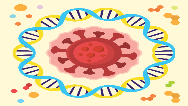
5.1. Covid-19
The novel coronavirus disease (SARS-CoV-2 or COVID-19) first emerged in Wuhan city, China in December 2019. COVID-19, due to its rapid spread worldwide, affects a large portion of the world’s population. Unfortunately, this new type of viral disease has led to more than millions of infected cases and deaths worldwide [[83], [84], [85], [86]]. In a study reported by Pu et al. [87], deep learning technology and high-resolution computed tomography images were used as potential diagnostic biomarkers to investigate parameters associated with COVID-19. The findings showed that unique image biomarkers might not distinguish all COVID-19 infiltrates from community-acquired pneumonia. However, the automated image analysis can identify a very low fraction of COVID-19 cases because unique image features can accurately distinguish a sizable subsect of non-COVID-19 cases [87]. Biomarkers of myocardial injury or cardiovascular disease, particularly cTnI, cardiac troponin T (cTnT), and D-dimer, have revealed their potential for predicting, prognosis, and treatment of COVID-19 [83,88,89]. The increasing levels of serum amyloid A [90], C-reactive protein [91], urea and creatinine (Renal biomarkers) [92], ferritin [93], and lactate dehydrogenase [94] have been used as biomarkers in the diagnosis of COVID-19. Very recently, Huyut et al. [95] used the changes in routine blood values (RBV) parameters for the diagnosis and prognosis of COVID-19 disease. This study contained three main steps: raw data collection, the performance of the LogNNet neural network with the selection of main features, and testing combinations of features. The RBV dataset consists of biochemical, immunological, and hematological parameters. The results of this study revealed that erythrocyte sedimentation rate, neutrophil count, C-reactive protein, white blood cell count, red blood cell count, urea, albumin, and calcium are the most important biomarkers for the diagnosis and prognosis of patients with severe or mild infection. The LogNNet model achieved an accuracy rate of 94.4% for the detection of the prognosis of the disease using 48 features [95]. In another interesting study, Velichko et al. [96] reported machine learning (ML) sensors as a practical option for the Internet of Things (IoT) in clinical decision support systems to determine the preliminary diagnosis of COVID-19 with an accuracy of up to 95% using RBV. The histogram-based gradient boosting (HGB) model was used successfully in determining the diagnosis of COVID-19 disease. The HGB model with the fastest and highest accuracy identified the 11 most important biomarkers consisting of low-density lipoprotein, cholesterol, high-density lipoprotein cholesterol, mean corpuscular hemoglobin concentration, triglyceride, amylase, uric acid, lactate dehydrogenase, creatine kinase-myocardial band, alkaline phosphatase and mean corpuscular hemoglobin to identify the disease with 100% accuracy in 6.39 s. These methods afford rapid, reliable, and economically cost-effective mobile tools for the diagnosis and prognosis of COVID-19 disease based on the RBVs at the time of hospital admission [96]. Antioxidant and oxidative parameter values play a vital role in the estimating of many diseases [97]. The autoregressive integrated moving average expert models were used for estimating the antioxidant and oxidant levels in the diagnosis and prognosis of COVID-19 disease, with the lymphocyte count and ferritin biomarkers with the most effect and white blood cell count and c-reactive protein with the most negligible impact on models [98]. Huyut et al. [99] investigated the importance of routine immunological, biochemical and hematological laboratory parameters to predict mortality in COVID-19 patients. The results revealed that C-reactive protein, erythrocyte sedimentation rate, troponin, D-dimer, ferritin, fibrinogen, leukocyte, and neutrophil levels were higher in the patients who died. In addition, the procalcitonin level was increased in dead patients than in survival patients [99]. The dysregulated and uncontrolled immune response leads to acute respiratory distress syndrome (ARDS) and cytokine storm, which is a major cause of pulmonary damage and mortality in COVID-19. Interleukin-6 (IL-6) is an important cytokine with 212 amino acids in its structure that plays a determining role in the progression of ARDS in COVID-19 patients. Therefore, the inhibition of IL-6 signaling can reduce lung damage and improve the clinical outcomes of patients with COVID-19. IL-6 is one of the main inflammatory agents in COVID-19 disease, and the combination of anakinra and studies has shown methylprednisolone can be used for the treatment of severe COVID-19 patients [100,101].
5.2. Cancer
Cancer, the most important cause of death worldwide, is a spatial and complex genetic disease marked by metastasis in vital organs of the body [102,103]. Cancer biomarkers play a significant role in enhancing our understanding and knowledge of the cancer process in clinical practice, allowing for the development of more effective diagnostics and the reduction of undesired systemic toxicity [104]. They have been proposed for use in assessing cancer risk, studying tumor-host interactions, and reflecting tumor load and cellular activity [105]. In recent years, the knowledge of cancer markers has advanced considerably [106].
Cancer biomarkers can be found in the circulation (whole blood, plasma, or serum), in secretions (urine, stools, sputum, or nipple discharge), or in other human biological fluids [107] and can therefore be easily assessed serially and noninvasively, or they can be tissue-derived and require either a biopsy or surgical resection [108]. An ideal tumor biomarker should have the following characteristics: (a) produced only by the tumor cells; (b) associated with tumor burden and provided with an adequate lead time; (c) detectable in preclinical or early stages in the blood (or other biological fluids of humans) of cancer patients (preferably in one cancer type only); (d) undetectable (or present at very low levels) in the blood (or other biological fluids) of healthy people or those with benign diseases; (e) easy to measure, even in small amounts and with little preparation, using a reliable test, affordable, and associated with high analytical specificity (the percentage of individuals without cancer who test negative for the biomarker) and to sensitivity (the percentage of individuals with cancer who test positive for the biomarker) [109,110].
Several biomarkers can be used for various cancers, including KRAS mutations as prognostic biomarkers in pancreatic cancer [111], procalcitonin as a biomarker for medullary thyroid cancer [112], serum microRNA-21 as a diagnostic biomarker in breast cancer [113], cigarette smoking as a biomarker in lung cancer [15], carcinoembryonic antigen in the colon, medullary thyroid, stomach cancer [15], and α-chain of haptoglobin (Hp-α) as a serum biomarker for ovarian cancer [114]. In addition, as a novel tumor biomarker, liquid biopsy is the most innovative tool in medical oncology. Proteins, metabolites, nucleic acids, and extracellular vesicles can all be identified in urine, which is a component of liquid biopsy [115].
5.3. Heart failure
Heart failure (HF) is caused by various cardiac and extracardiac pathophysiological mechanisms, leading to a complex clinical disease with numerous phenotypes. The diagnosis of HF can be difficult because the clinical findings are not necessarily specific and, therefore, of limited diagnostic value. It may be significant to develop immediate and particular tests to carry out a rapid “rule-out” of HF in the emergency department. Biomarkers have multiple roles such as diagnosing, monitoring therapy, assessing prognosis, and stratifying risk in cardiovascular disease in clinical management to offer an integrated approach [[116], [117], [118], [119], [120]]. Imaging biomarkers provide important insight into the functional and structural abnormalities of the heart, but the readout of these biomarkers is unable to identify subclinical and early stages of HF [121]. Protein biomarkers that are now being used to predict the prognosis of HF are either released from the heart, demonstrating the heart’s usefulness as a tissue-specific damage marker, or they are released from other cells as a systemic response to HF. The half-life of protein biomarkers, in addition to tissue specificity, is frequently the most important aspect of its potential utility as a biomarker. Natriuretic Peptides (NP), i.e., brain-type natriuretic peptide (BNP) and N-terminal prohormone of BNP, and cardiac troponin measurements have been included in the guidelines for HF treatment and diagnosis of the American Heart Association [122] and the European Society of Cardiology [123]. The predictive utility of NT-proBNP and BNP biomarkers in different HF settings, such as chronic or acute HF, has been investigated in various studies, providing strong evidence of their incremental value [[124], [125], [126], [127]]. In a study involving 122 patients who had acute decompensated heart failure and decreasing renal function, BNP levels were measured. A considerable reduction in BNP value of ≥40% throughout hospitalization i.e., from baseline to discharge had a positive prognostic value in reduced rehospitalization [128]. Other diagnostic biomarkers, such as those for oxidative stress (e.g., growth differentiation factor-15), cardiac remodeling (e.g., galectin-3), and inflammation (e.g., soluble ST2 receptor), could be added to help in HF treatment [129]. Protein-based biomarkers are currently the clinical gold standard for HF prediction because they provide many benefits including easy accessibility, comparably low costs, and simple handling. However, protein-based biomarkers might not be specific for HF, and hence, reliable prognostic indicators for HF are still scarce [130]. Recent advances in genetic analysis have opened up new avenues for studying the pathophysiology of cardiovascular disease and have opened the way for the creation of gene-based biomarkers. A novel approach for identifying DNA/RNA-based biomarkers is using omics technology, which can identify genome-wide (GW) and transcriptome-wide (TW) gene variation. Omics analysis allows for understanding the molecular mechanisms underlying the diseases as well as the identification of genetic variations that can help in the identification of HF patients who are at risk. It is possible to identify populations at risk for disease by locating a specific single nucleotide polymorphism (SNP) (via GW) or a variation in gene expression (through TW). However, it should be noted that due to the novelty and origin of these analyses, as well as the complexity of cardiovascular disease, only a small number of research has been specifically conducted on HF. Using genome-wide association studies, some research groups simultaneously identified the 9p21 risk locus [[131], [132], [133]]. The locus encodes different transcripts of the long non-coding RNA ANRIL [134]. Recent studies show that the ratio of circular to linear ANRIL, which affects basic biological functions, is related to the risk of coronary artery disease and may be used as a biomarker [135]. Following the identification of the 9p21 locus, numerous genetic loci were identified that altogether account for around 25% of the estimated heritability of cardiovascular disease [135]. By improving diagnosis and prognosis and therefore improving patient treatment, novel biomarkers for HF may support the widely used traditional ones. There is a growing interest in the multi-marker approaches due to their benefit over single biomarkers to improve risk stratification and increase the diagnostic accuracy in HF. Otherwise, further investigation is required to determine the ideal biomarker combination for the management of HF treatment.
5.4. Neurological diseases
Biomarkers have important roles in the prevention, diagnosis, prognosis, and treatment of brain diseases and neurological and neuropsychiatric disorders such as Alzheimer, Parkinson, stroke, Huntington, and, epilepsy [136,137]. Plasma, urine, cerebrospinal fluid, saliva, blood, and others can be used as various sources to obtain candidate biomarkers to diagnosis, help the progression, and treatment of a variety of brain disorders and diseases [138]. 4-hydroxy-2,3-noenal, angiogenin and Cystatin-C are candidate protein biomarkers for the diagnosis or progression of motor neuron disease [[139], [140], [141]]. The metabolite N-acetyl aspartate [142], metabolite myoinositol [143], soluble glycoprotein V [144], and clusterin [128] are other types of biomarkers that have been used for neurological diseases. The blood biomarkers of different brain injuries, such as fibrinogen, D-dimer, and troponin, could potentially offer tools to diagnose, manage and treat brain injury in COVID-19 patients [145]. There are particular difficulties in finding clinically useful biomarkers for neurological diseases. These challenges include the historically low availability of relevant tissues from the site of pathology, the complexity and molecular heterogeneity of the brain, and a clear deficiency of the glaring lack of models for functional validation of potential biomarkers.
The combination of biomarker identification methods using modern genomics, proteomics, and metabolomic technologies, however, promises to make significant progress in overcoming these challenges.
5.5. Lung diseases
Lung or respiratory diseases are clinical problems affecting the lungs, such as asthma, Pleural effusion, chronic obstructive pulmonary disease, pneumonia, lung cancer, tuberculosis, and many other lung disorders [146]. Malondialdehyde, a lipid peroxidation product, is reliable, cheap, and user-friendly biomarker for diagnosing lung diseases [147]. YKL-40, a glycoprotein with three amino acids tyrosine (Y), lysine (K), and leucine (L) in the N-terminal and with a molecular weight of 40 kDa is proved as a biomarker for detecting pleural effusion [148]. MRI and CT lung biomarkers can be used in vivo evaluation of lung biomechanics to identify lung abnormalities [149]. Nitric oxide, carbon monoxide, acetone, isoprene, methanol, hydrogen cyanide, hydrogen peroxide, ethyl butyrate, sulfides, and nitrates are exhaled volatile and non-volatile compounds which can be considered possible biomarkers for lung diseases [150].
5.6. Kidney diseases
Kidneys act to filter blood to create urine, remove toxins, and control the volume of different human body fluids [151]. Microalbumin, N-acetyl-β-glucosaminidase, fatty acid-binding proteins, and cysteine-rich proteins have been successfully used as kidney disease biomarkers. Hepcidin-25 is an iron-binding protein associated with acute kidney injury that is used as a novel kidney biomarker in serum and urine specimens for major adverse kidney events after cardiac surgery [152]. A novel synthetic antibody of polypyrrole/Multiwall carbon nanotubes on a carbon screen-printed electrode was designed to detect the kidney biomarker cystatin C at the point of care [153]. Recently, FDA approved a panel of six urine creatinine-normalized biomarkers, including clusterin, cystatin C, kidney injury molecule 1, N-acetyl-β-d-glucosaminidase, neutrophil gelatinase-associated lipocalin, and osteopontin for the detection of preclinical ASO-induced kidney pathology [154]. The level of d-Serine in plasma and urine is commonly used as a dual biomarker for the reflection of kidney function and diseases [155].
5.7. Liver diseases
The liver is a vital organ in the human body that has many important tasks, including digesting food, distributing nutrients, converting food into energy and storing it, and helping to filter toxic substances and remove poisons from the bloodstream. There are many liver diseases, such as hepatitis, fatty liver disease, cancer, cirrhosis, hemochromatosis, Wilson’s disease, fascioliasis, chronic liver failure, and autoimmune conditions [156]. Liver diseases can be occurred by a variety of factors and conditions, including viruses, drugs, drinking, and poisons. Symptoms of liver diseases vary, depending on the underlying cause and severity of the disease, and can be easily confused with other diseases, but they often include yellow skin and eyes, dark urine, swelling of the abdomen and legs, easy bruising, and vomiting. The diagnosis of liver diseases can often be difficult; however, the using of biomarkers can be helpful in diagnosing and following liver diseases [157]. The hepatic injury can be determined by liver function tests, including albumin, alanine aminotransferase, lactate dehydrogenase, and aspartate transaminase associated with alkaline phosphatase and gamma-glutamyl transferase [158]. Alanine aminotransferase is a surrogate biomarker that is highly specific for liver diseases and is detectable in the bloodstream [159]. Hyaluronic acid, bilirubin, cytokines, laminin, and fibroblast growth factor 21) can be used as potential biomarkers of liver disease [160,161].
5.8. Gastrointestinal diseases
Gastrointestinal diseases refer to diseases from the mouth to the anus consisting of all the organs of the human digestive system [162]. Intermediate products of metabolism are valuable biomarkers for the diagnosis of gastrointestinal diseases using non-invasive methods [163]. Volatile organic compounds such as acetone, ammonia, ethanol, indole, carbon disulfide, 2,3-butanedione, and acetic acid can be used as biomarkers. These compounds with low molecular weight are produced in the gastrointestinal tract and can be transported in the blood flow and reach the lungs and appear in breath and finally by GC-MS technique were analyzed [164]. Calprotectin, as a noninvasive biomarker, is primarily clinically useful for active inflammatory bowel disease (IBD) and digestive disorders [165]. Lactoferrin, a laboratory biomarker, is used for clostridium difficile infection detection [166]. MicroRNAs [167], electronic-nose such as surface acoustic wave, carbon black polymer composite, carbon black polymer composite, and metal oxide semiconductors [168] have been used to identify gastrointestinal diseases. The fatty acid-binding protein has also been evaluated as a diagnostic biomarker in Gastrointestinal Diseases [169]. Urinary metabolomics, such as tricarboxylic acid and amino acids, are different in IBD patients and health groups and can be used as noninvasive biomarkers for gastrointestinal diseases [170].
Most of the currently investigated potential biomarkers have not yet achieved validation and approval for use in real-life clinical practice for screening or diagnoses of particular gastrointestinal diseases; however, the future of biomarkers looks promising. Although some studies indicate great diagnostic accuracy of different biomarkers, the heterogeneity of the findings raises questions in our minds. Therefore, more extensive research and clinical trials using standardized procedures created by multicenter consortiums are required.
5.9. Skeletal muscle and bone diseases
Skeletal muscle may be damaged during life by exercise, contraction, chemical insult, defects in the immune system, or muscle degeneration [171]. MRI and proton magnetic resonance spectroscopy have been used to characterize and evaluate a rare degenerative skeletal muscle disease, namely GNE myopathy [172]. Also, MRI, as an emerging diagnostic tool and biomarker, can be used to evaluate changes in the fat and fibrous tissue content of muscles [173]. Pyridinolines, deoxypyridinolines, and osteocalcin were measured to predict future bone diseases using clinical outcomes. Plasma interleukin-6 has been identified as a marker of inflammation for the prediction of long-term changes in growth failure, joint contractures, and hip dysplasia [174]. Fourier-transform infrared Spectroscopy as a biomarker of primary mitochondrial myopathies and other mitochondrial diseases is available. This tool is a fast, non-invasive, non-destructive, sensitive, and specific diagnostic biomarker that requires small amounts of samples [175]. The use of amino acids, especially cysteine, methionine, taurine, and glutathione, as an essential part of skeletal muscles, can be used for the prevention and diagnosis of skeletal muscles [176].
6. Biomarker development process
The process of developing a biomarker involves several iterative steps that begin with the discovery of the biomarker in healthy and diseased samples. To ensure that rational, evidence-based biomarker creation maintains clinical and scientific necessity, an approach for developing them that incorporates collaborative regulatory science involving many different fields is required. The field is changing quickly as a result of ongoing and recent explosive growth in computation, analysis, and measurement. The biomarker development process comprises consecutive phases: pre-analytical and analytical validation, clinical validation, regulatory approval, and demonstration of clinical utility. The pre-analytical phase standardizes indicators and analyzes quality indicators such as process, storage, and sample collection. Analyzing the biomarker’s performance parameters to make sure the test is repeatable, dependable, and has the right level of specificity and sensitivity is commonly known as the analytical validation phase of biomarker development. A biomarker is connected to clinical and biological endpoints through qualification, a graded evidential process. Fig. 7 shows a schematic representation of the biomarker development process.

Framework for the development of biomarkers. To find potential biomarkers, tissue extracts or biological fluids can be used in the discovery process. Urine, synovial fluid, cerebral fluid, blood, saliva, and tissue extracts are a few examples of the numerous biofluids that might act as the discovery matrix for biomarkers. During the discovery process, biomarker qualifying in samples (either in vitro or in vivo from animals or humans) gives confidence that the biomarkers may have clinical utility. For qualifying companion diagnostics or techniques associated with the development and regulatory approval of a therapeutic or drug, both the European Medicines Agency and the FDA have formal approval procedures.
However, there are certain challenges in developing biomarkers, such as:
- ✓The scientific basis for some biomarkers cannot always be verified, creating difficulties in the qualification and validation of biomarkers in the future. Moreover, it is important to avoid incorrectly interpreting biomarker measurements and connecting a biomarker to a disease.
- ✓The cost of developing a biomarker may increase due to longer clinical trials or more testing requirements.
- ✓It often takes a lot of time and resources to develop and qualify biomarkers. For qualification purposes rather than as part of an individual drug regulatory approval, more convincing evidence of a favorable benefit-risk analysis is typically needed.
7. Statistical methods for assessment and evaluation of new biomarkers
After biomarker discovery, its evaluation is necessary, especially for diagnostic biomarkers. Statistical methods are used for the assessment and evaluation of new biomarkers. Biomarkers evaluation need an appropriate statistical analysis for the representation of valid conclusions of new biomarker before being introduced into clinical practice [177,178]. The obtained datasets can be analyzed by the classical statistical and multivariate methods. The classical statistical methods are used for the identification of biomarkers by monovariate statistical tests so that each biomarker is considered independent of the others (such as t-test or Wilcoxon-Mann Whitney test etc.) [179]. These methods involve the evaluation of the variables that show a different behavior between two groups of samples for example control vs. pathological or control vs. drug treated. The classical statistical methods suffer from some weaknesses, such as a lack of statistical power, lack of interpretable results, and omitting of complex relationships between variables [180]. Although classical statistical methods based on univariate and bivariate tests are easy to use and interpretation of the results, are not sufficient to extract and analyze all of the ’omic’ data sets and should only be used as an exploratory or complementary secondary method [181]. The multivariate statistical analyses (MVAs) are based on comparing the relationships existing among two or more groups of candidate biomarkers. These methods are often used and are more effective and very useful for the identification of biomarkers pools with diagnostic or prognostic purposes according to robustness, sensitivity, and specificity properties [182]. Usually, these methods belong to unsupervised pattern recognition (clustering methods) or supervised classification methods. In the field of biomarkers identification, there are several unsupervised methods adopted, such as Principal Component Analysis (PCA), Nonnegative PCA, robust-PCA, Canonical Correlation Analysis (CCA), Sparse Canonical Correlation Analysis (SCCA), Hierarchical cluster analysis (HCA), K-means and Multidimensional scaling (MDS). Also, several methods were presented for supervised analysis such as the soft independent model of class analogy (SIMCA), Ranking PCA, Partial least squares (PLS), Random forests (RF), Classification and Regression Tree (CART), Linear Discriminant Analysis (LDA) and Support Vector Machines (SVM) [[183], [184], [185]].
Machine learning algorithms as a method family of supervised classification methods were used by Li et al. [186] for the prediction of mortality in confirmed COVID-19 cases. The obtained results of this study showed that the logistic regression model (LR-5) is effective in predicting the death of patients with COVID-19 with a high area under the curve (AUC) [186]. Abbasimehr et al. [187] reported the performance of deep learning models for COVID-19 time series forecasting using the long short-term memory (LSTM), gated recurrent units (GRU), and convolutional neural network (CNN) models. According to the results of the experiments, combining the three deep learning models improves the performance of LSTM, CNN, and GRU. This novel approach with minimum error can be utilized by governments to make accurate decisions in controlling the pandemic. In the past several years, numerous attempts have been made to use various supervised machine learning (ML) models to predict the diagnosis and mortality of COVID-19 [98,188,189]. Using the feature dataset consisting of the routine blood values (28 RBV parameters) and age variable to the various supervised ML models, the pressure on COVID-19 infection status (severely or mildly infected) can be reduced at the admission time. The findings of this research showed that the locally weighted learning (LWL), K-star (K*), Naive Bayes (NB), and K-nearest neighbor (KNN) have the highest overall accuracy and the highest area under the receiver operating characteristic curve (AUC) values [85].
In summary, a wide range of different statistical methods are used for the diagnosis, prognosis, and treatment of Covid-19 and other diseases, such as multivariate for pancreatic cancer [190], multivariate statistical and data mining analyses for sensorineural hearing loss or different types of vestibular disorders [184], meta-analysis techniques for Covid-19 [191], Bayesian model averaging (BMA) to predict acute kidney injury progression status [192] and rank product statistical method for multiple myeloma aggressiveness [193].
8. Summary and future outlook
In summary, the biomarkers are a part of a relatively novel and ideal clinical tool in the diagnosis, prognosis, and treatment of various diseases. There are considerable benefits of using biomarkers to study various aspects of diseases, drug development, and monitoring the potential effects of therapeutic interventions. In comparison to current measurements, biomarkers are expected to provide tests with greater sensitivity and specificity, improve the decision-making process, and facilitate the development of therapies.
To improve healthcare and produce cutting-edge therapeutics, numerous efforts have been made to explore the biomarker frontier in search of new and/or better biomarkers. However, because the processes that lead to disease pathogenesis are frequently complex, it is easier to identify useful biomarkers for assessing drug response, making diagnoses, and tracking the development of diseases that better understand the underlying abnormalities associated with the disease and the mechanism of drug action. For clinical and basic pharmacologists, as well as other people involved in the identification of biomarkers, accumulating this information is a challenge.
Making the difference between a potential biomarker and a reliable biomarker that can be universally used to guide critical clinical and commercial choices is one of the main challenges in the field of biomarkers.
An effective biomarker must influence clinical evaluation to improve patient care. Clinical decisions that are based on real test results must be more beneficial than those that are based on false negatives or false positives. Biomarkers should reduce costs and negative effects while preventing deaths in the setting of risk management. The effectiveness of a biomarker is determined by comparing it with an ideal biomarker and exploring its properties. Promising biomarkers have characteristics similar to those of ideal markers.
A biomarker should ideally have the following characteristics:
- •The first one is clinical relevance. The biomarker must provide evidence that supports some rational basis for its use. The evidence reflects some measurements or changes in physiological or pathological processes over a relatively short time;
- •High sensitivity and specificity for evaluating treatment effects;
- •Reliability, the ability to measure the biomarker analytically. There’s a need to detect changes in the biomarker with acceptable accuracy, precision, robustness, and reproducibility;
- •Practicality, defined as noninvasiveness or just modest invasiveness to avoid discomfort and inconvenience for healthy volunteers or patients;
- •Simplicity means accessible equipment and low cost. Simplicity facilitates widespread acceptance for use in drug development and clinical practice.
Although many new biomarkers have been discovered, only a small number of them are clinically useful. The difficulties and problems faced in biomarker validation and discovery are numerous and should not be underestimated when developing strategies and conducting experimental studies. To obtain promising results, researchers should use annotated clinical specimens, appropriate control groups, many samples, and standardized sample handling. In the future, integrating biomarkers identified with emerging high-throughput techniques into medical practice will be required to achieve ‘personalization’ of treatment and disease prevention.
References
1. De Gramont A., Watson S., Ellis L.M., Rodón J., Tabernero J., De Gramont A., Hamilton S.R. Pragmatic issues in biomarker evaluation for targeted therapies in cancer. Nat. Rev. Clin. Oncol. 2015;12:197–212. doi: 10.1038/nrclinonc.2014.202. [PubMed] [CrossRef] [Google Scholar]
2. Beasley V.R., Levengood J.M. Elsevier Inc.; 2012. Principles of Ecotoxicology, Veterinary Toxicology; pp. 831–855. [Google Scholar]
3. Godfrey A., Vandendriessche B., Bakker J.P., Fitzer‐Attas C., Gujar N., Hobbs M., Liu Q., Northcott C.A., Parks V., Wood W.A. Fit‐for‐purpose biometric monitoring technologies: leveraging the laboratory biomarker experience. Clin. Transl. Sci. 2021;14:62–74. doi: 10.1111/cts.12865. [PMC free article] [PubMed] [CrossRef] [Google Scholar]
4. Group B.D.W., Atkinson A.J., Jr., Colburn W.A., DeGruttola V.G., DeMets D.L., Downing G.J., Hoth D.F., Oates J.A., Peck C.C., Schooley R.T. Biomarkers and surrogate endpoints: preferred definitions and conceptual framework. Clin. Pharmacol. Ther. 2001;69:89–95. doi: 10.1067/mcp.2001.113989. [PubMed] [CrossRef] [Google Scholar]
5. Mayeux R. Biomarkers: potential uses and limitations. NeuroRx. 2004;1:182–188. doi: 10.1602/neurorx.1.2.182. [PMC free article] [PubMed] [CrossRef] [Google Scholar]
6. Wan-Ibrahim W.I., Singh V.A., Hashim O.H., Abdul-Rahman P.S. Biomarkers for bone tumors: discovery from genomics and proteomics studies and their challenges. Mol. Med. 2015;21:861–872. doi: 10.2119/molmed.2015.00183. [PMC free article] [PubMed] [CrossRef] [Google Scholar]
7. Mert D.G., Terzi H. Mean platelet volume in bipolar disorder: the search for an ideal biomarker. Neuropsychiatric Dis. Treat. 2016;12:2057. doi: 10.2147/NDT.S112374. [PMC free article] [PubMed] [CrossRef] [Google Scholar]
8. Antoniou M., Kolamunnage-Dona R., Wason J., Bathia R., Billingham C., Bliss J., Brown L., Gillman A., Paul J., Jorgensen A. Biomarker-guided trials: challenges in practice. Contemp. Clin. Trials Commun. 2019;16 doi: 10.1016/j.conctc.2019.100493. [PMC free article] [PubMed] [CrossRef] [Google Scholar]
9. Landeck L., Kneip C., Reischl J., Asadullah K. Biomarkers and personalized medicine: current status and further perspectives with special focus on dermatology. Exp. Dermatol. 2016;25:333–339. doi: 10.1111/exd.12948. [PubMed] [CrossRef] [Google Scholar]
10. Califf R.M. Biomarker definitions and their applications. Exp. Biol. Med. 2018;243:213–221. doi: 10.1177/1535370217750088. [PMC free article] [PubMed] [CrossRef] [Google Scholar]
11. García-Gutiérrez M.S., Navarrete F., Sala F., Gasparyan A., Austrich-Olivares A., Manzanares J. Biomarkers in psychiatry: concept, definition, types and relevance to the clinical reality. Front. Psychiatr. 2020;11:432. doi: 10.3389/fpsyt.2020.00432. [PMC free article] [PubMed] [CrossRef] [Google Scholar]
12. Porter K. Effect of homologous bone marrow injections in x-irradiated rabbits. Br. J. Exp. Pathol. 1957;38:401–412. [PMC free article] [PubMed] [Google Scholar]
13. Mundkur B.D. Evidence excluding mutations, polysomy, and polyploidy as possible causes of non-Mendelian segregations in Saccharomyces. Ann. Mo. Bot. Gard. 1949;36:259–280. doi: 10.2307/2394394. [CrossRef] [Google Scholar]
14. Aronson J.K. Biomarkers and surrogate endpoints. Br. J. Clin. Pharmacol. 2005;59:491. doi: 10.1111/j.1365-2125.2005.02435.x. [PMC free article] [PubMed] [CrossRef] [Google Scholar]
15. Aronson J.K., Ferner R.E. Biomarkers—a general review. Curr. Protoc. 2017;76:9. doi: 10.1002/cpph.19. [PubMed] [CrossRef] [Google Scholar]
16. MacNamara J., Eapen D.J., Quyyumi A., Sperling L. Novel biomarkers for cardiovascular risk assessment: current status and future directions. Future Cardiol. 2015;11:597–613. doi: 10.2217/fca.15.39. [PubMed] [CrossRef] [Google Scholar]
17. Gil F., Pla A. Biomarkers as biological indicators of xenobiotic exposure. J. Appl. Toxicol. 2001;21:245–255. doi: 10.1002/jat.769. [PubMed] [CrossRef] [Google Scholar]
18. Wishart D.S., Bartok B., Oler E., Liang K.Y., Budinski Z., Berjanskii M., Guo A., Cao X., Wilson M. MarkerDB: an online database of molecular biomarkers. Nucleic Acids Res. 2021;49:D1259–D1267. doi: 10.1093/nar/gkaa1067. [PMC free article] [PubMed] [CrossRef] [Google Scholar]
19. Dhama K., Latheef S.K., Dadar M., Samad H.A., Munjal A., Khandia R., Karthik K., Tiwari R., Yatoo M., Bhatt P. Biomarkers in stress related diseases/disorders: diagnostic, prognostic, and therapeutic values. Front. Mol. Biosci. 2019;91 doi: 10.3389/fmolb.2019.00091. [PMC free article] [PubMed] [CrossRef] [Google Scholar]
20. Mondello S., Hayes R.L. Biomarkers. Handb. Clin. Neurol. 2015;127:245–265. [PubMed] [Google Scholar]
21. Firestein G.S. A biomarker by any other name. Nat. Clin. Pract. 2006;2:635. doi: 10.1038/ncprheum0347. [PubMed] [CrossRef] [Google Scholar]
22. Jain K.K., Jain K.K. Springer; 2010. The Handbook of Biomarkers. [Google Scholar]
23. Frank R., Hargreaves R. Clinical biomarkers in drug discovery and development. Nat. Rev. Drug Discov. 2003;2:566–580. doi: 10.1038/nrd1130. [PubMed] [CrossRef] [Google Scholar]
24. Sharma V., Ricketts H.C., Steffensen F., Goodfellow A., Cowan D.C. Obesity affects type 2 biomarker levels in asthma. J. Asthma. 2022:1–8. doi: 10.1080/02770903.2022.2051548. [PubMed] [CrossRef] [Google Scholar]
25. Naylor S. Biomarkers: current perspectives and future prospects. Expert Rev. Mol. Diagn. 2003;3:525–529. doi: 10.1586/14737159.3.5.525. [PubMed] [CrossRef] [Google Scholar]
26. Pospelova M., Krasnikova V., Fionik O., Alekseeva T., Samochernykh K., Ivanova N., Trofimov N., Vavilova T., Vasilieva E., Topuzova M. Potential molecular biomarkers of central nervous system damage in breast cancer survivors. J. Clin. Med. 2022;11:1215. doi: 10.3390/jcm11051215. [PMC free article] [PubMed] [CrossRef] [Google Scholar]
27. Picó C., Serra F., Rodríguez A.M., Keijer J., Palou A. Biomarkers of nutrition and health: new tools for new approaches. Nutrients. 2019;11:1092. doi: 10.3390/nu11051092. [PMC free article] [PubMed] [CrossRef] [Google Scholar]
28. Rosenwald A., Wright G., Chan W.C., Connors J.M., Campo E., Fisher R.I., Gascoyne R.D., Muller-Hermelink H.K., Smeland E.B., Giltnane J.M. The use of molecular profiling to predict survival after chemotherapy for diffuse large-B-cell lymphoma. N. Engl. J. Med. 2002;346:1937–1947. doi: 10.1056/NEJMoa012914. [PubMed] [CrossRef] [Google Scholar]
29. Lukas A., Heinzel A., Mayer B. bioRxiv; 2019. Biomarkers for Capturing Disease Pathology as Molecular Process Hyperstructure. [CrossRef] [Google Scholar]
30. Patron J., Serra-Cayuela A., Han B., Li C., Wishart D.S. Assessing the performance of genome-wide association studies for predicting disease risk. PLoS One. 2019;14 doi: 10.1101/701086. [PMC free article] [PubMed] [CrossRef] [Google Scholar]
31. Karlson E.W., Chang S.-C., Cui J., Chibnik L.B., Fraser P.A., De Vivo I., Costenbader K.H. Gene–environment interaction between HLA-DRB1 shared epitope and heavy cigarette smoking in predicting incident rheumatoid arthritis. Ann. Rheum. Dis. 2010;69:54–60. doi: 10.1136/ard.2008.102962. [PMC free article] [PubMed] [CrossRef] [Google Scholar]
32. Corella D., Ordovás J.M. Biomarkers: background, classification and guidelines for applications in nutritional epidemiology. Nutr. Hosp. 2015;31:177–188. doi: 10.3305/nh.2015.31.sup3.8765. [PubMed] [CrossRef] [Google Scholar]
33. Sharifi-Rad J., Sureda A., Tenore G.C., Daglia M., Sharifi-Rad M., Valussi M., Tundis R., Sharifi-Rad M., Loizzo M.R., Ademiluyi A.O. Biological activities of essential oils: from plant chemoecology to traditional healing systems. Molecules. 2017;22:70. doi: 10.3390/molecules22010070. [PMC free article] [PubMed] [CrossRef] [Google Scholar]
34. Eggener S.E., Rumble R.B., Armstrong A.J., Morgan T.M., Crispino T., Cornford P., van der Kwast T., Grignon D.J., Rai A.J., Agarwal N. Molecular biomarkers in localized prostate cancer: ASCO guideline. J. Clin. Oncol. 2020;38:1474–1494. doi: 10.1200/jco.19.02768. [PubMed] [CrossRef] [Google Scholar]
35. Garner R., La Rocca M., Vespa P., Jones N., Monti M.M., Toga A.W., Duncan D. Imaging biomarkers of posttraumatic epileptogenesis. Epilepsia. 2019;60:2151–2162. doi: 10.1111/epi.16357. [PMC free article] [PubMed] [CrossRef] [Google Scholar]
36. Ziegler S.I. Positron emission tomography: principles, technology, and recent developments. Nucl. Phys. 2005;752:679–687. doi: 10.1016/j.nuclphysa.2005.02.067. [CrossRef] [Google Scholar]
37. Garvey C.J., Hanlon R. Computed tomography in clinical practice. Bmj. 2002;324:1077–1080. doi: 10.1136/bmj.324.7345.1077. [PMC free article] [PubMed] [CrossRef] [Google Scholar]
38. Ghantous C.M., Kamareddine L., Farhat R., Zouein F.A., Mondello S., Kobeissy F., Zeidan A. Advances in cardiovascular biomarker discovery. Biomedicines. 2020;8:552. doi: 10.3390/biomedicines8120552. [PMC free article] [PubMed] [CrossRef] [Google Scholar]
39. Young P.N., Estarellas M., Coomans E., Srikrishna M., Beaumont H., Maass A., Venkataraman A.V., Lissaman R., Jiménez D., Betts M.J. Imaging biomarkers in neurodegeneration: current and future practices. Alzheimer’s Res. Ther. 2020;12:1–17. doi: 10.1186/s13195-020-00612-7. [PMC free article] [PubMed] [CrossRef] [Google Scholar]
40. Dregely I., Prezzi D., Kelly‐Morland C., Roccia E., Neji R., Goh V. Imaging biomarkers in oncology: basics and application to MRI. J. Magn. Reson. Imag. 2018;48:13–26. doi: 10.1002/jmri.26058. [PMC free article] [PubMed] [CrossRef] [Google Scholar]
41. Weinreb J.C., Barentsz J.O., Choyke P.L., Cornud F., Haider M.A., Macura K.J., Margolis D., Schnall M.D., Shtern F., Tempany C.M. PI-RADS prostate imaging–reporting and data system: 2015, version 2. Eur. Urol. 2016;69:16–40. doi: 10.1016/j.eururo.2015.08.052. [PMC free article] [PubMed] [CrossRef] [Google Scholar]
42. Mercado C.L. Bi-rads update. Clin. Radiol. 2014;52:481–487. doi: 10.1016/j.rcl.2014.02.008. [PubMed] [CrossRef] [Google Scholar]
43. Tang A., Bashir M.R., Corwin M.T., Cruite I., Dietrich C.F., Do R.K., Ehman E.C., Fowler K.J., Hussain H.K., Jha R.C. Evidence supporting LI-RADS major features for CT-and MR imaging–based diagnosis of hepatocellular carcinoma: a systematic review. Radiology. 2018;286:29–48. doi: 10.1148/radiol.20171705544. [PMC free article] [PubMed] [CrossRef] [Google Scholar]
44. Mitchell D.G., Bruix J., Sherman M., Sirlin C.B. LI‐RADS (liver imaging reporting and data system): summary, discussion, and consensus of the LI‐RADS management working group and future directions. Hepatology. 2015;61:1056–1065. https://aasldpubs.onlinelibrary.wiley.com/doi/epdf/10.1002/hep.27304 [PubMed] [Google Scholar]
45. Erickson B.J., Bartholmai B. Computer-aided detection and diagnosis at the start of the third millennium. J. Digit. Imag. 2002;15:59–68. doi: 10.1007/s10278-002-0011-x. [PMC free article] [PubMed] [CrossRef] [Google Scholar]
46. Park J.-W., Kim J.H., Kim S.K., Kang K.W., Park K.W., Choi J.-I., Lee W.J., Kim C.-M., Nam B.H. A prospective evaluation of 18F-FDG and 11C-acetate PET/CT for detection of primary and metastatic hepatocellular carcinoma. J. Nucl. Med. 2008;49:1912–1921. doi: 10.2967/jnumed.108.055087. [PubMed] [CrossRef] [Google Scholar]
47. Drucker E., Krapfenbauer K. Pitfalls and limitations in translation from biomarker discovery to clinical utility in predictive and personalised medicine. EPMA J. 2013;4:1–10. doi: 10.1186/1878-5085-4-7. [PMC free article] [PubMed] [CrossRef] [Google Scholar]
48. Laterza O.F., Hendrickson R.C., Wagner J.A. Molecular biomarkers. Drug Inf. J. 2007;41:573–585. [Google Scholar]
49. Brocks J.J., Pearson A. Building the biomarker tree of life. Rev. Mineral. Geochem. 2005;59:233–258. doi: 10.2138/rmg.2005.59.10. [CrossRef] [Google Scholar]
50. Aizpurua-Olaizola O., Toraño J.S., Falcon-Perez J.M., Williams C., Reichardt N., Boons G.-J. Mass spectrometry for glycan biomarker discovery. Trends Anal. Chem. 2018;100:7–14. doi: 10.1016/j.trac.2017.12.015. [CrossRef] [Google Scholar]
51. Kailemia M.J., Park D., Lebrilla C.B. Glycans and glycoproteins as specific biomarkers for cancer. Anal. Bioanal. Chem. 2017;409:395–410. doi: 10.1007/s00216-016-9880-6. [PMC free article] [PubMed] [CrossRef] [Google Scholar]
52. Nadkarni G.N., Chauhan K., Rao V., Ix J.H., Shlipak M.G., Parikh C.R., Coca S.G. Effect of intensive blood pressure lowering on kidney tubule injury: findings from the ACCORD trial study participants. Am. J. Kidney Dis. 2019;73:31–38. doi: 10.1053/j.ajkd.2018.07.016. [PMC free article] [PubMed] [CrossRef] [Google Scholar]
53. Jungbauer C.G., Birner C., Jung B., Buchner S., Lubnow M., von Bary C., Endemann D., Banas B., Mack M., Böger C.A. Kidney injury molecule‐1 and N‐acetyl‐ß‐d‐glucosaminidase in chronic heart failure: possible biomarkers of cardiorenal syndrome. Eur. J. Heart Fail. 2011;13:1104–1110. doi: 10.1093/eurjhf/hfr102. [PubMed] [CrossRef] [Google Scholar]
54. MacKay M.-A.B., Kravtsenyuk M., Thomas R., Mitchell N.D., Dursun S.M., Baker G.B., D-Serine Potential therapeutic agent and/or biomarker in schizophrenia and depression? Front. Psychiatr. 2019;10:25. doi: 10.3389/fpsyt.2019.00025. [PMC free article] [PubMed] [CrossRef] [Google Scholar]
55. Anderson S.T., Kidd L.J., Barton A.J., Greer R.M. Serum bone biomarkers osteocalcin and pyridinoline in mares during pregnancy and lactation, and in foals during early post-natal life. Res. Vet. Sci. 2018;118:34–40. doi: 10.1016/j.rvsc.2018.01.007. [PubMed] [CrossRef] [Google Scholar]
56. Carlomagno N., Incollingo P., Tammaro V., Peluso G., Rupealta N., Chiacchio G., Sandoval Sotelo M.L., Minieri G., Pisani A., Riccio E. Diagnostic, predictive, prognostic, and therapeutic molecular biomarkers in third millennium: a breakthrough in gastric cancer. BioMed Res. Int. 2017;2017 doi: 10.1155/2017/7869802. [PMC free article] [PubMed] [CrossRef] [Google Scholar]
57. Verber N.S., Shepheard S.R., Sassani M., McDonough H.E., Moore S.A., Alix J.J., Wilkinson I.D., Jenkins T.M., Shaw P.J. Biomarkers in motor neuron disease: a state of the art review. Front. Neurol. 2019;10:291. doi: 10.3389/fneur.2019.00291. [PMC free article] [PubMed] [CrossRef] [Google Scholar]
58. Sidhom K., Obi P.O., Saleem A. A review of exosomal isolation methods: is size exclusion chromatography the best option? Int. J. Mol. Sci. 2020;21:6466. doi: 10.3390/ijms21186466. [PMC free article] [PubMed] [CrossRef] [Google Scholar]
59. Lin L.-L., Huang H.-C., Juan H.-F. Discovery of biomarkers for gastric cancer: a proteomics approach. J. Proteome Res. 2012;75:3081–3097. doi: 10.1016/j.jprot.2012.03.046. [PubMed] [CrossRef] [Google Scholar]
60. Takamura T.-a., Tsuchiya T., Oda M., Watanabe M., Saito R., Sato-Ishida R., Akao H., Kawai Y., Kitayama M., Kajinami K. Circulating malondialdehyde-modified low-density lipoprotein (MDA-LDL) as a novel predictor of clinical outcome after endovascular therapy in patients with peripheral artery disease (PAD) Atherosclerosis. 2017;263:192–197. doi: 10.1016/j.atherosclerosis.2017.06.029. [PubMed] [CrossRef] [Google Scholar]
61. Manne U., Srivastava R.-G., Srivastava S. Keynote review: recent advances in biomarkers for cancer diagnosis and treatment, Drug Discov. Today Off. 2005;10:965–976. doi: 10.1016/S1359-6446(05)03487-2. [PubMed] [CrossRef] [Google Scholar]
62. Gupta R., Soni S. An overview on inflammatory biomarkers for diabetes mellitus. Madridge J. Diabetes. 2019;3:64–66. doi: 10.18689/mjd-1000112. [CrossRef] [Google Scholar]
63. Preedy V.R., Patel V.B. Springer; Netherlands: 2015. General Methods in Biomarker Research and Their Applications. [Google Scholar]
64. Davis M.A., Eldridge S., Louden C. Elsevier; 2013. Biomarkers: Discovery, Qualification and Application, Haschek and Rousseaux’s Handbook of Toxicologic Pathology; pp. 317–352. [Google Scholar]
65. Zhou C., Simpson K.L., Lancashire L.J., Walker M.J., Dawson M.J., Unwin R.D., Rembielak A., Price P., West C., Dive C. Statistical considerations of optimal study design for human plasma proteomics and biomarker discovery. J. Proteome Res. 2012;11:2103–2113. doi: 10.1021/pr200636x. [PMC free article] [PubMed] [CrossRef] [Google Scholar]
66. Rundle A., Ahsan H., Vineis P. Better cancer biomarker discovery through better study design. Eur. J. Clin. Invest. 2012;42:1350–1359. doi: 10.1111/j.1365-2362.2012.02727.x. [PMC free article] [PubMed] [CrossRef] [Google Scholar]
67. Gosho M., Nagashima K., Sato Y. Study designs and statistical analyses for biomarker research. Sensors. 2012;12:8966–8986. doi: 10.3390/s120708966. [PMC free article] [PubMed] [CrossRef] [Google Scholar]
68. Silajdžić E., Björkqvist M. A critical evaluation of wet biomarkers for Huntington’s disease: current status and ways forward. J. Huntingt. Dis. 2018;7:109–135. doi: 10.3233/JHD-170273. [PMC free article] [PubMed] [CrossRef] [Google Scholar]
69. Mamidi T.K.K., Wu J., Hicks C. Integrating germline and somatic variation information using genomic data for the discovery of biomarkers in prostate cancer. BMC Cancer. 2019;19:1–12. doi: 10.1186/s12885-019-5440-8. [PMC free article] [PubMed] [CrossRef] [Google Scholar]
70. Umelo I.A., Costanza B., Castronovo V. Innovative methods for biomarker discovery in the evaluation and development of cancer precision therapies. Cancer Metastasis Rev. 2018;37:125–145. doi: 10.1007/s10555-017-9710-0. [PubMed] [CrossRef] [Google Scholar]
71. Tringe S.G., Rubin E.M. Metagenomics: DNA sequencing of environmental samples. Nat. Rev. Genet. 2005;6:805–814. doi: 10.1038/nrg1709. [PubMed] [CrossRef] [Google Scholar]
72. Anderson N.L., Anderson N.G. Proteome and proteomics: new technologies, new concepts, and new words. Electrophoresis. 1998;19:1853–1861. doi: 10.1002/elps.1150191103. [PubMed] [CrossRef] [Google Scholar]
73. Pandey A., Mann M. Proteomics to study genes and genomes. Nature. 2000;405:837–846. doi: 10.1038/35015709. [PubMed] [CrossRef] [Google Scholar]
74. Tomasina J., Lheureux S., Gauduchon P., Rault S., Malzert-Fréon A. Nanocarriers for the targeted treatment of ovarian cancers. Biomaterials. 2013;34:1073–1101. doi: 10.1016/j.biomaterials.2012.10.055. [PubMed] [CrossRef] [Google Scholar]
75. Hudler P., Kocevar N., Komel R. Proteomic approaches in biomarker discovery: new perspectives in cancer diagnostics. Sci. World J. 2014;2014 doi: 10.1155/2014/260348. [PMC free article] [PubMed] [CrossRef] [Google Scholar]
76. Noronha V., Patil V.M., Joshi A., Menon N., Chougule A., Mahajan A., Janu A., Purandare N., Kumar R., More S. Gefitinib versus gefitinib plus pemetrexed and carboplatin chemotherapy in EGFR-mutated lung cancer. J. Clin. Oncol. 2020;38:124–136. doi: 10.1200/jco.19.01154. [PubMed] [CrossRef] [Google Scholar]
77. Costa D., Valente A.J., Queiroz J. Stimuli-responsive polyamine-DNA blend nanogels for co-delivery in cancer therapy. Colloids Surf., B. 2015;132:194–201. doi: 10.1016/j.colsurfb.2015.04.064. [PubMed] [CrossRef] [Google Scholar]
78. Corella D., Coltell O., Macian F., Ordovás J.M. Advances in understanding the molecular basis of the mediterranean diet effect. Rev. Food Sci. Technol. (Mysore) 2018;9:227–249. doi: 10.1146/annurev-food-032217-020802. [PubMed] [CrossRef] [Google Scholar]
79. Ballereau S., Glaab E., Kolodkin A., Chaiboonchoe A., Biryukov M., Vlassis N., Ahmed H., Pellet J., Baliga N., Hood L. Springer; 2013. Functional Genomics, Proteomics, Metabolomics and Bioinformatics for Systems Biology, Systems Biology; pp. 3–41. [Google Scholar]
80. Weckwerth W., Morgenthal K. Metabolomics: from pattern recognition to biological interpretation. Drug Discov. Today. 2005;10:1551–1558. doi: 10.1016/S1359-6446(05)03609-3. [PubMed] [CrossRef] [Google Scholar]
81. Horgan R.P., Kenny L.C. ‘Omic’technologies: genomics, transcriptomics, proteomics and metabolomics. Obstet. Gynaecol. 2011;13:189–195. doi: 10.1576/toag.13.3.189.27672. [CrossRef] [Google Scholar]
82. Villas-Boas S.G., Nielsen J., Smedsgaard J., Hansen M.A., Roessner-Tunali U. John Wiley & Sons; 2007. Metabolome Analysis: an Introduction. [Google Scholar]
83. Aboughdir M., Kirwin T., Abdul Khader A., Wang B. Prognostic value of cardiovascular biomarkers in COVID-19: a review. Viruses. 2020;12:527. doi: 10.3390/v12050527. [PMC free article] [PubMed] [CrossRef] [Google Scholar]
84. Huyut M.T., Ilkbahar F. The effectiveness of blood routine parameters and some biomarkers as a potential diagnostic tool in the diagnosis and prognosis of Covid-19 disease. Int. Immunopharm. 2021;98 doi: 10.1016/j.intimp.2021.107838. [PMC free article] [PubMed] [CrossRef] [Google Scholar]
85. Huyut M. IRBM; 2022. Automatic Detection of Severely and Mildly Infected COVID-19 Patients with Supervised Machine Learning Models. [PMC free article] [PubMed] [CrossRef] [Google Scholar]
86. Zarei A., Fardood S.T., Moradnia F., Ramazani A. A review on coronavirus family persistency and considerations of novel type, COVID-19 features. Eurasian Chem. Commun. 2020:798–811. doi: 10.33945/SAMI/ECC.2020.7.7. [CrossRef] [Google Scholar]
87. Pu J., Leader J., Bandos A., Shi J., Du P., Yu J., Yang B., Ke S., Guo Y., Field J.B. Any unique image biomarkers associated with COVID-19? Eur. Radiol. 2020;30:6221–6227. doi: 10.1007/s00330-020-06956-w. [PMC free article] [PubMed] [CrossRef] [Google Scholar]
88. Guo T., Fan Y., Chen M., Wu X., Zhang L., He T., Wang H., Wan J., Wang X., Lu Z. Cardiovascular implications of fatal outcomes of patients with coronavirus disease 2019 (COVID-19) JAMA Cardiol. 2020;5:811–818. doi: 10.1001/jamacardio.2020.1017. [PMC free article] [PubMed] [CrossRef] [Google Scholar]
89. Inciardi R.M., Lupi L., Zaccone G., Italia L., Raffo M., Tomasoni D., Cani D.S., Cerini M., Farina D., Gavazzi E. Cardiac involvement in a patient with coronavirus disease 2019 (COVID-19) JAMA Cardiol. 2020;5:819–824. doi: 10.1001/jamacardio.2020.1096. [PMC free article] [PubMed] [CrossRef] [Google Scholar]
90. Ji W., Bishnu G., Cai Z., Shen X. MedRxiv; 2020. Analysis Clinical Features of COVID-19 Infection in Secondary Epidemic Area and Report Potential Biomarkers in Evaluation. [CrossRef] [Google Scholar]
91. Yip T.T., Chan J.W., Cho W.C., Yip T.-T., Wang Z., Kwan T.-L., Law S.C., Tsang D.N., Chan J.K., Lee K.-C. Protein chip array profiling analysis in patients with severe acute respiratory syndrome identified serum amyloid a protein as a biomarker potentially useful in monitoring the extent of pneumonia. Clin. Chem. 2005;51:47–55. doi: 10.1373/clinchem.2004.031229. [PMC free article] [PubMed] [CrossRef] [Google Scholar]
92. Henry B.M., Lippi G. Chronic kidney disease is associated with severe coronavirus disease 2019 (COVID-19) infection. Int. Urol. Nephrol. 2020;52:1193–1194. doi: 10.1007/s11255-020-02451-9. [PMC free article] [PubMed] [CrossRef] [Google Scholar]
93. Kaushal K., Kaur H., Sarma P., Bhattacharyya A., Sharma D.J., Prajapat M., Pathak M., Kothari A., Kumar S., Rana S. Serum ferritin as a predictive biomarker in COVID-19. A systematic review, meta-analysis and meta-regression analysis. J. Crit. Care. 2022;67:172–181. doi: 10.1016/j.jcrc.2021.09.023. [PMC free article] [PubMed] [CrossRef] [Google Scholar]
94. Han Y., Zhang H., Mu S., Wei W., Jin C., Tong C., Song Z., Zha Y., Xue Y., Gu G. Lactate dehydrogenase, an independent risk factor of severe COVID-19 patients: a retrospective and observational study. Aging (Albany N.Y.) 2020;12 doi: 10.18632/aging.103372. [PMC free article] [PubMed] [CrossRef] [Google Scholar]
95. Huyut M.T., Velichko A. Diagnosis and Prognosis of COVID-19 disease using routine blood values and LogNNet neural network. Sensors. 2022;22:4820. doi: 10.3390/s22134820. [PMC free article] [PubMed] [CrossRef] [Google Scholar]
96. Velichko A., Huyut M.T., Belyaev M., Izotov Y., Korzun D. Machine learning sensors for diagnosis of COVID-19 disease using routine blood values for Internet of Things application. Sensors. 2022;22:7886. doi: 10.3390/s22207886. [PMC free article] [PubMed] [CrossRef] [Google Scholar]
97. Cecchini R., Cecchini A.L. SARS-CoV-2 infection pathogenesis is related to oxidative stress as a response to aggression. Med. Hypotheses. 2020;143 doi: 10.1016/j.mehy.2020.110102. [PMC free article] [PubMed] [CrossRef] [Google Scholar]
98. Huyut M.T., Huyut Z. Forecasting of Oxidant/Antioxidant levels of COVID-19 patients by using Expert models with biomarkers used in the Diagnosis/Prognosis of COVID-19. Int. Immunopharm. 2021;100 doi: 10.1016/j.intimp.2021.108127. [PMC free article] [PubMed] [CrossRef] [Google Scholar]
99. Huyut M.T., Huyut Z., İlkbahar F., Mertoğlu C. What is the impact and efficacy of routine immunological, biochemical and hematological biomarkers as predictors of COVID-19 mortality? Int. Immunopharm. 2022;105 doi: 10.1016/j.intimp.2022.108542. [PMC free article] [PubMed] [CrossRef] [Google Scholar]
100. Elahi R., Karami P., Heidary A.H., Esmaeilzadeh A. An updated overview of recent advances, challenges, and clinical considerations of IL-6 signaling blockade in severe coronavirus disease 2019 (COVID-19) Int. Immunopharm. 2022 doi: 10.1016/j.intimp.2022.108536. [PMC free article] [PubMed] [CrossRef] [Google Scholar]
101. Atal S., Fatima Z. IL-6 inhibitors in the treatment of serious COVID-19: a promising therapy? Pharmaceut. Med. 2020;34:223–231. doi: 10.1007/s40290-020-00342-z. [PMC free article] [PubMed] [CrossRef] [Google Scholar]
102. Fattahi N., Shahbazi M.-A., Maleki A., Hamidi M., Ramazani A., Santos H.A. Emerging insights on drug delivery by fatty acid mediated synthesis of lipophilic prodrugs as novel nanomedicines. J. Contr. Release. 2020;326:556–598. doi: 10.1016/j.jconrel.2020.07.012. [PubMed] [CrossRef] [Google Scholar]
103. Kalave S., Hegde N., Juvale K. Applications of nanotechnology-based approaches to overcome multi-drug resistance in cancer. Curr. Pharmaceut. Des. 2022;28:3140–3157. doi: 10.2174/1381612828666220401142300. [PubMed] [CrossRef] [Google Scholar]
104. Dadar M., Dhama K., Iqbal H., Munjal A., Khandia R., Karthik K., Sachan S., Latheef S.K., Samad H.A., Joshi S.K. Molecular signatures of biomarkers in cancer development, diagn osis, and its prognostic accuracy. Curr. Biomark. 2016;6:89–96. doi: 10.2174/2468422807666170210164253. [CrossRef] [Google Scholar]
105. Gion M., Daidone M. Circulating biomarkers from tumour bulk to tumour machinery: promises and pitfalls. Eur. J. Cancer. 2004;40:2613–2622. doi: 10.1016/j.ejca.2004.07.031. [PubMed] [CrossRef] [Google Scholar]
106. Bray F., Ferlay J., Soerjomataram I., Siegel R.L., Torre L.A., Jemal A. Global cancer statistics 2018: GLOBOCAN estimates of incidence and mortality worldwide for 36 cancers in 185 countries, CA. Cancer J. Clin. 2018;68:394–424. doi: 10.3322/caac.21492. [PubMed] [CrossRef] [Google Scholar]
107. Kulasingam V., Diamandis E.P. Strategies for discovering novel cancer biomarkers through utilization of emerging technologies. Nat. Clin. Pract. Oncol. 2008;5:588–599. doi: 10.1038/ncponc1187. [PubMed] [CrossRef] [Google Scholar]
108. Paoletti C., Hayes D.F. Molecular testing in breast cancer. Annu. Rev. Med. 2014;65:95–110. doi: 10.1146/annurev-med-070912-143853. [PubMed] [CrossRef] [Google Scholar]
109. Duffy M.J. Tumor markers in clinical practice: a review focusing on common solid cancers. Med. Princ. Pract. 2013;22:4–11. doi: 10.1159/000338393. [PMC free article] [PubMed] [CrossRef] [Google Scholar]
110. Scatena R. Springer; 2015. Advances in Cancer Biomarkers: from Biochemistry to Clinic for a Critical Revision. [Google Scholar]
111. Li T., Zheng Y., Sun H., Zhuang R., Liu J., Liu T., Cai W. K-Ras mutation detection in liquid biopsy and tumor tissue as prognostic biomarker in patients with pancreatic cancer: a systematic review with meta-analysis. Med. Oncol. 2016;33:1–16. doi: 10.1007/s12032-016-0777-1. [PubMed] [CrossRef] [Google Scholar]
112. Giovanella L., Garo M.L., Ceriani L., Paone G., Campenni’ A., D’Aurizio F. Procalcitonin as an alternative tumor marker of medullary thyroid carcinoma. J. Clin. Endocrinol. 2021;106:3634–3643. doi: 10.1210/clinem/dgab564. [PubMed] [CrossRef] [Google Scholar]
113. Li S., Yang X., Yang J., Zhen J., Zhang D. Serum microRNA-21 as a potential diagnostic biomarker for breast cancer: a systematic review and meta-analysis. Clin. Exp. Med. 2016;16:29–35. doi: 10.1007/s10238-014-0332-3. [PubMed] [CrossRef] [Google Scholar]
114. Ye B., Cramer D.W., Skates S.J., Gygi S.P., Pratomo V., Fu L., Horick N.K., Licklider L.J., Schorge J.O., Berkowitz R.S. Haptoglobin-α subunit as potential serum biomarker in ovarian cancer: identification and characterization using proteomic profiling and mass spectrometry. Clin. Cancer Res. 2003;9:2904–2911. [PubMed] [Google Scholar]
115. Fernández-Lázaro D., García Hernández J.L., García A.C., Córdova Martínez A., Mielgo-Ayuso J., Cruz-Hernández J.J. Liquid biopsy as novel tool in precision medicine: origins, properties, identification and clinical perspective of cancer’s biomarkers. Diagnostics. 2020;10:215. doi: 10.3390/diagnostics10040215. [PMC free article] [PubMed] [CrossRef] [Google Scholar]
116. Esteve-Pastor M.A., Roldan V., Rivera-Caravaca J.M., Ramirez-Macias I., Lip G.Y., Marin F. The use of biomarkers in clinical management guidelines: a critical appraisal. Thromb. Haemostasis. 2019;119:1901–1919. doi: 10.1055/s-0039-1696955. [PubMed] [CrossRef] [Google Scholar]
117. DeKosky S.T., Kochanek P.M., Valadka A.B., Clark R.S., Chou S.H.-Y., Au A.K., Horvat C., Jha R.M., Mannix R., Wisniewski S.R. Blood biomarkers for detection of brain injury in COVID-19 patients. J. Neurotrauma. 2021;38:1–43. doi: 10.1089/neu.2020.7332. [PMC free article] [PubMed] [CrossRef] [Google Scholar]
118. Inai K. Biomarkers for heart failure and prognostic prediction in patients with Fontan circulation. Pediatr. Int. 2022;64 doi: 10.1111/ped.14983. [PubMed] [CrossRef] [Google Scholar]
119. Heslop C.L., Frohlich J.J., Hill J.S. Myeloperoxidase and C-reactive protein have combined utility for long-term prediction of cardiovascular mortality after coronary angiography. J. Am. Coll. Cardiol. 2010;55:1102–1109. doi: 10.1016/j.jacc.2009.11.050. [PubMed] [CrossRef] [Google Scholar]
120. Rivara M.B., Yeung C.K., Robinson-Cohen C., Phillips B.R., Ruzinski J., Rock D., Linke L., Shen D.D., Ikizler T.A., Himmelfarb J. Effect of coenzyme Q10 on biomarkers of oxidative stress and cardiac function in hemodialysis patients: the CoQ10 biomarker trial. Am. J. Kidney Dis. 2017;69:389–399. doi: 10.1053/j.ajkd.2016.08.041. [PMC free article] [PubMed] [CrossRef] [Google Scholar]
121. Members A.T.F., McMurray J.J., Adamopoulos S., Anker S.D., Auricchio A., Böhm M., Dickstein K., Falk V., Filippatos G., Fonseca C. ESC guidelines for the diagnosis and treatment of acute and chronic heart failure 2012: the task force for the diagnosis and treatment of acute and chronic heart failure 2012 of the European society of Cardiology. Developed in collaboration with the heart failure association (HFA) of the ESC. Eur. Heart J. 2012;33:1787–1847. doi: 10.1093/eurheartj/ehs104. [PubMed] [CrossRef] [Google Scholar]
122. van Kimmenade R.R., Januzzi J.L., Jr. Emerging biomarkers in heart failure. Clin. Chem. 2012;58:127–138. doi: 10.1373/clinchem.2011.165720. [PubMed] [CrossRef] [Google Scholar]
123. De Caterina R., Dean V., Dickstein K., Filippatos G., Tendera M., Widimsky P., Zamorano J.L. ESC Guidelines for the diagnosis and treatment of acute and chronic heart failure 2008. Eur. J. Heart Fail. 2008;10:933–989. doi: 10.1016/j.ejheart.2008.08.005. [PubMed] [CrossRef] [Google Scholar]
124. Masson S., Latini R., Anand I.S., Barlera S., Angelici L., Vago T., Tognoni G., Cohn J.N., Investigators V.-H. Prognostic value of changes in N-terminal pro-brain natriuretic peptide in val-HeFT (valsartan heart failure trial) J. Am. Coll. Cardiol. 2008;52:997–1003. doi: 10.1016/j.jacc.2008.04.069. [PubMed] [CrossRef] [Google Scholar]
125. Bettencourt P., Azevedo A., Pimenta J., Friões F., Ferreira S., Ferreira A. N-terminal–pro-brain natriuretic peptide predicts outcome after hospital discharge in heart failure patients. Circulation. 2004;110:2168–2174. doi: 10.1161/01.CIR.0000144310.04433.BE. [PubMed] [CrossRef] [Google Scholar]
126. Hartmann F., Packer M., Coats A.J., Fowler M.B., Krum H., Mohacsi P., Rouleau J.L., Tendera M., Castaigne A., Anker S.D. Prognostic impact of plasma N-terminal pro–brain natriuretic peptide in severe chronic congestive heart failure: a substudy of the Carvedilol Prospective Randomized Cumulative Survival (COPERNICUS) trial. Circulation. 2004;110:1780–1786. doi: 10.1161/01.CIR.0000143059.68996.A7. [PubMed] [CrossRef] [Google Scholar]
127. Logeart D., Thabut G., Jourdain P., Chavelas C., Beyne P., Beauvais F., Bouvier E., Solal A.C. Predischarge B-type natriuretic peptide assay for identifying patients at high risk of re-admission after decompensated heart failure. J. Am. Coll. Cardiol. 2004;43:635–641. doi: 10.1016/j.jacc.2003.09.044. [PubMed] [CrossRef] [Google Scholar]
128. Stolfo D., Stenner E., Merlo M., Porto A., Moras C., Barbati G., Aleksova A., Buiatti A., Sinagra G. Prognostic impact of BNP variations in patients admitted for acute decompensated heart failure with in-hospital worsening renal function. Heart Lung Circ. 2017;26:226–234. doi: 10.1016/j.hlc.2016.06.1205. [PubMed] [CrossRef] [Google Scholar]
129. Yancy C.W., Jessup M., Bozkurt B., Butler J., Casey D.E., Jr., Colvin M.M., Drazner M.H., Filippatos G.S., Fonarow G.C., Givertz M.M. ACC/AHA/HFSA focused update of the 2013 ACCF/AHA guideline for the management of heart failure: a report of the American college of Cardiology/American heart association task force on clinical practice guidelines and the heart failure society of America. J. Am. Coll. Cardiol. 2017;70:776–803. doi: 10.1161/CIR.0000000000000509. [PubMed] [CrossRef] [Google Scholar]
130. Liu Z., Ma C., Gu J., Yu M. Potential biomarkers of acute myocardial infarction based on weighted gene co-expression network analysis. Biomed. Eng. Online. 2019;18:1–12. doi: 10.1186/s12938-019-0625-6. [PMC free article] [PubMed] [CrossRef] [Google Scholar]
131. Samani N.J., Erdmann J., Hall A.S., Hengstenberg C., Mangino M., Mayer B., Dixon R.J., Meitinger T., Braund P., Wichmann H.-E. Genomewide association analysis of coronary artery disease. N. Engl. J. Med. 2007;357:443–453. doi: 10.1056/NEJMoa072366. [PMC free article] [PubMed] [CrossRef] [Google Scholar]
132. McPherson R., Pertsemlidis A., Kavaslar N., Stewart A., Roberts R., Cox D.R., Hinds D.A., Pennacchio L.A., Tybjaerg-Hansen A., Folsom A.R. A common allele on chromosome 9 associated with coronary heart disease. Science. 2007;316:1488–1491. doi: 10.1126/science.1142447. [PMC free article] [PubMed] [CrossRef] [Google Scholar]
133. Helgadottir A., Thorleifsson G., Manolescu A., Gretarsdottir S., Blondal T., Jonasdottir A., Jonasdottir A., Sigurdsson A., Baker A., Palsson A. A common variant on chromosome 9p21 affects the risk of myocardial infarction. Science. 2007;316:1491–1493. doi: 10.1126/science.1142842. [PubMed] [CrossRef] [Google Scholar]
134. Holdt L.M., Stahringer A., Sass K., Pichler G., Kulak N.A., Wilfert W., Kohlmaier A., Herbst A., Northoff B.H., Nicolaou A. Circular non-coding RNA ANRIL modulates ribosomal RNA maturation and atherosclerosis in humans. Nat. Commun. 2016;7:1–14. doi: 10.1038/ncomms12429. [PMC free article] [PubMed] [CrossRef] [Google Scholar]
135. Erdmann J., Kessler T., Munoz Venegas L., Schunkert H. A decade of genome-wide association studies for coronary artery disease: the challenges ahead. Cardiovasc. Res. 2018;114:1241–1257. doi: 10.1093/cvr/cvy084. [PubMed] [CrossRef] [Google Scholar]
136. Dunckley T., Coon K.D., Stephan D.A. Discovery and development of biomarkers of neurological disease. Drug Discov. Today. 2005;10:326–334. doi: 10.1016/S1359-6446(04)03353-7. [PubMed] [CrossRef] [Google Scholar]
137. Murcko R., Marchi N., Bailey D., Janigro D. Research Square; 2022. Diagnostic Pharmacokinetics: How Brain-Derived Biomarkers Distribute through the Human Body, and How This Affects Their Diagnostic Significance-The Case of S100B. [PMC free article] [PubMed] [CrossRef] [Google Scholar]
138. Agoston D.V., Shutes-David A., Peskind E.R. Biofluid biomarkers of traumatic brain injury. Brain Inj. 2017;31:1195–1203. doi: 10.1080/02699052.2017.1357836. [PubMed] [CrossRef] [Google Scholar]
139. Simpson E., Henry Y., Henkel J., Smith R., Appel S.H. Increased lipid peroxidation in sera of ALS patients: a potential biomarker of disease burden. Neurology. 2004;62:1758–1765. doi: 10.1212/WNL.62.10.1758. [PubMed] [CrossRef] [Google Scholar]
140. Pasinetti G., Ungar L., Lange D., Yemul S., Deng H., Yuan X., Brown R., Cudkowicz M., Newhall K., Peskind E. Identification of potential CSF biomarkers in ALS. Neurology. 2006;66:1218–1222. doi: 10.1212/01.wnl.0000203129.82104.07. [PubMed] [CrossRef] [Google Scholar]
141. Cronin S., Greenway M., Ennis S., Kieran D., Green A., Prehn J., Hardiman O. Elevated serum angiogenin levels in ALS. Neurology. 2006;67:1833–1836. doi: 10.1212/01.wnl.0000244466.46020.47. [PubMed] [CrossRef] [Google Scholar]
142. Klunk W., Panchalingam K., Moossy J., McClure R., Pettegrew J. N‐acetyl‐L‐aspartate and other amino acid metabolites in Alzheimer’s disease brain: a preliminary proton nuclear magnetic resonance study. Neurology. 1992;42:1578. doi: 10.1212/WNL.42.8.1578. [PubMed] [CrossRef] [Google Scholar]
143. Miller B.L., Moats R., Shonk T., Ernst T., Woolley S., Ross B. Alzheimer disease: depiction of increased cerebral myo-inositol with proton MR spectroscopy. Radiology. 1993;187:433–437. doi: 10.1148/radiology.187.2.8475286. [PubMed] [CrossRef] [Google Scholar]
144. Wolff V., Aleil B., Giroud M., Lorenzini J.-L., Meyer N., Wiesel M.-L., Cazenave J.-P., Lanza F.o. Soluble platelet glycoprotein V is a marker of thrombosis in patients with ischemic stroke. Stroke. 2005;36:E17–E19. doi: 10.1161/01.STR.0000155738.02753.4d. [PubMed] [CrossRef] [Google Scholar]
145. Güntert A., Campbell J., Saleem M., O’Brien D.P., Thompson A.J., Byers H.L., Ward M.A., Lovestone S. Plasma gelsolin is decreased and correlates with rate of decline in Alzheimer’s disease. J. Alzheim. Dis. 2010;21:585–596. doi: 10.3233/JAD-2010-100279. [PubMed] [CrossRef] [Google Scholar]
146. Bernard A., Gonzalez-Lorenzo J., Siles E., Trujillano G., Lauwerys R. Early decrease of serum Clara cell protein in silica-exposed workers. Eur. Respir. J. 1994;7:1932–1937. doi: 10.1183/09031936.94.07111932. [PubMed] [CrossRef] [Google Scholar]
147. Jafari M., Solhi E., Tagi S., Hasanzadeh M., Jouyban-Gharamaleki V., Jouyban A., Shadjou N. Non-invasive quantification of malondialdehyde biomarker in human exhaled breath condensate using self-assembled organic-inorganic nanohybrid: a new platform for early diagnosis of lung disease. J. Pharm. Biomed. Anal. 2019;164:249–257. doi: 10.1016/j.jpba.2018.10.048. [PubMed] [CrossRef] [Google Scholar]
148. Hussain S.J., Selvaraj J., Mohapatra M.M., Rajendiran S. Clinical utility of pleural fluid YKL-40 as a marker of malignant pleural effusion. Curr. Probl. Cancer. 2019;43:354–362. doi: 10.1016/j.currproblcancer.2018.10.001. [PubMed] [CrossRef] [Google Scholar]
149. Young H.M., Eddy R.L., Parraga G. MRI and CT lung biomarkers: towards an in vivo understanding of lung biomechanics. Clin. Biomech. 2019;66:107–122. doi: 10.1016/j.clinbiomech.2017.09.016. [PubMed] [CrossRef] [Google Scholar]
150. Kharitonov S.A., Barnes P.J. Biomarkers of some pulmonary diseases in exhaled breath. Biomarkers. 2002;7:1–32. doi: 10.1080/13547500110104233. [PubMed] [CrossRef] [Google Scholar]
151. Baker P.R. Nutrition Management of Inherited Metabolic Diseases, Springer; 2015. Pathophysiology of Inherited Metabolic Disease; pp. 35–45. [Google Scholar]
152. Cho S.Y., Hur M. Hepcidin-25 as a novel kidney biomarker for cardiac surgery-associated acute kidney injury. J. Lab. Med. 2021;41:355. doi: 10.3343/alm.2021.41.4.355. [PMC free article] [PubMed] [CrossRef] [Google Scholar]
153. Gomes R.S., Gomez-Rodríguez B.A., Fernandes R., Sales M.G.F., Moreira F.T., Dutra R.F. Plastic antibody of polypyrrole/multiwall carbon nanotubes on screen-printed electrodes for cystatin C detection. Biosensors. 2021;11:175. doi: 10.3390/bios11060175. [PMC free article] [PubMed] [CrossRef] [Google Scholar]
154. Sandelius Å., Basak J., Hölttä M., Sultana S., Hyberg G., Wilson A., Andersson P., Söderberg M. Urinary kidney biomarker panel detects preclinical antisense oligonucleotide-induced tubular toxicity. Toxicol. Pathol. 2020;48:981–993. doi: 10.1177/0192623320964391. [PubMed] [CrossRef] [Google Scholar]
155. Hesaka A., Sakai S., Hamase K., Ikeda T., Matsui R., Mita M., Horio M., Isaka Y., Kimura T. D-Serine reflects kidney function and diseases. Sci. Rep. 2019;9:1–8. doi: 10.1038/s41598-019-41608-0. [PMC free article] [PubMed] [CrossRef] [Google Scholar]
156. Watanabe T. Springer; 2016. Physiological and Pathological Interactions between Liver and Kidney, the Liver in Systemic Diseases; pp. 221–249. [Google Scholar]
157. Fu S., Wu D., Jiang W., Li J., Long J., Jia C., Zhou T. Molecular biomarkers in drug-induced liver injury: challenges and future perspectives. Front. Pharmacol. 2020:1667. doi: 10.3389/fphar.2019.01667. [PMC free article] [PubMed] [CrossRef] [Google Scholar]
158. Dufour D.R., Lott J.A., Nolte F.S., Gretch D.R., Koff R.S., Seeff L.B. Diagnosis and monitoring of hepatic injury. II. Recommendations for use of laboratory tests in screening, diagnosis, and monitoring. Clin. Chem. 2000;46:2050–2068. doi: 10.1093/clinchem/46.12.2050. [PMC free article] [PubMed] [CrossRef] [Google Scholar]
159. Arsik I., Frediani J.K., Frezza D., Chen W., Ayer T., Keskinocak P., Jin R., Konomi J.V., Barlow S.E., Xanthakos S.A. Alanine aminotransferase as a monitoring biomarker in children with nonalcoholic fatty liver disease: a secondary analysis using TONIC trial data. Children. 2018;5:64. doi: 10.3390/children5060064. [PMC free article] [PubMed] [CrossRef] [Google Scholar]
160. Gressner O.A., Gao C. Monitoring fibrogenic progression in the liver. Clin. Chim. Acta. 2014;433:111–122. doi: 10.1016/j.cca.2014.02.021. [PubMed] [CrossRef] [Google Scholar]
161. Paquette M., Gauthier D., Chamberland A., Prat A., Rolfe E.D.L., Rasmussen J.J., Kaduka L., Seidah N.G., Bernard S., Christensen D.L. Circulating PCSK9 is associated with liver biomarkers and hepatic steatosis. Clin. Biochem. 2020;77:20–25. doi: 10.1016/j.clinbiochem.2020.01.003. [PMC free article] [PubMed] [CrossRef] [Google Scholar]
162. Kong F., Singh R. Disintegration of solid foods in human stomach. J. Food Sci. 2008;73:R67–R80. doi: 10.1111/j.1750-3841.2008.00766.x. [PubMed] [CrossRef] [Google Scholar]
163. Rondanelli M., Perdoni F., Infantino V., Faliva M.A., Peroni G., Iannello G., Nichetti M., Alalwan T.A., Perna S., Cocuzza C. Volatile organic compounds as biomarkers of gastrointestinal diseases and nutritional status. J. Anal. Chem. 2019 doi: 10.1155/2019/7247802. [PMC free article] [PubMed] [CrossRef] [Google Scholar]
164. Amann A., de Lacy Costello B., Miekisch W., Schubert J., Buszewski B., Pleil J., Ratcliffe N., Risby T. The human volatilome: volatile organic compounds (VOCs) in exhaled breath, skin emanations, urine, feces and saliva. J. Breath Res. 2014;8 doi: 10.1088/1752-7155/8/3/034001. [PubMed] [CrossRef] [Google Scholar]
165. McMahon C.W., Chhabra R. The role of fecal calprotectin in investigating digestive disorders. J. Lab Precis. Med. 2018;3:1–6. doi: 10.1590/S1807-59322009000500009. [CrossRef] [Google Scholar]
166. Shane A.L., Mody R.K., Crump J.A., Tarr P.I., Steiner T.S., Kotloff K., Langley J.M., Wanke C., Warren C.A., Cheng A.C. Infectious Diseases Society of America clinical practice guidelines for the diagnosis and management of infectious diarrhea. Clin. Infect. Dis. 2017;65:e45–e80. doi: 10.1093/cid/cix669. [PMC free article] [PubMed] [CrossRef] [Google Scholar]
167. Kumstel S., Janssen-Peters H., Abdelrahman A., Tang G., Xiao K., Ernst N., Wendt E.H.U., Palme R., Seume N., Vollmar B. MicroRNAs as systemic biomarkers to assess distress in animal models for gastrointestinal diseases. Sci. Rep. 2020;10:1–14. doi: 10.1038/s41598-020-73972-7. [PMC free article] [PubMed] [CrossRef] [Google Scholar]
168. Wilson A.D. Application of electronic-nose technologies and VOC-biomarkers for the noninvasive early diagnosis of gastrointestinal diseases. Sensors. 2018;18:2613. doi: 10.3390/s18082613. [PMC free article] [PubMed] [CrossRef] [Google Scholar]
169. Ho S.S., Keenan J.I., Day A.S. The role of gastrointestinal-related fatty acid-binding proteins as biomarkers in gastrointestinal diseases. Dig. Dis. Sci. 2020;65:376–390. doi: 10.1007/s10620-019-05841-x. [PubMed] [CrossRef] [Google Scholar]
170. Sarosiek I., Schicho R., Blandon P., Bashashati M. Urinary metabolites as noninvasive biomarkers of gastrointestinal diseases: a clinical review. World J. Gastrointest. Oncol. 2016;8:459. doi: 10.4251/wjgo.v8.i5.459. [PMC free article] [PubMed] [CrossRef] [Google Scholar]
171. Tabebordbar M., Wang E.T., Wagers A.J. Skeletal muscle degenerative diseases and strategies for therapeutic muscle repair. Annu. Rev. Pathol. 2013;8:441–475. doi: 10.1146/annurev-pathol-011811-132450. [PubMed] [CrossRef] [Google Scholar]
172. Liu C.-Y., Yao J., Kovacs W.C., Shrader J.A., Joe G., Ouwerkerk R., Mankodi A.K., Gahl W.A., Summers R.M., Carrillo N. Skeletal muscle magnetic resonance biomarkers in GNE myopathy. Neurology. 2021;96:e798–e808. doi: 10.1212/WNL.0000000000011231. [PMC free article] [PubMed] [CrossRef] [Google Scholar]
173. Dahlqvist J.R., Widholm P., Leinhard O.D., Vissing J. MRI in neuromuscular diseases: an emerging diagnostic tool and biomarker for prognosis and efficacy. Ann. Neurol. 2020;88:669–681. doi: 10.1002/ana.25804. [PubMed] [CrossRef] [Google Scholar]
174. Lund T.C., Doherty T.M., Eisengart J.B., Freese R.L., Rudser K.D., Fung E.B., Miller B.S., White K.K., Orchard P.J., Whitley C.B. Biomarkers for prediction of skeletal disease progression in mucopolysaccharidosis type I. JIMD rep. 2021;58:89–99. doi: 10.1002/jmd2.12190. [PMC free article] [PubMed] [CrossRef] [Google Scholar]
175. Gervasoni J., Primiano A., Marini F., Sabino A., Biancolillo A., Calvani R., Picca A., Marzetti E., Persichilli S., Urbani A. Fourier-transform infrared spectroscopy of skeletal muscle tissue: expanding biomarkers in primary mitochondrial myopathies. Genes. 2020;11:1522. doi: 10.3390/genes11121522. [PMC free article] [PubMed] [CrossRef] [Google Scholar]
176. Rehman T., Shabbir M.A., Inam‐Ur‐Raheem M., Manzoor M.F., Ahmad N., Liu Z.W., Ahmad M.H., Siddeeg A., Abid M., Aadil R.M. Cysteine and homocysteine as biomarker of various diseases. Food Sci. Nutr. 2020;8:4696–4707. doi: 10.1002/fsn3.1818. [PMC free article] [PubMed] [CrossRef] [Google Scholar]
177. Soussi S., Collins G.S., Jüni P., Mebazaa A., Gayat E., Le Manach Y. Evaluation of biomarkers in critical care and perioperative MedicineA clinician’s overview of traditional statistical methods and machine learning algorithms. Anesthesiology. 2021;134:15–25. doi: 10.1097/ALN.0000000000003600. [PubMed] [CrossRef] [Google Scholar]
178. Parikh C.R., Thiessen-Philbrook H. Key concepts and limitations of statistical methods for evaluating biomarkers of kidney disease. JASN is J. Am. Soc. Nephrol. 2014;25:1621–1629. doi: 10.1681/ASN.2013121300. [PMC free article] [PubMed] [CrossRef] [Google Scholar]
179. Robotti E., Manfredi M., Marengo E. Biomarkers discovery through multivariate statistical methods: a review of recently developed methods and applications in proteomics. J. Proteonomics Bioinf. 2014;3 doi: 10.4172/jpb.S3-003. [CrossRef] [Google Scholar]
180. Strasak A.M., Zaman Q., Pfeiffer K.P., Göbel G., Ulmer H. Statistical errors in medical research-a review of common pitfalls. Swiss Med. Wkly. 2007;137 doi: 10.4414/smw.2007.11587. [PubMed] [CrossRef] [Google Scholar]
181. Vetter T.R., Mascha E.J. Unadjusted bivariate two-group comparisons: when simpler is better. Anesth. Analg. 2018;126:338–342. doi: 10.1213/ANE.0000000000002636. [PubMed] [CrossRef] [Google Scholar]
182. Marengo E., Robotti E., Bobba M., Milli A., Campostrini N., Righetti S.C., Cecconi D., Righetti P.G. Application of partial least squares discriminant analysis and variable selection procedures: a 2D-PAGE proteomic study. Anal. Bioanal. Chem. 2008;390:1327–1342. doi: 10.1007/s00216-008-1837-y. [PubMed] [CrossRef] [Google Scholar]
183. Karp N.A., Griffin J.L., Lilley K.S. Application of partial least squares discriminant analysis to two‐dimensional difference gel studies in expression proteomics. Proteomics. 2005;5:81–90. doi: 10.1002/pmic.200400881. [PubMed] [CrossRef] [Google Scholar]
184. Smith P.F., Zheng Y. Applications of multivariate statistical and data mining analyses to the search for biomarkers of sensorineural hearing loss, tinnitus, and vestibular dysfunction. Front. Neurol. 2021;12 doi: 10.3389/fneur.2021.627294. [PMC free article] [PubMed] [CrossRef] [Google Scholar]
185. Smolinska A., Blanchet L., Buydens L.M., Wijmenga S.S. NMR and pattern recognition methods in metabolomics: from data acquisition to biomarker discovery: a review. Anal. Chim. Acta. 2012;750:82–97. doi: 10.1016/j.aca.2012.05.049. [PubMed] [CrossRef] [Google Scholar]
186. Li S., Lin Y., Zhu T., Fan M., Xu S., Qiu W., Chen C., Li L., Wang Y., Yan J. Development and external evaluation of predictions models for mortality of COVID-19 patients using machine learning method. Neural Comput. Appl. 2021:1–10. doi: 10.1007/s00521-020-05592-1. [PMC free article] [PubMed] [CrossRef] [Google Scholar]
187. Abbasimehr H., Paki R., Bahrini A. A novel approach based on combining deep learning models with statistical methods for COVID-19 time series forecasting. Neural Comput. Appl. 2022;34:3135–3149. doi: 10.1007/s00521-021-06548-9. [PMC free article] [PubMed] [CrossRef] [Google Scholar]
188. Cabitza F., Campagner A., Ferrari D., Di Resta C., Ceriotti D., Sabetta E., Colombini A., De Vecchi E., Banfi G., Locatelli M. Development, evaluation, and validation of machine learning models for COVID-19 detection based on routine blood tests. Clin. Chem. Lab. Med. 2021;59:421–431. doi: 10.1101/2020.10.02.20205070. [PubMed] [CrossRef] [Google Scholar]
189. Brinati D., Campagner A., Ferrari D., Locatelli M., Banfi G., Cabitza F. Detection of COVID-19 infection from routine blood exams with machine learning: a feasibility study. J. Med. Syst. 2020;44:1–12. doi: 10.1007/s10916-020-01597-4. [PMC free article] [PubMed] [CrossRef] [Google Scholar]
190. Marengo E., Robotti E. Biomarkers for pancreatic cancer: recent achievements in proteomics and genomics through classical and multivariate statistical methods. World J. Gastroenterol. 2014;20 doi: 10.3748/wjg.v20.i37.13325. [PMC free article] [PubMed] [CrossRef] [Google Scholar]
191. Maishman T., Schaap S., Silk D.S., Nevitt S.J., Woods D.C., Bowman V.E. Statistical methods used to combine the effective reproduction number, R (t), and other related measures of COVID-19 in the UK. Stat. Methods Med. Res. 2022;31:1757–1777. doi: 10.1177/096228022211095. [PMC free article] [PubMed] [CrossRef] [Google Scholar]
192. Kerr K.F., Morenz E.R., Roth J., Thiessen-Philbrook H., Coca S.G., Parikh C.R. Developing biomarker panels to predict progression of acute kidney injury after cardiac surgery. Kidney Int. Rep. 2019;4:1677–1688. doi: 10.1016/j.ekir.2019.08.017. [PMC free article] [PubMed] [CrossRef] [Google Scholar]
193. Pestana C.F., Sousa L., Carneiro E., Barahona F., Caetano J., Lopes R., Ferreira B., João C. Statistical methods for the study of extracellular vesicles content and their potential as biomarkers of multiple myeloma aggressiveness. J. Statist. Health Decision. 2020;2:47–48. doi: 10.34624/jshd.v2i2.19953. [CrossRef] [Google Scholar