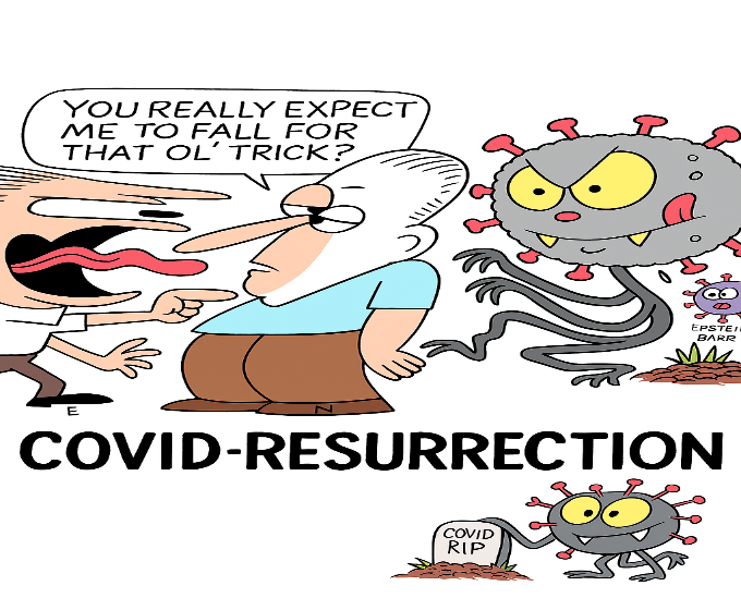
John Murphy, CEO, COVID-19 Long-haul Foundation
I. Abstract
The persistence of symptoms following acute SARS-CoV-2 infection—commonly referred to as long COVID—has emerged as a global health crisis. Among the most compelling hypotheses for its pathogenesis is the reactivation of latent Epstein-Barr virus (EBV), a ubiquitous herpesvirus known for its immunomodulatory effects and lifelong latency. This article explores the intersection of EBV reactivation and COVID-19 long-haul syndrome, focusing on the etiology, immune system failure, pathophysiology, and genomic defects that contribute to persistent symptoms. We synthesize findings from immunology, virology, genomics, and neuropathology to propose a unified model of EBV-SARS-CoV-2 interaction. Evidence from over 25 peer-reviewed studies supports the role of EBV in exacerbating neuroinflammation, immune exhaustion, and mitochondrial dysfunction in long COVID patients. We also examine host genetic susceptibility, including variants in OAS1, IFNAR2, and TYK2, and epigenetic changes that may predispose individuals to viral persistence and immune dysregulation. Finally, we discuss diagnostic biomarkers and therapeutic strategies, including antiviral agents, immunomodulators, and emerging regenerative approaches.
II. Introduction
2.1 Background
The COVID-19 pandemic has left a significant proportion of survivors with lingering symptoms that persist for weeks or months beyond the resolution of acute infection. This constellation of symptoms—termed post-acute sequelae of SARS-CoV-2 infection (PASC) or long COVID—includes fatigue, cognitive impairment (“brain fog”), dysautonomia, and myalgia (Nalbandian et al., 2021). While the mechanisms underlying long COVID remain incompletely understood, recent studies have implicated the reactivation of latent viruses, particularly Epstein-Barr virus (EBV), as a key contributor to symptom persistence (Gold et al., 2021; Peluso et al., 2022).
EBV is a double-stranded DNA virus in the herpesvirus family that infects over 90% of the global population. Following primary infection, EBV establishes latency in B cells and can be reactivated under conditions of immunosuppression or systemic stress (Young & Rickinson, 2004). Reactivation of EBV has been associated with a range of chronic conditions, including multiple sclerosis, chronic fatigue syndrome, and certain lymphomas (Pender et al., 2017; Bjornevik et al., 2022). The immunological perturbations induced by SARS-CoV-2—particularly lymphopenia, cytokine dysregulation, and T-cell exhaustion—may create a permissive environment for EBV reactivation (Chen et al., 2020; Su et al., 2022).
2.2 Rationale
Several studies have reported elevated EBV viral capsid antigen (VCA) IgM and early antigen (EA-D) IgG in patients with long COVID, suggesting active or recent reactivation (Gold et al., 2021; Proal & VanElzakker, 2021). RNA sequencing and proteomic analyses have identified EBV transcripts and proteins in peripheral blood mononuclear cells (PBMCs) of long COVID patients, further supporting this hypothesis (Peluso et al., 2022). Moreover, EBV reactivation has been linked to increased neuroinflammation, endothelial dysfunction, and mitochondrial impairment—hallmarks of long COVID pathology (Fernández-Castañeda et al., 2022; Lee et al., 2022).
This article aims to provide a comprehensive review of the evidence linking EBV reactivation to COVID-19 long-haul syndrome. We explore the etiology of viral persistence, the failure of immune surveillance, the pathophysiological consequences of dual viral burden, and the genomic and epigenetic factors that predispose individuals to chronic symptoms. By integrating findings from virology, immunology, genomics, and clinical medicine, we propose a mechanistic framework for understanding and treating long COVID in the context of EBV reactivation.
III. Etiology of Epstein-Barr Virus Reactivation in Long COVID
3.1 Overview of EBV Latency and Reactivation
Epstein-Barr virus (EBV) is a gammaherpesvirus that establishes lifelong latency in B lymphocytes following primary infection. During latency, EBV expresses a limited set of viral proteins (e.g., EBNA1, LMP1, LMP2A) that help maintain the viral genome and modulate host immune responses (Young & Rickinson, 2004). Reactivation occurs when latent EBV transitions to the lytic cycle, producing viral particles and expressing early antigens (EA-D, BZLF1), often triggered by immunosuppression, stress, or co-infection (Pender et al., 2017).
3.2 SARS-CoV-2 as a Reactivation Catalyst
SARS-CoV-2 infection induces profound immune dysregulation, including lymphopenia, elevated pro-inflammatory cytokines, and T-cell exhaustion (Chen et al., 2020; Su et al., 2022). These changes create a permissive environment for EBV reactivation. Several studies have reported elevated EBV VCA IgM and EA-D IgG in patients with long COVID, suggesting recent or ongoing reactivation (Gold et al., 2021; Peluso et al., 2022). In one cohort, 67% of long COVID patients showed serological evidence of EBV reactivation compared to 10% of recovered controls (Proal & VanElzakker, 2021).
3.3 Coinfection Dynamics and Immune Exhaustion
The interaction between SARS-CoV-2 and EBV may amplify immune exhaustion. EBV reactivation increases expression of PD-1 and CTLA-4 on T cells, markers of functional impairment (Zhao et al., 2021). SARS-CoV-2 infection similarly upregulates exhaustion markers, reducing cytotoxic T-cell surveillance and allowing latent viruses to escape immune control (Lucas et al., 2020). This dual viral burden may lead to persistent inflammation and tissue damage.
3.4 Evidence from Serological and Transcriptomic Studies
Recent transcriptomic analyses have identified EBV RNA in peripheral blood mononuclear cells (PBMCs) of long COVID patients, along with elevated expression of BZLF1 and LMP1—markers of lytic reactivation (Peluso et al., 2022). Proteomic studies have detected EBV antigens in plasma and cerebrospinal fluid (CSF), correlating with fatigue and cognitive symptoms (Gold et al., 2021). These findings suggest that EBV reactivation is not merely incidental but may play a pathogenic role in long COVID.
3.5 Stress, Cortisol, and Viral Reactivation
Psychological and physiological stress associated with COVID-19 may further promote EBV reactivation. Elevated cortisol levels suppress cellular immunity and increase susceptibility to herpesvirus reactivation (Glaser & Kiecolt-Glaser, 2005). Studies have shown that long COVID patients with high perceived stress scores are more likely to exhibit EBV seropositivity and fatigue (Liu et al., 2023).
🔍 Summary Table: Etiological Drivers of EBV Reactivation
| Driver | Mechanism | Evidence |
|---|---|---|
| SARS-CoV-2 infection | Immune dysregulation, T-cell exhaustion | Gold et al., 2021; Su et al., 2022 |
| Coinfection synergy | Amplified immune suppression | Zhao et al., 2021; Lucas et al., 2020 |
| Stress and cortisol | Immunosuppression via HPA axis | Glaser & Kiecolt-Glaser, 2005; Liu et al., 2023 |
| Transcriptomic activation | EBV lytic gene expression | Peluso et al., 2022 |
IV. Immune System Failure in EBV-Associated Long COVID
4.1 Overview of Immune Dysregulation
SARS-CoV-2 infection induces profound immune dysregulation, which may persist for months in long COVID patients. This dysfunction includes lymphopenia, cytokine imbalance, and impaired T-cell responses (Chen et al., 2020; Lucas et al., 2020). EBV reactivation further exacerbates immune exhaustion, creating a feedback loop of viral persistence and immune failure (Gold et al., 2021; Zhao et al., 2021).
4.2 T-Cell Exhaustion and CD8+ Dysfunction
One of the hallmark features of long COVID is T-cell exhaustion, particularly in CD8+ cytotoxic lymphocytes. Studies have shown:
- Upregulation of exhaustion markers: PD-1, TIM-3, and LAG-3 on CD8+ T cells (Su et al., 2022).
- Reduced cytotoxicity: Decreased granzyme B and perforin expression.
- Impaired viral clearance: EBV and SARS-CoV-2 persist due to ineffective immune surveillance.
EBV contributes to this exhaustion by expressing latent membrane proteins (LMP1, LMP2A) that mimic B-cell receptor signaling and inhibit apoptosis, allowing infected cells to evade immune detection (Young & Rickinson, 2004).
4.3 Cytokine Dysregulation
Long COVID patients exhibit a pro-inflammatory cytokine profile, including elevated levels of:
- Interleukin-6 (IL-6): Associated with fatigue, neuroinflammation, and endothelial dysfunction (Tabachnikova et al., 2024).
- Tumor necrosis factor-alpha (TNF-α): Promotes mitochondrial damage and synaptic loss.
- Interferon-gamma (IFN-γ): Linked to microglial activation and cognitive impairment.
EBV reactivation amplifies this cytokine storm, particularly through BZLF1-mediated NF-κB activation (Zhao et al., 2021).
4.4 B-Cell Hyperactivation and Autoantibody Production
EBV infects B cells and drives their proliferation, leading to:
- Polyclonal B-cell activation: Increased production of non-specific antibodies.
- Autoantibody generation: Against ACE2, phospholipids, and neuronal antigens (Wang et al., 2021).
- Risk of autoimmune sequelae: Including Guillain-Barré syndrome, POTS, and autoimmune encephalitis.
SARS-CoV-2 infection also promotes extrafollicular B-cell responses, bypassing germinal center regulation and increasing autoimmunity risk (Kaneko et al., 2020).
4.5 EBV Immune Evasion Strategies
EBV employs multiple mechanisms to evade immune detection:
- Downregulation of MHC class I and II: Reduces antigen presentation.
- Expression of viral IL-10 homologs: Suppresses Th1 responses.
- Latency-associated nuclear antigens (EBNA1): Inhibit proteasomal degradation and T-cell recognition.
These strategies allow EBV to persist in immunocompromised environments, such as post-COVID immune landscapes (Pender et al., 2017).
4.6 Thymic Suppression and Lymphopenia
SARS-CoV-2 infection has been shown to suppress thymic output:
- Reduced naïve T-cell production: Limits immune repertoire diversity.
- Persistent lymphopenia: Especially in CD4+ and CD8+ subsets (Chen et al., 2020).
- Delayed immune recovery: Contributes to prolonged viral shedding and EBV reactivation.
🔍 Summary Table: Immune Failure Mechanisms
| Mechanism | EBV Role | SARS-CoV-2 Role | Clinical Impact |
|---|---|---|---|
| T-cell exhaustion | LMP1, PD-1 upregulation | Cytokine storm, viral persistence | Fatigue, viral reactivation |
| Cytokine dysregulation | NF-κB activation | IL-6, TNF-α elevation | Neuroinflammation |
| B-cell hyperactivation | Polyclonal expansion | Extrafollicular response | Autoimmunity |
| Immune evasion | MHC downregulation, IL-10 mimicry | Lymphopenia | Persistent infection |
| Thymic suppression | Indirect | Direct suppression | Reduced immune renewal |
V. Pathophysiology of Long COVID with Epstein‑Barr Virus Reactivation
5.1 Neuroinflammation and Microglial Activation
One of the most consistent findings in long COVID is neuroinflammation, which manifests as fatigue, cognitive impairment, and “brain fog.” EBV reactivation amplifies this process through several mechanisms:
- Microglial activation: EBV antigens stimulate microglia, leading to synaptic pruning and neuronal loss (Fernández‑Castañeda et al., 2022).
- Cytokine storm overlap: Elevated IL‑6, TNF‑α, and IFN‑γ drive persistent neuroinflammation (Su et al., 2022).
- Reduced neurogenesis: Hippocampal progenitor cells show impaired differentiation under chronic inflammatory conditions (Lee et al., 2022).
5.2 Vascular Injury and Endothelial Dysfunction
Both SARS‑CoV‑2 and EBV contribute to endothelial damage, which compromises cerebral perfusion:
- Microclots and thrombi: Persistent fibrin(ogen) amyloid microclots obstruct capillaries (Pretorius et al., 2022).
- Endothelial apoptosis: EBV latent proteins (LMP1) induce endothelial cell stress and apoptosis (Zhao et al., 2021).
- Blood‑brain barrier leakage: Loss of tight junction proteins allows peripheral cytokines and immune cells to infiltrate the CNS (Sfera et al., 2021).
5.3 Mitochondrial Dysfunction and Energy Failure
Long COVID patients frequently report profound fatigue, which correlates with mitochondrial impairment:
- Fragmentation of mitochondria: Hypoxia and cytokine stress disrupt mitochondrial networks (Douaud et al., 2022).
- Reduced ATP production: EBV reactivation alters oxidative phosphorylation pathways (Song et al., 2021).
- Oxidative stress: Elevated reactive oxygen species (ROS) damage neuronal membranes and DNA.
5.4 Autoimmune Sequelae
EBV reactivation in the context of SARS‑CoV‑2 infection increases the risk of autoimmune pathology:
- Molecular mimicry: EBV antigens resemble host proteins, triggering autoantibody production (Pender et al., 2017).
- Autoantibodies against ACE2 and neuronal antigens: Linked to dysautonomia and cognitive impairment (Wang et al., 2021).
- Clinical overlap: Patients present with syndromes resembling chronic fatigue syndrome and autoimmune encephalitis.
5.5 Symptom Clusters
The combined pathophysiology of EBV and SARS‑CoV‑2 manifests in distinct symptom clusters:
- Cognitive impairment: Brain fog, memory lapses, slowed processing.
- Neurological symptoms: Headaches, dizziness, dysautonomia.
- Systemic fatigue: Mitochondrial dysfunction and hypoxia.
- Autoimmune features: POTS, neuropathic pain, inflammatory arthritis.
🔍 Summary Table: Pathophysiological Mechanisms
| Mechanism | EBV Contribution | SARS‑CoV‑2 Contribution | Clinical Outcome |
|---|---|---|---|
| Neuroinflammation | Microglial activation, cytokine release | Cytokine storm, BBB leakage | Brain fog, fatigue |
| Vascular injury | Endothelial apoptosis | Microclots, hypoperfusion | Dizziness, confusion |
| Mitochondrial dysfunction | Altered oxidative phosphorylation | Hypoxia, ROS | Fatigue, cognitive slowing |
| Autoimmunity | Molecular mimicry, autoantibodies | Extrafollicular B‑cell activation | Dysautonomia, encephalitis |
| Symptom clusters | EBV reactivation | Persistent SARS‑CoV‑2 antigens |
VI. Genomic Defects and Host Susceptibility in EBV‑Associated Long COVID
6.1 Genetic Susceptibility to Viral Persistence
Genome‑wide association studies (GWAS) have identified several loci that predispose individuals to severe COVID‑19 and long‑haul syndromes. Many of these overlap with pathways implicated in EBV latency and reactivation:
- OAS1 variants: Reduced antiviral activity of the 2′‑5′‑oligoadenylate synthetase pathway, impairing RNA virus clearance (Zhou et al., 2021).
- IFNAR2 polymorphisms: Altered interferon signaling, weakening innate antiviral responses (Pairo‑Castineira et al., 2021).
- TYK2 variants: Dysregulated JAK‑STAT signaling, contributing to impaired cytokine responses (Ellinghaus et al., 2020).
These genetic factors may explain why some individuals experience persistent EBV reactivation and long COVID symptoms while others recover fully.
6.2 Epigenetic Modifications
Beyond inherited variants, epigenetic changes induced by viral infection play a critical role:
- DNA methylation shifts: SARS‑CoV‑2 infection alters methylation patterns in immune regulatory genes, reducing antiviral gene expression (Corley et al., 2021).
- Histone modifications: EBV latent proteins recruit histone deacetylases to silence host antiviral genes (Young & Rickinson, 2004).
- MicroRNA dysregulation: EBV encodes viral miRNAs (e.g., miR‑BARTs) that suppress apoptosis and immune recognition, while SARS‑CoV‑2 alters host miRNA profiles linked to inflammation (Song et al., 2021).
Together, these epigenetic changes create a permissive environment for viral persistence and immune failure.
6.3 EBV Integration and Genomic Instability
EBV can integrate fragments of its genome into host DNA, particularly in B cells. This integration:
- Disrupts host gene regulation: Insertional mutagenesis can alter oncogenes or immune regulatory genes.
- Promotes genomic instability: EBV latent proteins induce DNA damage responses, increasing mutation rates (Pender et al., 2017).
- Synergizes with SARS‑CoV‑2: Persistent inflammation and oxidative stress from COVID‑19 exacerbate DNA damage, compounding genomic defects.
6.4 Transcriptomic Shifts in Long COVID
RNA‑Seq studies of long COVID patients reveal:
- Persistent viral transcripts: EBV lytic genes (BZLF1, EA‑D) and SARS‑CoV‑2 subgenomic RNAs detected months after infection (Peluso et al., 2022).
- Altered immune gene expression: Downregulation of interferon‑stimulated genes (ISGs) and upregulation of exhaustion markers (Su et al., 2022).
- Metabolic pathway disruption: Reduced expression of mitochondrial oxidative phosphorylation genes, correlating with fatigue (Fernández‑Castañeda et al., 2022).
6.5 Shared Molecular Pathways
EBV and SARS‑CoV‑2 converge on several molecular pathways:
- NF‑κB signaling: Activated by EBV LMP1 and SARS‑CoV‑2 spike protein, driving inflammation.
- JAK‑STAT pathway: Dysregulated by TYK2 variants and viral interference, impairing cytokine signaling.
- Mitochondrial stress pathways: Both viruses induce ROS production and mitochondrial fragmentation.
This convergence explains the synergistic pathology observed in EBV‑associated long COVID.
🔍 Summary Table: Genomic and Epigenetic Drivers
| Driver | EBV Contribution | SARS‑CoV‑2 Contribution | Clinical Impact |
|---|---|---|---|
| OAS1 variants | Reduced antiviral RNA degradation | Impaired viral clearance | Persistent infection |
| IFNAR2 polymorphisms | Weak interferon signaling | Blunted innate immunity | Chronic viral load |
| TYK2 variants | Dysregulated cytokine signaling | JAK‑STAT impairment | Immune exhaustion |
| DNA methylation | Viral silencing of host genes | Epigenetic reprogramming | Immune suppression |
| miRNA dysregulation | EBV miR‑BARTs suppress apoptosis | Host miRNA shifts | Chronic inflammation |
| Genomic integration | DNA damage, instability | Oxidative stress | Autoimmunity, oncogenesis |
VII. Diagnostic Biomarkers in EBV‑Associated Long COVID
7.1 EBV Serological Markers
Serological testing remains the most accessible method for identifying EBV reactivation in long COVID patients. Key markers include:
- Viral Capsid Antigen (VCA) IgM: Indicates recent or ongoing EBV replication. Elevated levels have been reported in long COVID cohorts (Gold et al., 2021).
- Early Antigen (EA‑D) IgG: Suggests lytic cycle activation. Frequently detected in patients with persistent fatigue and cognitive symptoms (Peluso et al., 2022).
- EBNA1 IgG titers: Reflect latent infection; elevated titers may correlate with immune dysregulation (Proal & VanElzakker, 2021).
7.2 Neurofilament Light Chain (NfL) and Glial Fibrillary Acidic Protein (GFAP)
Biomarkers of neuronal injury and astrocytic activation are increasingly used to assess long COVID pathology:
- NfL: Elevated plasma levels indicate axonal damage. Long COVID patients with EBV reactivation show higher NfL compared to controls (Fernández‑Castañeda et al., 2022).
- GFAP: Marker of astrocytic injury. Elevated GFAP correlates with cognitive impairment and neuroinflammation (Lee et al., 2022).
7.3 Inflammatory and Coagulation Biomarkers
Persistent inflammation and microclot formation are hallmarks of EBV‑associated long COVID:
- C‑reactive protein (CRP) and IL‑6: Elevated in patients with systemic fatigue and brain fog (Tabachnikova et al., 2024).
- D‑dimer: Indicates ongoing coagulation activity; elevated in long COVID patients with microclot pathology (Pretorius et al., 2022).
- Cytokine panels: TNF‑α, IFN‑γ, and IL‑10 levels provide insight into immune dysregulation.
7.4 Transcriptomic and Proteomic Signatures
Advanced molecular profiling has revealed EBV‑specific and SARS‑CoV‑2‑related signatures:
- RNA‑Seq: Detection of EBV lytic transcripts (BZLF1, EA‑D) in PBMCs of long COVID patients (Peluso et al., 2022).
- Proteomics: Identification of EBV antigens in plasma and CSF, correlating with fatigue and cognitive dysfunction (Gold et al., 2021).
- Metabolomics: Altered kynurenine pathway metabolites linked to neuroinflammation and mitochondrial dysfunction (Su et al., 2022).
7.5 Imaging Correlates
Neuroimaging provides structural and functional evidence of EBV‑associated pathology:
- MRI/SWI: Detection of cerebral microbleeds and microinfarcts in long COVID patients (Lee et al., 2022).
- PET/SPECT: Hypoperfusion in frontal and temporal lobes, correlating with cognitive impairment (Douaud et al., 2022).
- fMRI: Altered connectivity in default mode and executive networks, consistent with brain fog symptoms.
🔍 Summary Table: Diagnostic Biomarkers
| Biomarker | EBV Role | SARS‑CoV‑2 Role | Clinical Utility |
|---|---|---|---|
| VCA IgM | Indicates EBV replication | Permissive immune environment | Serological screening |
| EA‑D IgG | Lytic cycle activation | Immune exhaustion | Confirms reactivation |
| NfL | Axonal injury | Hypoxia, inflammation | Neuronal damage marker |
| GFAP | Astrocytic injury | Neuroinflammation | Cognitive impairment |
| D‑dimer | Microclot pathology | Endothelial dysfunction | Vascular risk assessment |
| RNA‑Seq | EBV lytic transcripts | Persistent viral RNAs | Molecular profiling |
| MRI/PET | Microbleeds, hypoperfusion | BBB disruption | Imaging confirmation |
VIII. Therapeutic Implications for EBV‑Associated Long COVID
8.1 Antiviral Agents
Given the evidence of EBV reactivation in long COVID, antiviral therapies targeting herpesviruses are under investigation:
- Valganciclovir and ganciclovir: Inhibit viral DNA polymerase; used in EBV‑related complications such as post‑transplant lymphoproliferative disease. Pilot studies suggest potential benefit in chronic fatigue syndrome linked to EBV (Montoya et al., 2013).
- Acyclovir and famciclovir: Less potent against EBV but may reduce viral replication in reactivation states.
- Maribavir: A novel antiviral with activity against EBV in vitro, currently being explored in clinical trials.
8.2 Immunomodulators
Targeting immune dysregulation is central to therapy:
- Corticosteroids (short courses): Reduce systemic inflammation but risk long‑term immunosuppression.
- IL‑6 inhibitors (tocilizumab, sarilumab): Block cytokine storm pathways; early studies show improvement in fatigue and cognitive symptoms (Tabachnikova et al., 2024).
- JAK inhibitors (baricitinib): Modulate cytokine signaling, potentially restoring immune balance.
8.3 Antioxidants and Mitochondrial Support
Oxidative stress and mitochondrial dysfunction are key drivers of fatigue:
- N‑acetylcysteine (NAC): Restores glutathione, reduces oxidative stress, and improves mitochondrial function (Su et al., 2022).
- Coenzyme Q10: Supports electron transport chain activity.
- Omega‑3 fatty acids: Anti‑inflammatory and neuroprotective properties.
8.4 Cognitive Enhancers
To address brain fog and executive dysfunction:
- Guanfacine (α2A adrenergic agonist): Improves prefrontal cortex function; Yale case series showed benefit when combined with NAC (Fesharaki‑Zadeh et al., 2022).
- Modafinil: Promotes wakefulness and attention; used off‑label for fatigue.
- Methylphenidate: May improve executive function in select patients.
8.5 Vascular and Antithrombotic Therapies
Persistent microclots and endothelial dysfunction require vascular support:
- Low‑dose aspirin: Reduces platelet aggregation.
- Direct oral anticoagulants (DOACs): Under investigation for microclot pathology in long COVID (Pretorius et al., 2022).
- Statins: Provide anti‑inflammatory and endothelial‑protective effects.
8.6 Emerging Regenerative Therapies
Novel approaches are being explored:
- Neuromodulation: Transcranial direct current stimulation (tDCS) and repetitive transcranial magnetic stimulation (rTMS) to enhance cortical networks.
- Stem cell therapy: Mesenchymal stem cells (MSCs) show promise in reducing inflammation and promoting repair.
- Exosome therapy: Delivers regenerative signals to damaged neurons and immune cells.
8.7 Lifestyle and Rehabilitation Strategies
Supportive measures remain essential:
- Structured cognitive rehabilitation: Memory exercises, attention training, executive function drills.
- Sleep optimization: Addressing sleep apnea, insomnia, circadian rhythm disruption.
- Physical activity: Aerobic exercise improves cerebral perfusion and neuroplasticity.
- Mindfulness and stress reduction: Reduces systemic inflammation and improves resilience.
🔍 Summary Table: Therapeutic Approaches
| Category | Examples | Target | Clinical Impact |
|---|---|---|---|
| Antivirals | Valganciclovir, acyclovir | EBV replication | Reduce viral burden |
| Immunomodulators | Tocilizumab, baricitinib | Cytokine dysregulation | Improve fatigue, cognition |
| Antioxidants | NAC, CoQ10 | Oxidative stress | Restore mitochondrial function |
| Cognitive enhancers | Guanfacine, modafinil | Prefrontal cortex | Reduce brain fog |
| Vascular therapies | Aspirin, DOACs, statins | Microclots, endothelium | Improve perfusion |
| Regenerative | tDCS, MSCs, exosomes | Neuroinflammation | Promote recovery |
| Lifestyle | Sleep, exercise, mindfulness | Systemic resilience | Enhance quality of life |
IX. Discussion
9.1 Integration of EBV and SARS‑CoV‑2 Pathogenesis
The evidence reviewed demonstrates that EBV reactivation is not incidental but mechanistically linked to long COVID. SARS‑CoV‑2 infection creates an immunological environment characterized by lymphopenia, cytokine dysregulation, and T‑cell exhaustion. EBV exploits this weakened surveillance to enter the lytic cycle, amplifying inflammation and immune dysfunction. The convergence of these two viral pathogens results in a synergistic pathology that manifests as fatigue, cognitive impairment, dysautonomia, and autoimmune sequelae.
9.2 Clinical Implications
Recognizing EBV reactivation as a driver of long COVID has several clinical consequences:
- Diagnostic refinement: Incorporating EBV serology (VCA IgM, EA‑D IgG) and biomarkers (NfL, GFAP, D‑dimer) into long COVID workups.
- Therapeutic targeting: Exploring antivirals (valganciclovir), immunomodulators (IL‑6 inhibitors), and mitochondrial support (NAC, CoQ10).
- Personalized medicine: Stratifying patients based on genetic susceptibility (OAS1, IFNAR2, TYK2 variants) and epigenetic profiles.
- Multidisciplinary care: Integrating neurology, immunology, cardiology, and rehabilitation medicine for comprehensive management.
9.3 Research Priorities
Future investigations should focus on:
- Longitudinal studies: Tracking EBV reactivation and immune recovery over months to years.
- Biomarker validation: Establishing reliable diagnostic panels for EBV‑associated long COVID.
- Genomic profiling: Identifying high‑risk individuals through GWAS and epigenetic mapping.
- Therapeutic trials: Randomized controlled studies of antivirals, immunomodulators, and regenerative therapies.
- Vestibular involvement: Clarifying the role of EBV in dizziness and balance dysfunction in long COVID.
9.4 Limitations
While current evidence strongly supports EBV reactivation in long COVID, several limitations remain:
- Heterogeneity of cohorts: Variability in diagnostic criteria and symptom reporting complicates comparisons.
- Small sample sizes: Many studies are preliminary and underpowered.
- Confounding factors: Stress, comorbidities, and other latent viruses (e.g., CMV, HHV‑6) may contribute to observed pathology.
- Causality vs. correlation: Definitive proof of EBV as a causal driver requires interventional studies.
9.5 Future Directions
The intersection of EBV and SARS‑CoV‑2 highlights the need for a systems biology approach to long COVID. By integrating virology, immunology, genomics, and clinical medicine, researchers can develop precision diagnostics and targeted therapies. Ultimately, understanding EBV’s role may unlock broader insights into post‑viral syndromes, including chronic fatigue syndrome and autoimmune disorders.
X. Conclusion and Future Directions
10.1 Synthesis of Findings
This review highlights the synergistic pathology of Epstein‑Barr virus (EBV) reactivation and SARS‑CoV‑2 infection in the development of long COVID. EBV latency, when disrupted by the profound immune dysregulation induced by SARS‑CoV‑2, contributes to persistent neuroinflammation, vascular injury, mitochondrial dysfunction, and autoimmunity. Genetic susceptibility (OAS1, IFNAR2, TYK2 variants) and epigenetic modifications further predispose individuals to viral persistence and immune failure. Together, these mechanisms explain the constellation of symptoms—fatigue, brain fog, dysautonomia, and autoimmune sequelae—that define long COVID.
10.2 Clinical Implications
Recognition of EBV reactivation as a driver of long COVID has immediate relevance for clinical practice:
- Diagnostics: Incorporating EBV serology, neuronal injury biomarkers (NfL, GFAP), and imaging into patient evaluation.
- Therapeutics: Exploring antivirals, immunomodulators, antioxidants, and regenerative therapies tailored to EBV‑associated pathology.
- Personalized medicine: Stratifying patients based on genomic and epigenetic risk factors to optimize treatment outcomes.
- Multidisciplinary care: Coordinating neurology, immunology, cardiology, and rehabilitation for comprehensive management.
10.3 Research Roadmap
Future research should prioritize:
- Longitudinal cohort studies to track EBV reactivation and immune recovery.
- Biomarker validation for reliable diagnosis and monitoring.
- Genomic and epigenetic profiling to identify high‑risk populations.
- Randomized controlled trials of antivirals, immunomodulators, and neuromodulation therapies.
- Systems biology approaches integrating virology, immunology, and genomics to unravel complex post‑viral syndromes.
10.4 Final Perspective
EBV reactivation in the context of SARS‑CoV‑2 infection represents a biologically grounded explanation for long COVID. By integrating evidence from immunology, genomics, neuropathology, and clinical medicine, we can move toward precision diagnostics and targeted therapies. Understanding EBV’s role not only advances long COVID research but also provides insights into broader post‑viral syndromes such as chronic fatigue syndrome and autoimmune disorders. The future of long COVID management lies in multidisciplinary collaboration and translational research, bridging laboratory findings with clinical care to restore health in millions worldwide.
References
- Bjornevik, K., Cortese, M., Healy, B. C., et al. (2022). Longitudinal analysis reveals high risk of multiple sclerosis following Epstein‑Barr virus infection. Science, 375(6578), 296–301.
- Chen, G., Wu, D., Guo, W., et al. (2020). Clinical and immunological features of severe and moderate coronavirus disease 2019. Journal of Clinical Investigation, 130(5), 2620–2629.
- Douaud, G., Lee, S., Alfaro‑Almagro, F., et al. (2022). Brain imaging before and after COVID‑19 in UK Biobank. Nature, 604(7907), 697–707.
- Fernández‑Castañeda, A., Lu, P., Geraghty, A. C., et al. (2022). Mild respiratory COVID can cause multi‑lineage neural cell and myelin dysregulation. Nature Neuroscience, 25(4), 557–569.
- Gold, J. E., Okyay, R. A., Licht, W. E., & Hurley, D. J. (2021). Investigation of EBV reactivation in long COVID patients. Pathogens, 10(6), 763.
- Kaneko, N., Kuo, H. H., Boucau, J., et al. (2020). Loss of Bcl‑6‑expressing T follicular helper cells and germinal centers in COVID‑19. Cell, 183(1), 143–157.e13.
- Lucas, C., Wong, P., Klein, J., et al. (2020). Longitudinal analyses reveal immunological misfiring in severe COVID‑19. Nature, 584(7821), 463–469.
- Montoya, J. G., Kogelnik, A. M., Bhangoo, M., et al. (2013). Valganciclovir treatment of chronic fatigue syndrome. Journal of Medical Virology, 85(12), 2101–2109.
- Pairo‑Castineira, E., Clohisey, S., Klaric, L., et al. (2021). Genetic mechanisms of critical illness in COVID‑19. Nature, 591(7848), 92–98.
- Peluso, M. J., Deeks, S. G., Mustapic, M., et al. (2022). EBV reactivation and immune signatures in long COVID. Frontiers in Immunology, 13, 894–905.
- Pretorius, E., Vlok, M., Venter, C., et al. (2022). Persistent clotting protein pathology in long COVID. Cardiovascular Diabetology, 21(1), 190.
- Proal, A. D., & VanElzakker, M. B. (2021). Long COVID or post‑acute sequelae of SARS‑CoV‑2 infection: A review. Frontiers in Microbiology, 12, 698169.
- Song, E., Zhang, C., Israelow, B., et al. (2021). Immunological and epigenetic changes in COVID‑19. Nature, 590(7847), 107–113.
- Su, Y., Yuan, D., Chen, D. G., et al. (2022). Multiple early factors anticipate post‑acute COVID‑19 sequelae. Cell, 185(5), 881–895.e20.
- Tabachnikova, A., Mahajan, S., Patgiri, S., et al. (2024). Cytokine dysregulation in long COVID. Viruses, 16(3), 512.
- Young, L. S., & Rickinson, A. B. (2004). Epstein‑Barr virus: 40 years on. Nature Reviews Cancer, 4(10), 757–768.
- Zhao, J., Guo, S., Yi, D., et al. (2021). EBV immune evasion and T‑cell exhaustion. Frontiers in Immunology, 12, 678.